Introduction
Small-scale mechanical testing (SSMT) at the micro- and nanoscales has become an integral part of materials science and is widely used as a tool to evaluate either a material’s mechanical properties changes due to microstructural alterations or to obtain new insights about its physics. It has become common to utilize small-scale mechanical testing even for engineering applications, for evaluating coatings, irradiated samples, individual grains, welds, and joints, as examples.
For better engineering performance and to better understand materials behavior, there is a currently a push to test materials under in operando conditions, such as temperature, high strain rate, or hydrogen environment to assess property changes of the materials under these environments. In addition, expanding the testing conditions to these areas allows new insights to be gleaned into multidimensional environmental space. This article adumbrates current efforts in using small-scale mechanical testing in extreme environments to push the envelope on what is possible using SSMT.
High and low temperatures
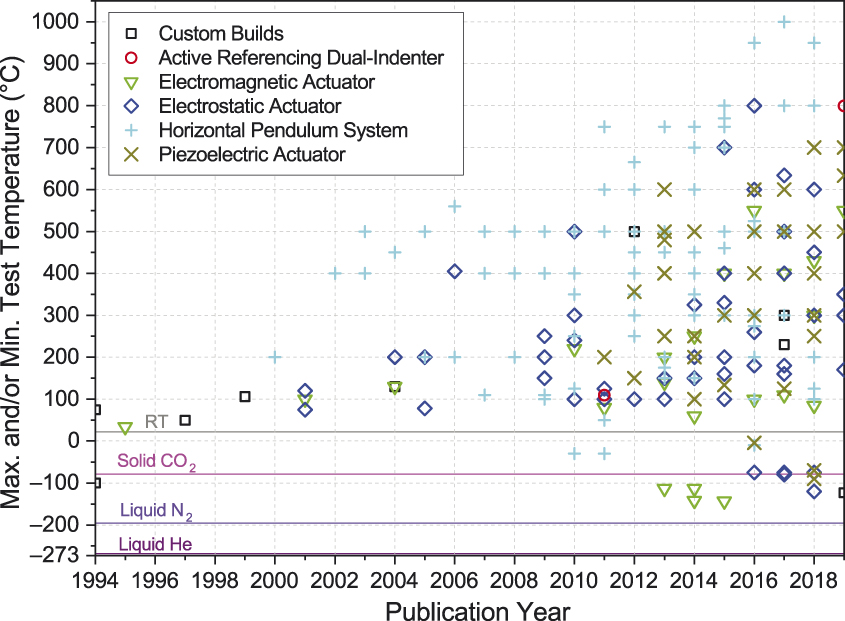
Variable temperature nanoindentation is one of the more developed capabilities in the area of small-scale mechanical testing under extreme conditions. As illustrated in Figure 1,1,Reference Wheeler, Armstrong, Heinz and Schwaiger2 several commercially available nanoindentation systems have been brought to the market in the last few decades, which are capable of operating at variable temperatures from approximately–140Reference Lee, Jafary-Zadeh, Chen, Zhang and Greer3 up to 1000°C,Reference Beake and Smith4–Reference Gibson, Schröders, Zehnder and Korte-Kerzel9 including in situ in the scanning electron microscope.Reference Wheeler and Michler10–Reference Smolka, Motz, Detzel, Robl and Griesser12 These developments offer new measurement capabilities that enrich our scientific understanding of fundamental material response, including strain rate and temperature effects on deformation mechanisms at the micro- and nanoscale.Reference Prasitthipayong, Vachhani, Tumey, Minor and Hosemann13 This allows testing relevant technological materials under real operating conditions, widening the applicability of these techniques to important industrial applications, such as the hard-coating industry, high-temperature metallic alloys, and corrosion layers.Reference Monclús and Molina-Aldareguia14,Reference Rebelo de Figueiredo, Abad, Harris, Czettl, Mitterer and Hosemann15
Maximum or minimum published small-scale mechanical testing temperatures for various system configurations in recent years.1,Reference Wheeler, Armstrong, Heinz and Schwaiger2

The major difficulties faced when testing under variable temperature conditions include instrument stability, accounting for thermal gradients, management of thermal drift, and chemical stability, not only of the specimen itself, but also of the indenter tip material. Several recent reviews discuss these issues in detail.Reference Wheeler, Armstrong, Heinz and Schwaiger2,Reference Monclús and Molina-Aldareguia14 The key developments that have allowed pushing the temperature limits are the heating/cooling of the indenter tip and the specimen to ensure thermal equilibrium at the contactReference Everitt, Davies and Smith16 and the development of more advanced environmental control, either by enclosing the instrument in a purging chamber filled with argon or by using a high-vacuum chamber, as is found in situ within an electron microscope. Measurement temperatures as high as 1000°C have been reported in the case of standalone high-temperature, vacuum indentation instruments.Reference Harris, Beake, Armstrong and Davies8,Reference Gibson, Schröders, Zehnder and Korte-Kerzel9
However, variable temperature nanoindentation is far from being a well-established technique and further developments are required to increase reliability and reproducibility and to facilitate the widespread use of the technique. More automated approaches to continuously monitor and correct for thermal drift, and more appropriate indenter tip materials are still needed. Diamond is the standard material choice for the tip and can be safely used up to around 550°C in vacuum. At temperatures above 550°C, diamond starts to oxidize; it can also react with carbide-forming metals, such as steels, Ti or Co.Reference Wheeler and Michler17
As an alternative to diamond, cubic boron nitride (cBN) can be used, but its hardness decreases quickly with temperature, and it can react with some elements at high temperatures to form more stable borides.Reference Wheeler, Armstrong, Heinz and Schwaiger2 cBN is also nonconductive, which limits its use in the electron microscope. Nevertheless, even if more stable indenter tip materials are developed, the harsh conditions encountered at the indentation contact will inevitably lead to quick blunting of the tip due to tip-sample reactions or deposition of sample material,Reference Monclús, Lotfian and Molina-Aldareguía18 limiting the lifetime of the expensive indenter tips to performing just a few indents.
Future developments thus call for dramatically new approaches, where the high-temperature nanoindentation indenters can be mass produced, such as using lithography techniques, and treated as affordable consumables that can be thrown away and exchanged automatically after performing just a few indents. Alternative geometries may be necessary for extreme temperatures—spherical or flat punch/microcompression geometries to reduce the influence of blunting or using mutual indentation approaches (crossed cylinders or wedges of the same material) to prevent reactions between dissimilar tip/sample materials.
While high- and low-temperature capabilities for SSMT are now established and growing, the larger materials science community is still limited to commercial systems restricting testing to ambient conditions. The remaining instrumental difficulties will still need to be addressed in order to make variable temperature testing gain widespread usage. Without a doubt, this new tool in the materials science toolbox will provide many valuable insights into materials physics and engineering applications for years to come.
High strain rates
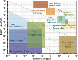
High strain rates are commonplace in everyday life. High rates can occur simply from the impact of dropping an object onto a hard surface, a ballistic impact from a projectile, or during an automobile crash. In order to design structures resistant to these occurrences, it is important to understand the deformation behavior of materials at these high strain rates. Conventionally, this is accomplished at large scales using high-speed universal testing frames for dynamic tension testingReference Dalla Torre, Van Swygenhoven and Victoria19 or a Kolsky (Split Hopkinson) bar apparatus,Reference Ramesh and Sharpe20 which uses pressure waves to measure deformation at strain rates from 102 to 103 s–1 (Figure 2). Higher rates can be accessed by using a miniaturized Kolsky bar, dynamic/ballistic indentationReference Tirupataiah and Sundararajan21 or a pressure-shear plate impact test.Reference Klopp, Clifton and Shawki22 However, in order to study the high strain-rate behavior of individual microstructural features, there is considerable interest in developing micromechanical means of high strain-rate testing.
Schematic strain rate and sample length-scale relationships for various mechanical testing techniques.Reference Dalla Torre, Van Swygenhoven and Victoria19–Reference Klopp, Clifton and Shawki22,Reference Constantinides, Tweedie, Savva, Smith and Vliet26,Reference Sudharshan Phani and Oliver28–Reference Hassani-Gangaraj, Veysset, Nelson and Schuh30

The primary technique used for small-scale strain-rate investigation is nanoindentation. The three-sided, Berkovich pyramidal geometry typically used for nanoindentation has variable strain-rate testing intrinsically incorporated, due to the increasing contact area with increased penetration depth. Maintaining a constant strain rate during nanoindentation requires a proportionally increasing displacement or loading rate:  $\dot \varepsilon = \frac{{\dot h}}{h} \approx \frac{{\dot p}}{{2p}}$, where ε is strain, h is displacement, and P is load.Reference Cheng and Cheng23 Using this relationship, indentation has been widely used to probe strain-rate sensitivity using monotonicReference Schwaiger, Moser, Dao, Chollacoop and Suresh24 (single strain rate) and jumpReference Maier, Mueller, Backes, Höppel and Göken25 tests (multiple strain rates during a single test). The typical range of strain rates interrogated using these tests is 10–3 s–1 up to 101 s–1. Even lower strain-rate regimes can be probed using indentation creep tests, where the load is kept constant while the slowly increasing penetration depth is measured. Together, this range of strain rates, while quite wide already, is primarily limited by the system’s mechanical design and data acquisition/control electronics.
To probe higher strain rates with nanoindentation, a few systems have been developed. MicroMaterials Ltd.Reference Constantinides, Tweedie, Savva, Smith and Vliet26 has developed a nanoimpact system where the indenter is accelerated to perform ballistic impact on the surface of a sample, achieving very high strain ratesReference Zehnder, Peltzer, Gibson and Korte-Kerzel27 during the initial penetration and being rapidly decelerated during the indentation. A similar test, using step loading to accelerate the indenter, has been developed by Nanomechanics Inc./KLA Tencor,Reference Sudharshan Phani and Oliver28 allowing hardness measurements at very high strain rates (4000 s–1) by accounting for the dynamic behavior of the system.
Another technique from Alemnis AG,Reference Guillonneau, Mieszala, Wehrs, Schwiedrzik, Grop, Frey, Philippe, Breguet, Michler and Wheeler29 using high-speed piezoelectric transducers, allows rapid displacement and load sensing to be achieved in both indentation and microcompression conditions over a wide range of strain rates (10-4–103 s–1). This has the benefit over ballistic impact of producing controlled levels of displacement at the specified rates. For example, a micropillar could be precisely deformed to 2% strain and then sectioned for later investigation by transmission electron microscopy.
A third strategy is also being pursued to allow investigation of materials properties at the highest rates of 107–109 s–1. This involves laser-induced particle impact testing (LIPIT), where a ∼10–50-µm particle of a material is rapidly accelerated to speeds on the order of 100–1000 m/s by laser-induced expansion of a thin film.Reference Hassani-Gangaraj, Veysset, Nelson and Schuh30 The particle then impacts locally on a target substrate, producing an indentation or an adhering splat. The incident and rebound velocities are measured by a high-speed camera, allowing for the work of indentation to be determined from the impression volume and coefficient of restitution similarly to the ballistic techniques previously described.Reference Zehnder, Peltzer, Gibson and Korte-Kerzel27
Together, all of these techniques have greatly extended the range of strain rates accessible by SSMT to ranges similar to, and in some cases higher than, their large-scale counterparts. Combining variable strain-rate methods with high-/low-temperature methods from the previous sections creates enormous potential to probe material plasticity and activation behavior,Reference Wheeler, Armstrong, Heinz and Schwaiger2 and some researchersReference Somekawa and Schuh31 have already begun extending this to the high-strain-rate regimes. Future efforts are expected to allow routine SSMT at even higher strain rates and bridge the gaps with conventional testing to achieve a holistic understanding of material deformation over a wide, continuous range of length and time scales.
Hydrogen environments
Hydrogen has a deleterious effect on the mechanical properties of metals in the form of a severe degradation of strength and toughness, commonly known as hydrogen embrittlement (HE). Traditionally, macroscale mechanical tests have been used to study the effect of dissolved hydrogen on the mechanical behavior of metals and alloys. However, the response of a hydrogen-charged macroscale sample to a mechanical load is affected by discrete spatiotemporal events caused by H interaction with the material. At this scale, the effects of these discrete events can only be seen as their average aggregate behavior, due to their large number, so only the sum of the H effect on mechanical properties in the form of reduced ductility is registered. Certainly, useful information and design parameters can be extracted from macroscale tests; however, a mechanistic understanding of the HE phenomenon requires new in situ environmental SSMT tools with higher spatial and temporal resolution to resolve and elucidate individual HE events.Reference Iannuzzi, Barnoush and Johnsen32
When the size of the tested material is reduced, the most challenging task is to retain the H atoms in such small dimensions. Except for special alloys and metals,Reference Tal-Gutelmacher, Gemma, Volkert and Kirchheim33,Reference Nibur, Bahr and Somerday34 it is impossible to stop hydrogen outgassing from a small sample, which can also have a significant effect on mechanical behavior.Reference Wheeler and Clyne35 Therefore, small-scale mechanical testing of HE must be combined with in situ H charging. As the most versatile method of H charging, electrochemical techniques are used and combined with small-scale testing. Recently, commercial electrochemical cells have become available for performing in situ electrochemical nanoindentation. The most crucial and challenging aspect of this type of testing is maintaining local control of the electrochemistry at the surface to maintain a constant chemical potential or H charging level during a test. Even though electrochemical H charging might be thought of as protecting the metal surface through cathodic protection, for sensitive metals such as low alloy steels, keeping the surface stable can be challenging. Local variations in the electrical field and pH at the surface can result in unacceptable levels of corrosion and loss of surface integrity for small-scale mechanical testing. Hajilou et al.Reference Hajilou, Hope, Zavieh, Kheradmand, Johnsen and Barnoush36 developed a glycerol-based electrolyte to stabilize the surface electrochemistry to combat this problem. Wang et al.Reference Wang, Lu, Deng, Guo and Barnoush37 successfully applied this electrolyte to perform in situ electrochemical nanoindentation on a corrosion-sensitive TWIP (twinning-induced plasticity) steel. In another approach, Kim and Tasan used a miniaturized electrochemical permeation setup inside an SEM, where the hydrogen exit site was exposed to the electron beam for high-resolution SEM and in situ nanoindentation.Reference Kim and Tasan38 This approach ensures an intact surface for the whole testing period while at the same time raising some concern about the amount of hydrogen in the tested volume in the vicinity of the surface in contact with the vacuum.
Nanoindentation, as the most popular small-scale testing method, has been the main technique used in combination with electrochemical hydrogen charging to study the effects of hydrogen on mechanical properties.Reference Kim and Tasan38–Reference Barnoush and Vehoff40 To extend this electrochemical understanding, it is desirable to avoid the complex stress state underneath a nanoindenter tip by using an alternative micromechanical test geometry, microcompression or microcantilever bending. In these geometries, surface stability during the experiment is highly decisive for the validity of the test results. Additionally, the thermal stability and control of thermal drift becomes important due to longer experiment durations. Several researchers have performed electrochemical pillar compression tests during electrolytic charging.Reference Barnoush, Dake, Kheradmand and Vehoff41 However, the most conclusive works so far have been the electrochemical microcantilever bending tests performed in an electrolyte by Hajilou et al.,Reference Hajilou, Deng, Kheradmand and Barnoush42 where a clear change in the cracking process—enhanced crack nucleation and propagation—was directly observable in the presence of hydrogen.
Recent work has started to move beyond electrochemistry to investigate hydrogen embrittlement. Normally, the alternative to electrolytic charging with H is charging with H2. Since the process of H2 uptake in metals typically requires the slow process of H2 chemisorption and its thermal dissociation into H on the surface, tests are usually performed in high-pressure H2 or high pressure H2 combined with elevated temperature to enhance H2 uptake. Either of these is far more technically demanding for in situ testing than electrochemical charging. Thus, alternative approaches for enhancing H charging are attractive.
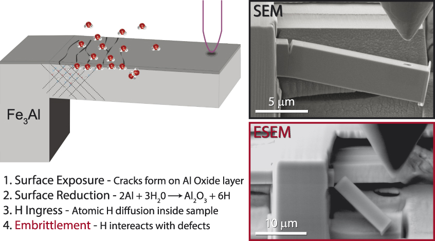
Takahashi et al. studied the fracture behavior of bicrystalline nickel-aluminide microcantilevers inside a high-voltage environmental transmission electron microscope (TEM).Reference Takahashi, Kondo, Asano, Arai, Higuchi, Yamamoto, Muto and Tanaka43 The tests were conducted either in vacuum or N2–20 vol% H2 gas (H2 partial pressure of 1 kPa). They used a high acceleration voltage of 1 MeV to facilitate the dissociation and ionization of gas molecules. Recently, Wan et al.Reference Wan, Deng and Barnoush44 used a low-pressure plasma to enhance the H uptake from H2 for in situ mechanical testing in an environmental scanning electron microscope (ESEM). Deng et al.Reference Deng and Barnoush45 and Rogne et al.Reference Rogne, Kheradmand, Deng and Barnoush46 used the high reactivity of FeAl and Fe3Al intermetallic alloys with water vapor, which yields H uptake through surface reduction of the water, to perform microcantilever bending tests in situ in an ESEM (Figure 3). They were able to observe H-enhanced crack propagation in real time. Testing in the SEM typically allows high-resolution visualization for positioning prior to environmental exposure, as well as precise measurements of the crack mouth opening or the use of digital image correlation for local strain measurement. However, upon increasing the chamber pressure in an ESEM, the imaging resolution is reduced (Figure 3), precluding high resolution, local visualization of the deformation processes, but enabling the acquisition of local mechanical behavior in the selected environment.
Schematic showing hydrogen ingress and embrittlement of an FeAl sample with in situ secondary electron micrographs on the right showing the relatively tough behavior in the high vacuum of the scanning electron microscope (SEM) (upper micrograph) in contrast with the hydrogen embrittlement behavior in the environmental SEM (ESEM) (lower micrograph).Reference Deng and Barnoush45

In situ SSMT in combination with electrochemical H charging is a powerful tool for investigating the underlying mechanisms of hydrogen embrittlement. In the future, we expect to see increasing application of these environmental techniques to address the HE problem. Future efforts are expected to combine local hydrogen measurements with small-scale mechanical testing using special, functionalized nanoindenter tips.
Radiation
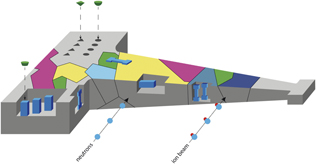
Nuclear environments are among the most challenging extreme environments for materials. Materials can be exposed to high temperatures, corrosion, and radiation simultaneously for extended periods of time, sometimes even for decades.Reference Zinkle and Was47,Reference Mansur, Rowcliffe, Nanstad, Zinkle, Corwin and Stoller48 Associated with these environments are materials degradation mechanisms that can lead to mechanical failure, such as radiation-induced embrittlement, increased yield strength, irradiation-induced creep, and radiation-induced stress corrosion cracking, to name a few. Small-scale mechanical testing has been increasingly utilized on materials exposed to these environments; the small volumes of radioactive material allow for increased safety of workers, to access specific regions of interest, to target specific phenomena and separate different radiation effects, and to directly assess mechanical property degradation using ion beam irradiations instead of neutrons. This final advantage has gained popularity due to its specificity and higher dose rates allowing shorter turnaround times.Reference Hosemann49 Numerous small-scale mechanical testing techniques are currently deployed for irradiated model or engineering materials, including nanoindentation,Reference Hardie and Roberts50–Reference Mao, Sun, Huang, Shiau, Garner, Freyer and P Wharry53 microcompression,Reference Shin, Jin, Sung, Kim, Choi and Oh54–Reference Pouchon, Chen, Ghisleni, Michler and Hoffelner56 and microtensile testing,Reference Reichardt, Ionescu, Davis, Edwards, Harrison, Hosemann and Bhattacharyya57–Reference Ding, Tian, Han, Li, Ma and Shan59 and these have been employed both at room temperature and at elevated temperatures, mostly ex situ, but also during irradiation (Figure 4).
Schematic cartoon of possible and used micromechancial test samples (microcompression, microtensile, microcantilever, nanoindentation) on a polycrystalline irradiated material. The shape of the tensile sample illustrates that neutron irradiation can penetrate large volumes while ion beam irradiations considered can only penetrate small volumes.

The main aim of this area of research is to assess the property changes at small scales after/during irradiation and to relate this to bulk property changes. While assessing the mechanical effects of radiation on materials on a phenomenological level allows learning about the importance of specific effects, the ultimate goal is to translate the small-scale results into bulk property assessments. This goal is also the main challenge, since scaling effects, dose gradients, implantation effects, surface effects, and microstructure effects all need to be addressed in order to accurately infer bulk properties. Recent studies have focused on indentation of neutron and ion-beam irradiated materials,Reference Saleh, Xu, Hurt, Ionescu, Daniels, Munroe, Edwards and Bhattacharyya60–Reference Pathak, Kalidindi, Weaver, Wang, Doerner and Mara62 and this has fostered a more general understanding of scaling effects associated with specific defects in the material. Indentation lends itself well to statistical testing and therefore allows investigation of numerous effects, such as dose gradients in ion-beam irradiated materials. In contrast, microcompression testing allows the assessment of phenomenological materials failures, such as highly localized slip from radiation-induced creep. K. Tai et al.Reference Özerinç, Averback and King63 recently undertook the difficult task of performing microcompression testing in situ during ion-beam irradiation to investigate radiation-induced creep as a function of dose rate. Reichardt et al.Reference Reichardt, Lupinacci, Frazer, Bailey, Vo, Howard, Jiao, Minor, Chou and Hosemann64 have addressed localized slip and therefore sudden failure in irradiated materials.
Most studies have previously focused on the stress-related aspects of irradiated materials deformation, but recently, strain-to-failure and ductility have been investigated utilizing microtensile testing and microcantilever testing.Reference Vo, Reichardt, Frazer, Bailey, Chou and Hosemann65,Reference Armstrong, Hardie, Gibson, Bushby, Edmondson and Roberts66 It was found that reduced strain-to-failure can be accurately assessed using these approaches, and further work in this area is greatly needed. Recently, grain-boundary failure due to transmutation products agglomerating at grain boundaries has gained interest and attention, but so far, only a limited number of studies have attempted to investigate grain-boundary failure using SSMT techniques.Reference Miura, Fujii and Fukuya58 This exciting field of research is currently wide open.
Despite the recent efforts, it is still vital to find a way to transfer the small-scale mechanical properties measured and phenomena observed to the macroscopic world, and this can only be achieved by pairing small-scale investigations with crystal plasticity modeling. Despite the challenges previously mentioned, utilizing small-scale mechanical testing for irradiated materials research will be a key component to fully understanding the materials degradation phenomena under irradiation and enabling the prediction and prevention of materials failures in service.
Summary
We have highlighted the numerous advances made toward deploying small-scale mechanical testing of materials in situ in extreme environments. Significant progress has been made, but in our opinion, the community is still only in the inception phase. New, refined, and synergistic approaches in coming years are expected to provide fundamental insights into materials behavior at extreme conditions.
Acknowledgments
A.B. acknowledges the Research Council of Norway for support to NTNU NanoLab through the Norwegian Micro- and Nano-Fabrication Facility, Norfab (197411/V30) and to the HyF-Lex Project (244068/E30). P.H. thanks the US Department of Energy (DOE) Office of Science program entitled “Fundamental Understanding of Transport Under Reactor Extremes for sponsoring part of this research, as well as the DOE nuclear energy university program.” J.M.-A. acknowledges financial support from the European Union’s Horizon 2020 Research and Innovation Programme under the Marie Skłodowska-Curie DYNACOMP Project (722096), from the Spanish Ministry of Science under Project CRACKTIAL (MAT2016–77189-R), and from Madrid region under Project S2018/NMT-4381-MAT4.0-CM. J.M.W. would like to thank D. Ghedalia of ETHZ for his assistance updating Figure 1, and C. Schuh and M. Hassani-Gangaraj of the Massachusetts Institute of Technology for the inspiration for Figure 2. J.M.W. is grateful for financial support through the Swiss National Science Foundation (SNSF Grant No. 200021_166094).

Afrooz Barnoush is a full professor in the Department of Mechanical and Industrial Engineering at the Norwegian University of Science and Technology, Norway. He obtained his PhD degree from Saarland University, Germany, in 2008. His current research focuses on the development of novel nano- and microscale examination methods to study environmental effects on mechanical properties, including hydrogen embrittlement. Barnoush can be reached by email at afrooz.barnoush@ntnu.no.

Peter Hosemann is the leader of the Nuclear Materials Group in the Department of Nuclear Engineering at the University of California, Berkeley, where he is currently department chair. He obtained his PhD degree from the Montanuniversität Leoben, Austria, in 2008. He completed doctoral research at Los Alamos National Laboratory. His research focuses on materials evaluation in extreme environments with a focus on nuclear environments and spearheads small-scale mechanical testing on nuclear materials. Hosemann can be reached by email at peterh@berkeley.edu.

Jon M. Molina-Aldareguia is the leader of the research program on Multiscale Characterization of Materials and Processes at IMDEA Materials Institute, Spain. He received his PhD degree at the University of Cambridge, UK, in 2002. His research focuses on micromechanical testing in a wide range of structural materials, from composites to light and high-temperature metallic alloys, with the aim of understanding deformation mechanisms and informing microstructurally based multiscale materials models. Molina-Aldareguia can be reached by email at jon.molina@imdea.org.

Jeffrey M. Wheeler is a senior scientist in the Laboratory for Nanometallurgy at ETH Zürich, Switzerland. He received his PhD degree in materials science and metallurgy from the University of Cambridge, UK, in 2009. His research interests lie in the areas of high-temperature nanoindentation and micromechanical testing, particularly in situ in the scanning electronic microscope. His current research focuses on the plasticity of strong materials at small length scales. Wheeler can be reached by email at jeff.wheeler@mat.ethz.ch.