Introduction
The Careless Creek Quarry (CCQ) is a multispecific bonebed occurring in strata of the Judith River Formation of eastern Wheatland County, central Montana, USA (Fiorillo, Reference Fiorillo1991; Fig. 1). Excavations conducted in the 1980s recovered approximately 1,500 vertebrate remains. These remains consisted of chondrychthian and teleost fishes, amphibians, lizards, crocodylians, pterosaurs, and a diverse assemblage of dinosaur clades that included theropods, ankylosaurs, pachycephalosaurs, ceratopsians, and hadrosaurids (Dodson, Reference Dodson1986; Fiorillo, Reference Fiorillo1989). The CCQ is an important fossil site because of the richness and diversity of vertebrate taxa preserved; it is becoming one of the most productive localities of the Judith River Formation (Fiorillo, Reference Fiorillo1991). In addition, it documented the co-occurrence of lambeosaurine and saurolophine hadrosaurids, as well as one of the earlier findings of juvenile material for this clade of ornithopods (Fiorillo, Reference Fiorillo1987, Reference Fiorillo1989).
Location of the Careless Creek Quarry (CCQ) in Wheatland County, south-central Montana, USA.

The Judith River Formation is 170–180 m thick in northwestern Montana (Rogers, Reference Rogers1998), and about 130 m of the lower-most part of the formation is exposed at Careless Creek Quarry (Fiorillo, Reference Fiorillo1991). CCQ is found in the lower third of the local section, approximately 30m above the Parkman Sandstone of the Pierre Shale (Fiorillo, Reference Fiorillo1991, fig. 2). This author reports radiometric ages approximately 78–79 Ma for bentonite layers that are low in the Judith River Formation cropping out in northern Montana. Congruent with those dates, recently Longrich (Reference Longrich2013, fig. 8) suggested a lower-middle Campanian age for the CCQ ceratopsian Avaceratops.
The CCQ was deposited in a river channel within the context of an aggrading coastal plain, and the bone assemblage was interpreted to have accumulated as a logjam (Fiorillo, Reference Fiorillo1991). We focus on the hadrosaurid remains from CCQ. The fossil bones were recovered as associated skeletal remains, possibly representing a group death event and subsequent transport of the carcasses (Fiorillo, Reference Fiorillo1991). A scenario such as a herd crossing a river in flood conditions has been pointed out as a possible explanation for this event (Fiorillo, Reference Fiorillo1987, Reference Fiorillo1991).
Hadrosaurids were among the most diverse and abundant large terrestrial herbivores of the Late Cretaceous (Lull and Wright, Reference Lull and Wright1942; Horner et al., Reference Horner, Weishampel and Forster2004), with a fossil record spanning the Santonian through the late Maastrichtian of Eurasia, the Americas, and Antarctica (Prieto-Márquez, Reference Prieto-Márquez2010a). Hadrosaurids are notorious for having complex dental batteries, expanded rostra, and hypertrophied nasal passages often associated with supracranial crests (Evans, Reference Evans2006; Prieto-Márquez, Reference Prieto-Márquez2010b). Fiorillo (Reference Fiorillo1989) reported the presence of Corythosaurus and Kritosaurus/Hadrosaurus in the CCQ on the basis of postcranial comparisons with data presented in Brett-Surman (Reference Brett-Surman1975; erroneously referenced as 1972). The monospecific genus Hadrosaurus is a pre-saurolophid hadrosaurid restricted to the Campanian of the east coast of North America (Leidy, Reference Leidy1858; Prieto-Márquez et al., Reference Prieto-Márquez, Weishampel and Horner2006; Prieto-Márquez, Reference Prieto-Márquez2011a), whereas the two known species of Kritosaurus come from upper Campanian rocks of southern North America (Brown, Reference Brown1910; Lucas et al., Reference Lucas, Spielmann, Sullivan, Hunt and Gates2006; Prieto-Márquez, Reference Prieto-Márquez2014a). It is therefore unlikely that Hadrosaurus or Kritosaurus are present in the CCQ sample. This and the fact that no mention was given of the characters supporting such taxonomic identities warranted a revision of the CCQ hadrosaurid material.
With the goal of addressing those shortcomings, the skeletal morphology of the hadrosaurid material from CCQ is described and figured for the first time, and its phylogenetic relationships are inferred via maximum parsimony analysis. We accompany this study with a number of comparative anatomical observations on ontogenetic variation of cranial and appendicular elements of the hadrosaurid skeleton using CCQ juvenile material. Based on the maximum number of specimens of the same side for the most abundant skeletal elements under consideration, a minimum of five individuals are represented in our study sample. This minimum number of five individuals comes from counting at least one juvenile, one large subadult or adult lambeosaurine (given by a single ischium), and at least three large subadult or adult saurolophines (given by three right pubes).
Materials
Repositories and institutional abbreviations
AMNH, American Museum of Natural History (FABR, Fossil Amphibians, Birds, and Reptiles collection), New York City, New York, USA; ANSP, Academy of Natural Sciences of Philadelphia, Philadelphia, USA; CMN, Canadian Museum of Nature, Ottawa, Ontario, Canada; MOR, Museum of the Rockies, Bozeman, Montana, USA; ROM, Royal Ontario Museum, Toronto, Ontario, Canada; TMP, Royal Tyrrell Museum of Paleontology, Drumheller, Alberta, Canada.
Systematic paleontology
Superorder Dinosauria Owen, Reference Owen1842
Order Ornithischia Seeley, Reference Seeley1887
Suborder Ornithopoda Marsh, Reference Marsh1881
Family Hadrosauridae Cope, Reference Cope1870
Hadrosaurid indeterminate
CCQ indeterminate hadrosaurid elements. (1, 2) Partial parasphenoid, presphenoid, and basisphenoid (ANSP 18325) in (1) ventral and (2) left lateral view. (3) Partial opisthotic-exoccipital complex (ANSP 17690) in right lateral view. (4) Fragment of left maxilla in lateral view. (5, 6) Partial left pterygoid (ANSP 17689) in (5) medial and (6) lateral views. (7, 8) Partial left dentary (ANSP 17671) in (7) lateral and (8) medial views. (9, 10) Partial surangular (ANSP 18322) in (9) ventral and (10) dorsal views. (11) Right lateral view of a sacrum (ANSP 17728).
CCQ juvenile hadrosaurid ANSP material compared to selected adult hadrosaurid specimens from elsewhere. (1, 2) Juvenile partial left dentary (ANSP 16242) in (1) lateral and (2) medial views. (3) Partial left scapula (ANSP 15979) in lateral view. (4, 5) Partial left dentary of Gryposaurus latidens (AMNH 5465) in (4) lateral and (5) medial views. (6) Partial left scapula of G. latidens (AMNH 5465) in lateral view. (7, 8) Cast of left juvenile humerus (ANSP 15979) in (7) craniomedial and (8) caudolateral views. (9) Juvenile left tibia (ANSP 15979) in medial view. (10, 11) Left humerus of Prosaurolophus maximus (TMP 84.1.1) in (10) craniomedial and (11) caudolateral views. (12) Left tibia of P. maximus (ROM 787) in medial view.
Referred material
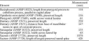
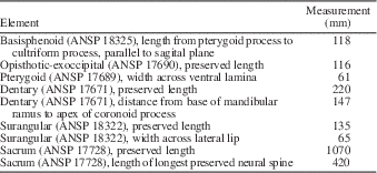
ANSP 15979, a juvenile specimen including cast of left humerus, left scapula, and left tibia; ANSP 15986, cast of partial juvenile right humerus; ANSP 16242, partial juvenile left dentary; ANSP 18325, partial fused basisphenoid, parasphenoid, and presphenoid; ANSP 17671, partial left dentary; ANSP 17689, partial pterygoid; ANSP 18326, fragmentary maxilla; ANSP 17690, fragmentary opisthotic-exoccipital; ANSP 18322, partial left surangular; ANSP 17728, sacrum; and ANSP 16957–16961, pedal phalanges (Table 1).
Selected measurements (in millimeters) of CCQ indeterminate hadrosaurid elements.
Skull elements
One of the only two neurocranial fragments represented in the CCQ sample, ANSP 18325, preserves most of the parasphenoid and the rostroventral extent of the basisphenoid including the right pterygoid process (Fig. 2.1, 2.2). The thick cultriform process of the parasphenoid is slightly expanded both proximally and distally. Dorsally, it is fused to the presphenoid, forming a gently concave wall. Proximally, the parasphenoid is continuous with the basisphenoid. The stout basipterygoid process projects caudolaterally and ventrally, forming a 150° angle (as measured in ventral view) with the long axis of the cultriform process of the parasphenoid. A ventromedian process was probably present, judging from the eroded median prominence seen in between the bases of the pterygoid processes.
The second hadrosaurid neurocranial remain consists of a partial opisthotic-exoccipital complex (ANSP 17695; Fig. 2.3). The element preserves the proximal extent of the opitshotic-exoccipital complex. It corresponds to the lateral surface of the caudal region of the braincase, caudal to the prootic, dorsal to the basioccipital, and ventral to the parietal. The bone projects caudodorsally and displays a prominent longitudinal crista otosphenoidalis. Proximoventrally, below that ridge, the element is rostrocaudally expanded, containing the metotic foramen rostrally and the exit for branches of cranial nerve 12 caudally.
The maxilla is solely represented by ANSP 18327, a fragment of the lateral wall of this facial element (Fig. 2.4). The bone preserves most of the ectopterygoid ridge and part of the lateral and rostral-most extent of the ectopterygoid shelf, as well as a large section of the surface ventral to the rostrodorsal region of the maxilla. The ectopterygoid ridge is heavily eroded and becomes gradually thicker caudally, as in all hadrosaurids (Prieto-Márquez, Reference Prieto-Márquez2008). The ventral jugal tubercle is prominent and occurs adjacent to the embayment present at the rostral region of the ectopterygoid shelf. The medial surface of this maxillary fragment preserves 21 eroded alveolar sulci.
The only palatal element recovered, ANSP 17689, is the ventral region of a left pterygoid (Fig. 2.5, 2.6). The ectopterygoid process projects ventrally and slightly caudally at the rostroventral region of the pterygoid. This process is rostrocaudally compressed and shows elliptical rostral and caudal surfaces. Caudodorsally, it is continuous with the ventral lamina of the pterygoid. This triangular lamina extends among the partially preserved quadrate wing, central buttress, and ecopterygoid process. The medial surface of the lamina is strongly concave whereas the lateral side is flat. Only parts of the ventral extent of the quadrate wing are preserved, rising from the proximal segment of the quadrate ramus.
Mandibular elements
The dentary ANSP 17671 preserves most of the coronoid process, the caudal half of the mandibular ramus, and heavily eroded fragments of the alveoli of the caudal region of the dental battery (Fig. 2.7, 2.8). The Meckelian fossa widely opens caudally and the coronoid process is rostrally inclined, as expected from a hadrosaurid (Prieto-Marquez, 2010b). The medial surface of the apex of the process bears an array of fine striations arranged radially, and its dorsal margin is incompletely preserved. The ventral border of the dentary shows only a very minor convex lateral profile.
The surangular ANSP 18322 is missing most of the rostral ascending flange and the entire retroarticular processes (Fig. 2.9, 2.10). The mediodorsal surface of the base of the ascending flange shows a large concavity that ends caudally into the rostral region of the central body of the surangular. The ventrolateral surface of this region of the ascending flange is occupied by an extensive V-shaped facet that would underlie the caudal region of the dentary by means of a scarf joint. On the medial side of the surangular, the mediodorsal edge of the ascending flange is caudally continuous with a prominent ridge. This ridge constitutes the dorsal border of the long and shallow articular facet for the angular and separates this facet from the articular surface for the splenial above. The thick D-shaped lateral lip of the surangular is caudally continuous with the glenoid facet for the quadrate.
Postcranial elements
The sacrum ANSP 17728 consists of eight fused vertebrae (Fig. 2.11). It is relatively complete, missing the distal region of most of the neural spines. The stout transverse processes show triangular distal facets. An extensive sacral rib fuses to the ventral margin of each transverse process and, ventrally, to the iliac bar. The latter extends parallel and slightly ventral to the sacral centra. Like the entire series of sacral vertebrae, the thick iliac bar is strongly curved, showing a maximum point of inflexion at the level of the third centrum. The neural spines are subrectangular laminae. Progressing along the vertebral series, these structures become slightly more inclined caudally. Distally, they become gradually wider and gently curved craniodorsally.
The five preserved pedal phalanges (not shown) possess the usual hadrosaurid morphology in being broad wedge-shaped, dorsoventrally compressed, and spade-shaped bones. The dorsal and plantar surfaces are gently convex and concave, respectively. No ridges are present on the plantar side of each phalanx. The proximal articular surfaces are elliptical and mediolaterally convex.
Remarks
The juvenile indeterminate hadrosaurid material (Fig. 3) is treated in the ‘Discussion’ section of this study, where we focus on those morphological attributes that are ontogenetically variable in a comparative context with adult hadrosaurid specimens.
Saurolophinae Brown, Reference Brown1914 (sensu Prieto-Márquez, Reference Prieto-Márquez2010b)
Kritosaurini Lapparent and Lavocat, Reference Lapparent and Lavocat1955 (sensu Prieto-Márquez, Reference Prieto-Márquez2014a)
?Gryposaurus sp.
CCQ kritosaurin cranial elements. (1–3) partial left prefrontal (ANSP 17692) in (1) dorsal, (2) lateral, and (3) medioventral views. (4–6) right postorbital (ANSP 18320) in (4) dorsal, (5) lateral, and (6) medial views. (7–9) Right lacrimal (ANSP 18328) in (7) caudal, (8) lateral, and (9) medial views. (10–12) Left squamosal (ANSP 18323) in (10) medial, (11) laterodorsal, and (12) caudal views.
CCQ kritosaurin appendicular elements. (1, 2) right humerus (ANSP 15981) in (1) caudolateral and (2) craniomedial views. (3) Left pubis (ANSP 17725) in lateral view. (4) Right pubis (ANSP 17724) in lateral view. (5) Right pubis (ANSP 17723) in lateral view. (6) Right pubis (ANSP 17726) in lateral view. (7) Right ilium (ANSP 17729) in lateral view.
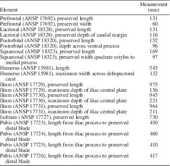
Selected measurements (in millimeters) of CCQ saurolophid elements. All elements are referable to kritosaurin saurolophines, except the lambeosaurine ischium ANSP 17727.
Referred material
ANSP 15981, right humerus; ANSP 17692, partial left prefrontal; ANSP 18320, right postorbital; ANSP 18323, left squamosal; ANSP 18324, fragmentary right squamosal; ANSP 18328, right lacrimal; ANSP 17723, 17724, 17726, and 17725, three right and one left pubes, respectively; and ANSP 17729, a right ilium. Although it could be argued that each individual bone in the saurolophine sample may correspond to a different species, we opted for choosing a most parsimonious hypothesis here, that is, that all saurolophine material belongs to the same taxon. As shown below, the anatomy of these elements is congruent with this hypothesis.
Prefrontal
The incomplete prefrontal ANSP 17692 preserves the main body that forms the rostrodorsal corner of the orbit. It lacks the ventral margin of the rostroventral process and part of the caudomedial process (Fig. 4.1–4.3). The orbital margin exhibits a subsquared profile, as in saurolophine hadrosaurids (Prieto-Márquez, Reference Prieto-Márquez2010b), and becomes gradually thicker caudally. Medial to the orbit, the dorsal surface of the prefrontal is greatly flattened and subtly concave mediolaterally. Rostrally, the prefrontal is abruptly deflected ventrally to form a broad rostroventral process that is extensively exposed laterally, another condition commonly present in saurolophines (Prieto-Márquez, Reference Prieto-Márquez2010b). This process faces both laterally and slightly rostrally. The medial side of the prefrontal is deeply excavated, containing a few irregularly shaped and poorly defined ridges (Fig. 4.3). The caudomedial process preserves part of the medial apex that would insert into the lateral margin of the frontal.
Lacrimal
The lacrimal, ANSP 18328, consists of a triangular bony lamina (Fig. 4.7–4.9). The wedge-shaped rostral process is missing the distal apex, as well as a few fragments of the proximodorsal border. The jugal notch occupies 40% of the preserved ventral margin of the lacrimal, adjacent to the caudoventral corner of the element. The notch becomes deeper caudally; its shallower rostral half shows a medially recessed surface in lateral view. The caudoventral corner of the lacrimal forms a prominent subtrihedral process. The ventral margin of the lacrimal displays a wide convexity just rostral to the jugal notch, as in Kritosaurus navajovius Brown, Reference Brown1910, Gryposaurus spp., Saurolophus spp., and Prosaurolophus maximus Brown, Reference Brown1916 (Bell, Reference Bell2011; McGarrity et al., Reference McGarrity, Campione and Evans2013; Prieto-Márquez, Reference Prieto-Márquez2014a; Prieto-Márquez et al., Reference Prieto-Márquez, Wagner, Bell and Chiappe2015). The caudodorsal region of the lacrimal forms a tall and subtriangular lamina. This caudodorsal process is gently deflected medially. Laterally, the process displays an extensive surface that would underlie the rostroventral process of the prefrontal. The medial side of the lacrimal is concave. Its caudal half is longitudinally excavated by a wide lacrimal canal. This broad groove gradually becomes deeper caudally and exits through a wide opening onto the caudal margin of the lacrimal.
Postorbital
The postorbital, exemplified by ANSP 18320, consists of three processes that extend rostrally, ventrally, and caudally from a triangular central region (Fig. 4.4–4.6). This configuration gives the postorbital a T-shaped lateral profile with a gently concave dorsal margin. In ANSP 18323, the distal segments of the ventral and caudal processes are missing. The rostral process of the postorbital is triangular in dorsal view and widens rostromedially to form a thick and crenulated medial margin for articulation with the frontal. Laterally, the rostral process contributes to the caudodorsal margin of the orbit by means of a relatively thin border that is ornamented with a series of narrow vertical indentations. The ventral process projects and wedges rostroventrally to meet the jugal, forming about half of the caudodorsal magin of the orbit. Caudodorsally, the caudal edge of the ventral process becomes continuous with that of the caudal process; both processes form an extremely wide rostrodorsal margin for the infratemporal fenestra. The caudal process of the postorbital is greatly compressed dorsoventrally and slightly inclined ventrally, so that its inner side faces ventromedially. The latter surface is excavated and would overlap part of the rostral process of the squamosal. The medioventral surface of the central body of the postorbital, dorsal to the ventral process, displays an eroded oval depression for reception of the lateral process of the laterosphenoid (Fig. 4.6).
Squamosal
The squamosal consists of a thick curved lamina that is deeply concave ventrally. From it, three processes project medially, rostrally, and ventrolaterally. In the most complete of the available squamosals, ANSP 18324 (Fig. 4.10–4.12), the bone is missing the distal tip of the rostral process, portions of the medial margin of the main squamosal body, and the distal segment of the postcotyloid process. The rostral process forms a long wedge that is dorsoventrally compressed. Its dorsal surface, particularly the lateral extent, is occupied by a large recessed facet that receives the caudal process of the postorbital. A robust subconical precotyloid process extends lateroventrally adjacent and lateral to the proximal region of the rostral process. This process is nearly as long as the quadrate cotylus is wide. The triangular surface present between the rostral margin of the proximal region of the precotyloid process and the lateral edge of the proximal region of the postorbital process is smooth and gently concave. The caudal margin of the precotyloid process forms the rostral boundary of the quadrate cotylus. The cotylus consists of a dorsoventrally compressed but deep oval depression on the lateral surface of the main body of the squamosal that receives the head of the quadrate. The caudal wall of the quadrate cotylus is continuous caudally with the large postcotyloid process. This process is dorsoventrally compressed and hook-like, extending lateroventrally from the caudolateral corner of the squamosal. Its rugose recessed caudoventral surface would contact extensively the proximal region of the paroccipital process of the fused opisthotic-exoccipital. The medial process of the squamosal extends medially and slightly dorsally. Distally, this process curves rostromedially to form the caudomedial corner of the supratemporal fenestra.
Humerus
The humerus ANSP 15981 combines relatively slender proportions with a substantially expanded dectopectoral crest (Fig. 5.1,5.2). Specifically, the length of the element is more than six times the width of the lateral surface of the proximal end. Among hadrosaurids, similarly elongate humeri are present in specimens of Gryposaurus notabilis (e.g., TMP 80.22.1) and Prosaurolophus maximus (e.g., MOR 454; Prieto-Márquez, Reference Prieto-Márquez2008, fig. H.18). The deltopectoral crest comprises slightly more than half of the length of the humerus and projects lateroventrally from the proximal half of the humerus; the ratio between the maximum width of the crest and the minimum diameter of the humeral shaft is 1.80 in ANSP 15981. This combination of overall proportions and development of the deltopectoral crest is unlike that seen in lambeosaurines, in which the ratio of humerus length to proximal width tends to be less than 4.9 and the ratio of deltopectoral crest width to minimum humeral circumference is greater than 1.90 (Prieto-Márquez, Reference Prieto-Márquez2008). The humeral shaft is elliptical in cross section and mediolaterally compressed. The distal end of the humerus is mediolaterally expanded to form the lateral ulnar and medial radial condyles. The radial condyle is wider and more craniocaudally expanded than the ulnar condyle. Deep and wide depressions separate the two condyles on the cranial and caudal surfaces of the distal end of the humerus.
Pubis
The pubis has a ventrally deflected subrectangular prepubic blade, with subparallel dorsal and ventral margins, and an angular craniodorsal corner (Fig. 5.3–5.6). This geometry is characteristic of kritosaurins such as Gryposaurus notabilis (e.g., ROM 764), Secernosaurus koerneri Brett-Surman, Reference Brett-Surman1979 (e.g., MACN RN 2) and saurolophins such as Prosaurolophus maximus (e.g., MOR 454), and probably, Saurolophus spp. (Prieto-Márquez, Reference Prieto-Márquez2008). The length of the proximal constriction is subequal to that of the prepubic blade. The tetrahedral iliac peduncle projects caudodorsally from the dorsal extent of the acetabular region of the pubis. The concave caudal surface of the iliac peduncle forms half of the acetabular margin of the pubis and is continuous with the ventral half of the proximal region of the ischiadic peduncle. The ischiadic peduncle is mediolaterally compressed and laterally offset relative to the proximal region of the postpubic process. The rod-like postpubic process is relatively robust and projects caudoventrally forming a 115° angle with the long axis of the prepubic process.
Ilium
ANSP 17729 (Fig. 5.7) is characterized by a remarkably shallow iliac plate. Specifically, the maximum depth:maximum length ratio of the iliac plate is 0.58. Relatively shallow (depth-to-width ratio less than 0.8) iliac plates are widespread among saurolophines except known brachylophosaurin ilia (Prieto-Márquez, Reference Prieto-Márquez2007). The preacetabular process of the ilium projects rostroventrally beyond the level of the pubic process, forming an angle of 147° with a line uniting the pubic and ischiadic processes. The dorsal margin of the preacetabular process is straight, whereas the ventral border displays a wide convexity. Two longitudinal ridges are present on the lateral side of the process: one ridge extends obliquely from mid-depth of the proximal region to merge with ventral margin at mid-length of the process; the other ridge occurs near the distal end of the process. The supraacetabular crest of ANSP 17729 shows an asymmetrical, caudally skewed V-shaped lateral profile. It greatly extends lateroventrally to overlap part of the ischiadic process. As in Gryposaurus notablis, G. latidens, Secernosaurus koerneri, and Willinaqake salitralensis Juárez Valieri, Haro, Fiorelli, and Calvo, Reference Juárez Valieri, Haro, Fiorelli and Calvo2010 (Juárez Valieri et al., Reference Juárez Valieri, Haro, Fiorelli and Calvo2010; Prieto-Márquez and Salinas, Reference Prieto-Márquez and Salinas2010), the length of the crest of ANSP 17729 is approximately three-fourths of the length of the iliac plate. The caudodorsal margin of the supraacetabular crest is continuous with the proximolateral surface of the postacetabular process, lacking a well-defined ridge. The postacetabular process of ANSP 17729 is subrectangular (slightly wider distally than proximally, as in G. latidens AMNH FABR 5465), medially inclined, and caudodorsally oriented. As preserved, the postacetabular process is 10% longer than the iliac plate. However, the process was probably slightly longer given that its caudal margin is incomplete. Such long postacetabular processes characterize species of Gryposaurus (Prieto-Márquez, Reference Prieto-Márquez2012) and Saurolophini (Prieto-Márquez et al., Reference Prieto-Márquez, Wagner, Bell and Chiappe2015).
Kritosaurin indeterminate
CCQ saurolophid pelvic elements. (1) Saurolophine left ilium (ANSP 17731) in lateral view. (2) Saurolophine right ilium (ANSP 17730) in lateral view. (3) Lambeosaurine left ischium (ANSP 17727) in lateral view.
Referred material
ANSP 17730 and 17731, right and left ilia, respectively (Table 2).
Remarks
Because these elements show similar size and morphology, and correspond to the left and right side of the pelvic girdle, we interpret them as coming from a single individual. These ilia are treated separately from the kritosaurin taxon mentioned previously because they differ from the ANSP 17729 ilium in possessing a more slender preacetabular process, a slightly deeper iliac plate, a less lateroventrally expanded supraacetabular crest, and a shorter and more medially inclined postacetabular process. Given that the morphology of ANSP 17730 and 17731 is less markedly saurolophine (and kritosaurin) when compared to that of ANSP 17729, we tested phylogenetically the affinities of this material by including it in the analysis as a separate operational taxonomic unit (see the following for a detailed account of supporting characters).
Lambeosaurine Parks, 1923
Lambeosaurine indeterminate
Referred Material
ANSP 17727, a left ischium.
Ischium
The only recovered ischium, ANSP 17727, lacks the pubic process and the ventral margin of the proximal region (Table 2). The iliac process extends craniodorsally, forming a 140º angle with the proximal-most dorsal margin of the ischiadic shaft. This process is relatively long, with a ratio between its proximodistal length and the width of its distal margin of 2.1; typically, this ratio ranges from 1.5 to 2 in lambeosaurines dinosaurs (Prieto-Márquez, Reference Prieto-Márquez2010b). The caudodorsal margin of the process is recurved, as in all lambeosaurines (Brett-Surman and Wagner, Reference Brett-Surman and Wagner2007). The articular surface of the iliac process is abraded. Ventrally from the iliac process, the proximal region of the ischium becomes thinner and laterally concave. The ischiadic shaft displays a gently sinusoidal lateral profile, and it is remarkably short and robust. Specifically, the shaft accounts for 65% of the total length of the ischium. Its minimum dorsoventral diameter is 10% of the total length of the shaft. Other lambeosaurines with relatively thick ischiadic shafts (i.e., ratio of minimum thickness to total length greater than 7.5%) include Corythosaurus spp., Lambeosaurus magnicristatus Sternberg, Reference Sternberg1935, Magnapaulia laticaudus Morris, Reference Morris1981, and Parasaurolophus cyrtocristatus Ostrom, Reference Ostrom1961 (Prieto-Márquez, Reference Prieto-Márquez2008; Prieto-Márquez et al., Reference Prieto-Márquez, Chiappe and Joshi2012). The distal half of the shaft is postdepositionally compressed mediolaterally. ANSP 17727 partially preserves a foot-like distal process, as expected in a lambeosaurine specimen (Brett-Surman and Wagner, Reference Brett-Surman and Wagner2007). The distal surface of this process is abraded, and the ventral tip has been eroded away.
Phylogenetic analysis
The phylogenetic position of the CCQ saurolophine material was inferred via maximum parsimony analysis. The taxonomic sample included 60 iguanodontian species (14 hadrosaurid outgroups, 23 Saurolophinae, and 21 Lambeosaurinae). The data set consisted of 273 equally weighted morphological characters (189 cranial and 84 postcranial; see Supplemental Data 1 and 2). Multistate characters containing states that are not mutually exclusive and following a natural morphocline were ordered. This criterion allows for ‘crediting’ shared intermediate states. An example of such characters is the position of the large maxillary foramen (character 85) that includes three states: exposed laterally and opening ventral to the mid-depth of the premaxillary shelf (0); exposed laterally and opening near the dorsal margin of the premaxillary shelf (1); not exposed laterally and opening on the dorsal surface of the premaxillary shelf (2). The optimal tree(s) search was conducted in TNT version 1.1 (Goloboff et al., Reference Goloboff, Farris and Nixon2008). A heuristic search of 10,000 replicates using random additional sequences was performed, followed by branch swapping by tree-bisection-reconnection holding ten trees per replicate. Decay indices (Bremer support) and Bootstrap proportions were computed using TNT. The Bootstrap analysis was set for 5,000 replicates using heuristic searches, in which each search was conducted using random additional sequences with branch-swapping by subtree pruning and regrafting and 25 replicates.
The analysis resulted in 116 most parsimonious trees (C.I.=0.42; R.I.=0.77; Fig. 7.1), with a score of 934 steps that was hit 8,968 times out of the 10,000 replicates. Exclusion of ANSP 17730-17731 from the data set reduced the number of most parsimonious trees to 16 (C.I.=0.43; R.I.=0.77; Fig. 7.2), with a score of 934 steps that was hit 9,853 times out of the 10,000 replicates. The CCQ saurolophine is deeply nested within Kritosaurini forming a polytomy with species of Gryposaurus and Rhinorex condrupus. It shares the following ambiguous synapomorphy with other kritosaurins except Naashoibitosaurus ostromi Hunt and Lucas, Reference Hunt and Lucas1993: lacrimal with convex ventral margin rostral to the jugal notch (convergent in Saurolophini and unknown in the Secernosaurus clade). The CCQ taxon also shares a broad supraacetabular crest being between 70% and 85% of the length of the iliac plate with all kritosaurines excluding N. ostromi and Kritosaurus (ilium not preserved in the latter two taxa). Characters shared with Gryposaurus and Rhinorex condrupus are (1) extension of the caudal ramus of the postorbital that overlaps the laterodorsal surface of the squamosal rostral to quadrate cotylus (postorbital not preserved in the Secernosaurus clade) and (2) ratio between the craniocaudal length of the postacetabular process and the craniocaudal length of the central plate of the ilium greater than 1.1 (convergent in Saurolophini).
Results of the maximum parsimony analysis for inferring the phylogenetic position of the CCQ saurolophine specimens within Hadrosauridae. (1) Strict consensus tree resulting from the analysis including the specimen represented by the ilia ANSP 17730 and 17731. (2) Insert of the Kritosaurini portion of the strict consensus tree resulting from the analysis including ANSP 17730 and 17731 as a specimen representing an operational taxonomic unit.
Inclusion of ANSP 17730 and 17729 within Saurolophinae is supported by the following synapomorphies: (1) absent or poorly demarcated caudodorsal margin of the lateroventral rim of the supraacetabular process and (2) supraacetabular crest at least slightly wider than half the width of the central iliac plate. In addition, nesting of this individual within saurolophines to the exclusion of Brachylophosaurini is supported by the possession of a relatively shallow iliac plate, with a ratio between this and the distance between the pubic peduncle and the caudodorsal prominence of the ischiadic peduncle less than 0.80. Finally, placement of the specimen within Kritosaurini is supported by a supraacetabular crest with a breadth lying between 70% and 85% of the width of the iliac plate; this condition is shared with the Gryposaurus and Secernosaurus clades.
Remarks
All three currently recognized species of the genus Gryposaurus lived in Laramidia (present-day western North America) during Campanian times (Fig. 8). The oldest of these species is G. latidens, from lower Campanian (about 80 Ma; Horner et al., Reference Horner, Schmitt, Jackson and Hanna2001) deposits of lithofacies 3 in the lower Two Medicine Formation of northwestern Montana, USA (Prieto-Márquez, Reference Prieto-Márquez2012). Lithofacies 3 is alluvial in nature and its fauna diversified in the context of an expanding coastal plain (Horner et al., Reference Horner, Schmitt, Jackson and Hanna2001). The other two species, G. notabilis and G. monumentensis Gates and Sampson, Reference Gates and Sampson2007, are late Campanian in age. In particular, G. notabilis came from the lower Dinosaur Park Formation of southern Alberta, Canada (Evans Reference Evans2007; Prieto-Márquez, Reference Prieto-Márquez2010c). The lower Dinosaur Park Formation was deposited in fluvial systems developed in alluvial wetland environments of a coastal plain influenced by a subtropical to warm-temperate monsoonal climate (Eberth and Currie, Reference Eberth and Currie2005). G. monumentensis was recorded in the Kaiparowits Formation of southern Utah (USA) corresponding to a humid alluvial plain containing marshes and other wetlands adjacent to rivers and streams (Gates and Sampson, Reference Gates and Sampson2007). A fourth integrant of the Gryposaurus clade recovered in our consensus tree (Fig. 7) is Rhinorex condrupus from late Campanian (74.5–75.2 Ma) strata of the Neslen Formation in east-central Utah (Gates and Scheetz, Reference Gates and Scheetz2015; Fig. 8). These strata document a marine-influenced environment (Gates and Scheetz, Reference Gates and Scheetz2015).
Biostratigraphical and geographical distribution of species of the North American kritosaurin genera Gryposaurus and Rhinorex. Literature sources for each taxon’s distribution and geochronological range are as follows: G. latidens (Prieto-Márquez, Reference Prieto-Márquez2012); G. notabilis (Evans, Reference Evans2007; Farke and Herrero, Reference Farke and Herrero2014 Gates et al., Reference Gates, Jinnah, Levitt and Getty2014); G. monumentensis (Gates and Sampson, Reference Gates and Sampson2007); and R. condrupus (Gates and Scheetz, Reference Gates and Scheetz2015). Depicted skulls are redrawn and/or modified based on the following: G. latidens (Horner, Reference Horner1992); G. notabilis (photographs of CMN 2278 by APM); G. monumentensis (Gates and Sampson, Reference Gates and Sampson2007); R. condrupus (Gates and Scheetz, Reference Gates and Scheetz2015).
It is most parsimonious to consider that the CCQ kritosaurin material may represent stratigraphical extensions of the fossil record of either G. latidens or G. notabilis (mainly due to geographical proximity compared to the other members of the clade) or a new species of Gryposaurus. In the absence of more diagnostic material, we tentatively refer the more informative bulk of the kritosaurin material from CCQ to ?Gryposaurus sp. The aforementioned paleoenvironemnts posited for the Gryposaurus species offer no additional clues for refining our identification of the CCQ kritosaurin in terms of potential habitat preferences as they all indicate similar depositional settings among known Gryposaurus and between these and the CCQ specimens.
Observations on hadrosaurid ontogenetic variation
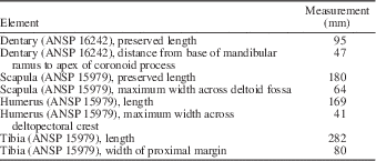
The hadrosaurid sample collected at CCQ includes a number of relatively small juvenile elements, of which we were able to observe a dentary, a scapula, two humeri, and a tibia (Fig. 3; Table 3). Comparisons between the dimensions of these elements and those of commonly large hadrosaurid specimens (Fig. 3) indicate that the CCQ juveniles are no larger than one-third of typical adult size. The absence of diagnostic characters that would allow intrageneric or intraspecific ontogenetic comparisons forced us to arbitrarily choose adult specimens from selected hadrosaurid taxa for such purposes.
Selected mandibular measurements (in millimeters) of CCQ juvenile hadrosaurid elements.
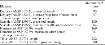
The dentary (ANSP 16242) preserves the coronoid process and a ventrally eroded caudal region of the mandibular ramus (Fig. 3.1, 3.2). Eight alveoli are preserved lacking all teeth. The coronoid process is nearly vertically oriented, in contrast to the strong rostral inclination typically seen in adult forms. Similarly, Maryanska and Osmólska (Reference Maryanska and Osmólska1981) noted that in the saurolophine Saurolophus angustirostris Rozhdestvensky, Reference Rozdestvensky1952, coronoid process is also less rostrally inclined in juveniles. While this may be a trend in hadrosaurids, at least in some hadrosaurid outgroups such as Bactrosaurus johnsoni Gilmore, Reference Gilmore1933, the coronoid process remains subvertically oriented throughout ontogeny (Prieto-Márquez, Reference Prieto-Márquez2011b). The apex of the coronoid process of ANSP 16242 is not rostrocaudally expanded, unlike those of adult hadrosaurid individuals (Fig. 3.4, 3.5).
The scapula (ANSP 15979) is nearly complete, missing only the distal extent of the blade (Fig. 3.3). The proximal constriction is 63% of the width of the articular margin of the scapula, wider than typically seen in adult hadrosaurids (e.g., in G. latidens AMNH FABR 5465, it is 54% the width of the proximal articular margin; Fig. 3.6). Narrower juvenile scapular ‘necks’ have been also previously noted in the saurolophine Edmontosaurus annectens Marsh, Reference Marsh1892 (Prieto-Márquez, Reference Prieto-Márquez2010b), the lambeosaurine Hypacrosaurus stebingeri Horner and Currie, Reference Horner and Currie1994, and some nonhadrosaurid hadrosauroids (Prieto-Márquez, Reference Prieto-Márquez2011b). Brett-Surman and Wagner (Reference Brett-Surman and Wagner2007) and Prieto-Márquez (Reference Prieto-Márquez2011b, Reference Prieto-Márquez2014b) reported an increase in the robustness and caudoventral elongation of the deltoid ridge in adult hadrosaurids and the basal hadrosauroid Bactrosaurus johnsoni, respectively. Such trend is not oberved here: the CCQ juvenile displays a relatively robust, long, and well-defined deltoid ridge (Fig. 3.3), more so than in some large adult specimens such G. latidens AMNH FABR 5465 (Fig. 3.6).
Differences between the CCQ juvenile humeri (best exemplified by the complete element ANSP 15979; Fig. 3.7, 3.8) and typical adult hadrosaurid humeri follow previous trends noted in the literature (Dilkes, Reference Dilkes2001; Godefroit et al., Reference Godefroit, Bolotsky and Van Itterbeek2004; Grigorescu and Csiki, Reference Grigorescu and Csiki2006; Brett-Surman and Wagner, Reference Brett-Surman and Wagner2007; Guenther, Reference Guenther2014; Prieto-Márquez, Reference Prieto-Márquez2014b). Specifically, the CCQ juveniles show overall less robust humeri, with relatively less expanded deltopectoral crests and less prominent distal condyles; however, Guenther (Reference Guenther2014) found no substantial ontogenetic increase in the development of the condyles in the saurolophine Maiasaura peeblesorum Horner and Makela, Reference Horner and Makela1979. These differences are evident when comparing ANSP 15979 with adult humeri such as, for example, that of Prosaurolophus maximus TMP 84.1.1 (Fig. 3.10, 3.11). Prieto-Márquez (Reference Prieto-Márquez2014b) noted the lack of an angular laterodistal corner of the deltopectoral crest in a juvenile Edmontosaurus annectens (LACM 23504). It is interesting to note that angularity of the lateroventral corner of the deltopectoral crest in the CCQ humerus is comparable to that of adult hadrosaurids such as the aforementioned P. maximus TMP 84.1.1.
In agreement with previous observations on the ontogenetic variation of hadrosauroid tibiae (Dilkes, Reference Dilkes1996; Grigorescu and Csiki, Reference Grigorescu and Csiki2006; Brett-Surman and Wagner, Reference Brett-Surman and Wagner2007; Prieto-Márquez, Reference Prieto-Márquez2011b, Reference Prieto-Márquez2014b; Guenther, Reference Guenther2014), the tibia of the juvenile ANSP 15979 shows a more slender shaft and caudolaterally less expanded cnemial crest and internal condyles (Fig. 3.9) than adult hadrosaurid specimens such as, for example, Prosaurolophus maximus ROM 787 (Fig. 3.12).
Although limited by the lack of lower taxonomic comparative context, contrasting these observations with those documented in the literature evidences that, while generally many osteological attributes appear to change in a consistent manner through hadrosaurid ontogeny, other character growth changes vary among taxa.
Acknowledgments
We thank T. Daeschler for providing high-resolution photographs of the pelvic bones and neonate specimens from the CCQ housed at the ANSP. We are grateful to T. Gates and an anonymous reviewer for insightful comments on an earlier draft of the manuscript. This study was funded by a Marie Curie Intra-European Fellowship for Career Development (European Commission) presented to APM.
Accessibility of supplemental data
Data available from the Dryad Digital Repository: http://datadryad.org/handle/doi:10.5061/dryad.4km97