Introduction
A number of studies suggest that patients with bipolar disorder have a super-sensitivity to light at night where plasma melatonin levels are suppressed to a greater degree than in patients with other affective disorders and healthy control subjects (Reference Lewy, Wehr, Goodwin and Newsome1–Reference Nathan, Burrows and Norman3). This sensitivity has been proposed to provide a useful endophenotypic marker of bipolar disorder, particularly in light of evidence that the occurrence of this sensitivity increases in those with high genetic loading for the bipolar affective disorder Reference Nurnberger, Berrettini, Tamarkin, Hamovit, Norton and Gershon(4).
In contrast, evidence from a number of follow-up studies did not support this sensitivity (Reference Whalley, Perini, Shering and Bennie5–Reference Lam, Berkowitz, Berga, Clark, Kripke and Gillin7). While methodological differences may account for the variance between studies, the most recent research by Nurnberger et al. Reference Nurnberger, Adkins and Lahiri(8) provides a possible explanation. In this research, no overall differences were observed between bipolar (29.8 ± 5.5%), unipolar (32.2 ± 6.2%) and healthy control subjects (34.6 ± 2.6%) in dark adjusted melatonin suppression by light when exposed to 500 lux of nocturnal light. When the bipolar group was sub-divided into types I and II, analysis of sensitivity estimates revealed a trend (p = 0.08) for bipolar I patients to have higher melatonin sensitivity to light (62 ± 15.6%) than matched healthy controls (40 ± 9.1%). Contrasting the sensitivity estimates of bipolar I patients to those tested by Lewy at 500 lux (61.5 ± 1.6%) Reference Lewy, Nurnberger and Wehr(2) and Nathan at 200 lux (43.5 ± 9%) Reference Nathan, Burrows and Norman(3) indicates that the bipolar I sub-group of patients may show the characteristic sensitivity.
Whalley et al. also proposed that this super-sensitivity may be an epiphenomena of the mood stabilising agents Reference Whalley, Perini, Shering and Bennie(5). The results of Lewy contradict this notion as this group also used drug-free (for 2 weeks) bipolar patients and found super-sensitivity Reference Lewy, Nurnberger and Wehr(2). Recent research investigating the effect of the mood stabilising agents lithium Reference Hallam, Olver, Horgan, McGrath and Norman(9) and sodium valproate Reference Hallam, Olver and Norman(10) in healthy controls indicates that rather than leading to super-sensitivity, these medications reduce sensitivity in healthy controls. This raises the possibility that the effect may also ameliorate sensitivity in patients possibly indicating a downstream effect of their mechanism of action.
Earlier studies on light sensitivity in bipolar disorder did not sub-divide patients into type I and II bipolar disorder. Whalley acknowledges that the lack of super-sensitivity in their study may reflect a skewed selection of less severely affected patients (all patients were euthymic for 3–6 months without medication). This raises questions about the reproducibility of the finding and sample selection in this population. To address this issue the selection of a homogenous group of patients with bipolar I disorder may indicate whether discordance between studies is an artefact of sample heterogeneity inherent in the identification of people with bipolar disorder, or may indicate that sensitivity is associated with genetic loading for the illness. Evidence for the latter hypothesis is provided by differences in one study finding an increased sensitivity to light in probands with one or both parents affected by the disorder. Control offspring had the lowest sensitivity Reference Nurnberger, Berrettini, Tamarkin, Hamovit, Norton and Gershon(4).
Previous research in healthy controls indicates that melatonin sensitivity to light is intensity dependent, with higher light levels being associated with increased melatonin suppression. McIntyre et al. showed this dose relationship among healthy volunteers, a finding that has been reproduced by a number of groups (Reference Aoki, Yamada, Ozeki, Yamane and Kato11–Reference Zeitzer, Dijk, Kronauer, Brown and Czeisler18). While these studies indicate a consistent change in sensitivity across various light intensities, variations have been reported in the healthy individuals used as controls in the bipolar studies. For example, at 500 lux of light, sensitivity in controls used have ranged from 0% Reference Lewy, Wehr, Goodwin and Newsome(1) to 28% Reference Lewy, Nurnberger and Wehr(2), 34% Reference Nurnberger, Adkins and Lahiri(8), 43% Reference Cummings, Berga and Cummings(19) and 50% Reference Whalley, Perini, Shering and Bennie(5).
The aim of this study was to examine whether a group of patients with a history of bipolar disorder type 1 show greater sensitivity to light than a group of healthy adult subjects. As one of the most consistent findings regarding melatonin sensitivity to light is that of intensity dependency, the study investigated whether this effect was consistent across doses of light. There were two reasons for investigating light sensitivity in patients and controls across intensities: (a) control of the sampling variability inherent in comparing results of previous studies conducted at different light intensities and (b) determining if super-sensitivity in the patients is a threshold phenomena (i.e. once a particular light level is surpassed, the patients show extreme sensitivity) or if this sensitivity follows the same linear dose relationship observed in controls in previous studies.
A further aim of this study was to more fully characterise the nature of melatonin changes observed in patients with bipolar disorder compared with healthy adult controls. To this end, a number of phases of the melatonin–light sensitivity profile were evaluated. Area under the plasma melatonin time curve was evaluated to determine if there were differences in total melatonin secretion during darkness. Few studies have investigated differences in the rebound of late-night melatonin concentration after lights off. Differential rebound between controls and patients was examined (i.e. the rate at which secretion of melatonin returns to the expected nocturnal concentration after light exposure). This measure might provide evidence of differences in the plasticity of the synthetic pathway, indicating potential differences in melatonin synthetic tone.
It was hypothesised that people with bipolar type I disorder would show greater sensitivity to light than healthy adult controls. It was further hypothesised that both controls and patients would show a dose-response relationship between increasing light intensities and melatonin suppression. It was further hypothesised that the dose-response curve in patients would show a left shift compared with the control curves, indicating greater sensitivity. As no evidence exists on rebound after light exposure in patients and controls, no hypotheses were generated on this response.
Method
Participants
All patients and participants attended a preliminary screening session in the Department of Psychiatry at Austin Hospital where they were interviewed and completed screening questionnaires and a medical examination with a psychiatrist (JO). All participants and patients who met inclusion criteria for the study provided written informed consent. The study was approved by the Austin Health Human Research and Ethics Committee (approval number H98/00450).
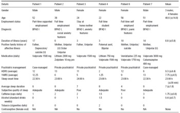
Seven patients (4 females, 3 males) were recruited for this study from a variety of referral sources. These included the Australian Twin Registry (n = 1), Internet depression support websites, (n = 4) and through referrals from North East Mobile Support Team (n = 2). All patients satisfied a Diagnostic and Stastitical Manual (DSM-IV) diagnosis of bipolar affective disorder with a positive history of both mania and depression. The diagnosis of bipolar disorder was derived from both a clinical interview with a consultant psychiatrist and the fully structured computer based World Health Organisation Composite International Diagnostic Interview (CIDI-Auto 2.1). The inclusion criteria for the patients were being aged between 18 and 60 years, satisfying a diagnosis of bipolar I disorder and being in a euthymic state at testing time. Euthymia was defined as a score on the Young Mania Rating Scale (YMRS) of 12 or less Reference Ketter, Warrington, Loebel, Kolb and Sui(20) and a score of less than 16 on the Hamilton Depression Rating Scale (HDRS) Reference Mottram, Wislson and Copeland(21). Patients were required to be on a stable medication regimen for at least 6 weeks preceding testing and have no other major physical illness. One patient was excluded following testing based on the emergence of depressive symptoms and prominent personality disorder issues. The demographic, circadian and treatment details of the study patients are depicted in Table 1.
Demographic details of patients with bipolar affective disorder in study

BPAD, Bipolar Affective Disorder; HDRS, Hamilton Depression Rating Scale; U, undiagnosed illness/patients subjective account; Young Mania Rating Scale.
Control sample
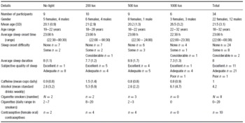
Thirty-four control participants (22 females, 12 males) were tested in this investigation. Participants were included if they were between the ages of 18 and 60 years and had no personal or family history of psychiatric illness. Participants with blindness, colour blindness or any serious visual deficit, any history of head trauma or seizure disorder and a history of adverse reactions to venipuncture procedures were excluded from the study. The final participant pool had a mean age of 21.5 years (±3.1) with an age range from 18 to 32 years. All participants were medication free for the trial duration (except the female oral contraceptive pill). Participants and patients abstained from alcohol for the 24 h preceding testing, caffeine for 12 h and cigarette smoking during testing. Detailed demographic and circadian details of the control group are depicted in Table 2.
Demographic details of healthy control subjects in each light condition

Materials and Procedure
Clinical scales and screening questionnaires. The CIDI-Auto 2.1 is a fully structured clinical interview with 14 sections. The lifetime version of the program was used to determine any lifetime history of mental illness. The CIDI interview took between 60 and 120 min for participants to complete. On visit one, a life history questionnaire, general health questionnaire Reference Goldberg and Hillier(22) and PRIME-MD Reference Spitzer, Williams and Kroenke(23) were completed along with a detailed psychiatric assessment and physical examination. At each visit, patients were asked about medication status and sleep parameters. The presence and severity of depressive symptoms was assessed using the HDRS Reference Hamilton(24). The presence and severity of mania symptoms was assessed at each visit using the YMRS Reference Young, Biggs, Ziegler and Meyer(25).
General testing procedure. Participants attended the Melatonin Laboratory in the Department of Psychiatry at the Austin Hospital on one to four testing nights separated by at least a 7-day recovery period. Participants had travel arranged for their trip to and from the hospital and were not permitted to drive to and from the testing sessions. Participants arrived in the department at 19:00 h and at 19:30 h a catheter was inserted into a forearm vein. At 20:00 h, the first blood sample was taken in normal room lighting conditions and participants were supplied with a standard evening meal. Blood samples were taken again at 21:00, 21:30, 22:00, 22:30, 23:00, 23:15, 23:30, 23:45, 24:00, 00:15, 00:30, 00:45, 01:00, 01:30, 02:00 and 02:30 h. The blood samples were immediately separated using lithium heparin tubes and stored at −20°C for the analysis. At all times in the testing room participants remained in recliner chairs. During the night participants were free to watch television or rest between blood samples. At 21:00 h, the room lights were extinguished and the background light intensity maintained at less than 10 lux, measured using a Topcon IM-3 photometer at participants eye level. There was a small television set placed at a distance of no less than 3 m from the participants (directly above the light box). Luminance from the television was measured at four times (21:00, 22:00, 23:00 and 01:00 h) on each testing night from directly in front at a distance of 2 m and did not exceed 6 lux on any testing occasion.
Participants were kept in darkness between 21:00 and 24:00 h. At 24:00, a darkness blood sample was taken before the light box was switched on for 1 h. In the no-light condition, the light box was not switched on between 24:00 and 01:00 but participants were regularly checked to ensure they were awake during this hour as they would be on the light nights. The light box was custom made (size 128 cm × 51 cm × 18 cm) and utilised a series of eight 97-W ‘true light' fluorescent tubes behind a diffuser screen. Variability in light intensity between 200, 500 and 1000 lux was obtained through altering the number of tubes illuminated.
Participants were positioned to be directly facing the light box during this time and looked at the light box, facilitated by positioning the television directly above. Photophobic behaviour such as reading or closing the eyes was not permitted and participants were instructed to ‘look directly at the light every few minutes for at least 30 s'. The experimenter returned at 5-min intervals during this period to check compliance. Light levels were measured at the corneal level at a horizontal angle of gaze for each individual and recorded to be at the appropriate intensity (±12 lux) at four (00:05, 00:15, 00:30 and 0045 h) times throughout the hour. At 01:00 the room was returned to virtual darkness (<8 lux of light). Participants were not permitted to stand or move during the light period in all four light conditions.
Melatonin analysis was performed in the Department of Obstetrics and Gynaecology, University of Adelaide using reagents from kits obtained from Buhlmann Laboratories (Allschwil, Switzerland) and a modified version of the standard assay methodology based on that previously published Reference Voultsious, Kennaway and Dawson(26). The assay utilised the Kennaway G280 melatonin antibody Reference Earl, D’Occhio, Kennaway and Seamark(27) in a [125I]-2-iodomelatonin-based Radioimmunoassay (RIA) Reference Vaughan(28). The assay range was between 4.3 and 430 pM with intra and inter assay coefficients of variation <10 and <14%, respectively, across the range of the standard curve. The antibody for the assay is highly specific and demonstrates <1% cross reactivity with other compounds in the melatonin synthetic pathway.
Statistical analysis. Melatonin suppression between 2400 and 0100 h provides a standardised measure of the change in melatonin secretion before and after light. Melatonin suppression between 24:00 and 01:00 h was determined using a previously devised formula Reference McIntyre, Norman, Burrows and Armstrong(14). The investigation of group differences (control vs. patient) in melatonin suppression by white light was performed using a univariate analysis of variance (ANOVA). This analysis further investigated whether either or both groups had significantly different levels of suppression based on light intensity (i.e. whether data formed a dose-response relationship). Data satisfied the assumption of normality and homogeneity based on the Kolmogorov-Smirnov test, hence non-transformed, parametric tests were utilised.
A regression model was created from the dose-response data from the controls and patients to provide a theoretical modelled relationship between light intensity and melatonin suppression. The dose-response data for patients and controls was linearised using a log-logit transformation and analysed by applying linear regression using Inplot© processing software.
An area under the plasma melatonin concentration–time curve was calculated using the trapezoidal method and Fig P™ (Biosoft) processing software. This calculation was used to provide an integrated measure of hourly plasma melatonin concentrations between 20:00 and 24:00 h (before lights). Area under the curve estimates was determined between 21:00 and 24:00 h (i.e. a 9-point estimate based on the acrophase). Data from the no-light group and three other control participants were excluded from this analysis, as a complete set of nine samples was not taken. A univariate ANOVA was conducted to determine if bipolar and control groups differed in the secretion of melatonin until midnight. Data satisfied the assumption of normality and homogeneity based on the Kolmogorov-Smirnov test, hence non-transformed, parametric analyses were utilised.
Melatonin post-light recovery levels provide a measure of the increase in melatonin following the extinction of lights at 01:00 h. Melatonin recovery was determined for 30 and 60 min following lights off in this study using the following formulae:

The results of these calculations were analysed using univariate ANOVAs with group (bipolar and control) and light level (0, 200, 500 and 1000 lux) as between subject factors. The results satisfied the assumption of normality and homogeneity based on the Kolmogorov-Smirnov test, hence non-transformed, parametric were utilised.
Results
Melatonin suppression by white light
Melatonin changes between 24:00 and 01:00 h were determined for patient and control groups in the absence of light were contrasted to assess the presence of any underlying reasons for variation at this time other than light. The results indicated no significant difference in melatonin changes between groups with no light, F(1,12) = 3.302, p = 0.097.
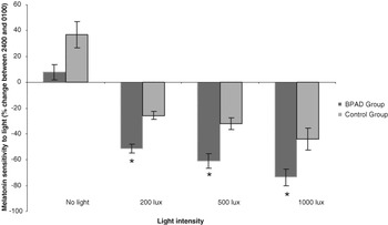
Melatonin suppression between 24:00 and 01:00 h was determined for each participant and patient tested at 200, 500 and 1000 lux. Light-response data for patient and control groups are shown in Fig. 1.
Melatonin sensitivity to light across different light intensities in bipolar and control groups (±SEM). The symbol *denotes significant difference between control and patient groups (at α = 0.05).

The results of the statistical analysis indicated that there was an overall difference in suppression between control and bipolar groups with the bipolar patients showing significantly more sensitivity to light than controls, F(1,37) = 34.4, p < 0.001. There was no statistically significant interaction between light intensity and diagnostic group, indicating this sensitivity in patients was consistently elevated across the light-response curve.
The results further indicated that melatonin sensitivity to light was dose dependent in both bipolar and control groups, F(5,37) = 5.800, p = 0.007. Post hoc tests using the Student Newman Keuls (SNK) method showed significantly more suppression at 1000 lux than 200 and 500 lux. Suppression by 200 and 500 lux of light did not differ significantly.
The regression model indicated that in both the patient and control groups light intensity predicted suppression (p < 0.001). The results of the analysis indicated a variance in the intercept of the patient and control groups but a similar (albeit slightly steeper) regression slope. In the patient group, the slope of the regression line was 0.584 ± 0.115 and the y-intercept 1.33 ± 0.308. In the control group, the slope of the regression line was 0.502 ± 0.112 and the y-intercept 1.63 ± 0.300. The regression coefficients for each group were 0.963 and 0.952, respectively.
Average melatonin secretion (melatonin area under the curve)
There was no statistically significant difference between groups with respect to melatonin secretion as tested using the Area under the plasma concentration time curve (AUC) measure F(1,38) = 1.177, p > 0.1. The average melatonin secretion was 294 (±60.2) pmol.h/l in patients and 403 (±70.4) pmol.h/l in the control group.
Recovery in secretion after light period
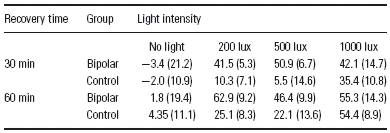
The average amount of melatonin increase 30 and 60 minutes after lights off at the different intensities investigated is shown in Table 3.
Recovery in melatonin levels after lights off for 30 and 60 min (% increase in melatonin since lights off and SEM)
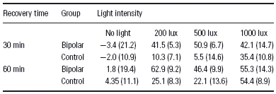
The results indicated that 30 min following lights off, melatonin concentrations significantly rebounded in all of the light conditions while there was no significant change in the control condition, (F(3,50) = 3.452, p < 0.05). Further, there is a significant difference between the groups (F(1,50) = 4.662, p < 0.05) with bipolar patients rebounding significantly more than control subjects.
After 60 min both groups showed considerable rebound in melatonin concentrations with a significant increase in melatonin levels in all but the control (no light) condition, F(3,50) = 5.558, p < 0.005. After 60 min the bipolar and control groups did not significantly differ in their recovery after the lights, F(1,50) = 2.579, p = 0.116.
Discussion
The results of this study support the hypothesis that patients with bipolar type I disorder show greater sensitivity to light than healthy adult controls. The greater sensitivity observed in patients occurred over a range of light intensities indicating a consistent super-sensitivity in this group. The data provide additional evidence of an abnormality in the photo-neuroendocrine system of patients with bipolar disorder. A number of previous studies have demonstrated similar such sensitivity in this patient group (Reference Lewy, Wehr, Goodwin and Newsome1–Reference Nurnberger, Berrettini, Tamarkin, Hamovit, Norton and Gershon4,Reference Nurnberger, Adkins and Lahiri8,Reference Leibenluft, Turner, Feldman-Naim, Schwartz, Wehr and Rosenthal29–Reference Hallam, Olver and Norman31). More specifically, this study indicates that abnormalities relating to sensitivity may occur upstream of the pineal gland as both the overall melatonin secretion and the ability to return to melatonin synthesis following late-night light (as measured at 30 and 60 min) were similar in patients and controls. Only melatonin sensitivity to light was changed. Alterations to either of these parameters would indicate overall changes in melatonin synthesis and secretion. Potentially light sensitivity is most likely to be affected as a result of changes at the level of the suprachiasmatic nucleus or the visual organs of the retinohypothalamic tract.
Clinically, the present results provide evidence of the major role that zeitgebers, particularly external lighting, play in disruption of the sleep/wake cycle of bipolar patients. Patients with bipolar disorder are particularly vulnerable to disruptions of the sleep wake cycle, which may be both a precipitant and a symptom of manic (and often depressive) episodes. More specifically, these results indicate that in education of bipolar patients about appropriate sleep hygiene rituals (such as in social rhythm therapy Reference Wirz-Justice, Quinto, Cagochen, Werth and Hock(32,Reference Frank, Swartz and Kupfer33)) should ideally include instruction on controlling lighting conditions. Owing to super-sensitivity to light, as shown here, exposure at night preceding sleep can disrupt (or suppress almost entirely if the environment does not become dark) the acrophase of melatonin secretion and associated sleep propensity.
The study has established a dose-response relationship between light intensity and melatonin suppression in both patients and controls. Regression analysis of transformed data revealed that both groups showed a similar (and predictable) response to light, but that the dose response in the patient group was more sensitive. With respect to previous research, at similar light intensities both patient and control groups show melatonin suppression similar to that observed previously. For the patient group, 200 lux of light in this study led to 51% suppression compared with 44% in our previous study Reference Nathan, Burrows and Norman(3). At 500 lux, patients in this study had 61% suppression, identical to that observed in patients in Lewy's study Reference Lewy, Nurnberger and Wehr(2) and the bipolar I sub-group in Nurnberger's study Reference Nurnberger and Foroud(6). At 1000 lux, the patients in the current study showed 73% sensitivity, which falls between the 61% sensitivity described at 500 lux Reference Lewy, Nurnberger and Wehr(2) and 88% sensitivity in a case study using 2000 lux Reference Lewy, Kern, Rosenthal and Wehr(34). In controls, this study again identified a clear dose-response curve, where increasing light intensity was associated with decreases in melatonin levels. There was robust agreement between the numerical values for melatonin suppression by light in this study and that in our previously reported study Reference McIntyre, Norman, Burrows and Armstrong(14,Reference Nathan, Wyndham, Burrows and Norman15). While there is some variation between studies in regard to the exact sensitivity values, a reproducible dose-response relationship is evident across studies.
A number of previous studies that report on sensitivity in healthy controls indicate heterogenous melatonin sensitivity levels in the normal population Reference Whalley, Perini, Shering and Bennie(5) possibly confounding patient/control group differences. The current study investigated sensitivity in 34 healthy adults with no family psychiatric history. In this group, four participants showed sensitivity similar to the patients group (within 9%) indicating super-sensitivity may occur in a small group of controls. As Lewy et al. have postulated that the existence of this super-sensitivity within healthy controls is consistent with the usefulness of the response as a potentialtrait marker (endophenotype) of illness as it may reflect a predisposition or vulnerability factor in phenotypically healthy individuals Reference Lewy, Nurnberger and Wehr(2).
Other measures of melatonin evaluated in the study indicated no overall differences between patients and controls for melatonin concentrations between 20:00 and 24:00 h. This result is likely attributable to wide inter-individual variation of melatonin secretion. This also accords with the evidence of complex and indirect relationships between overall melatonin levels and depressive disorders (Reference Beck-Friis, Ljunggren, Thoren, Von Rosen, Kjellman and Wetterberg35–Reference Leibenluft, Feldman-Naim, Turner, Schwartz and Wehr40). The study also examined a measure of melatonin rebound, i.e. the extent of recovery of melatonin concentrations 30 and 60 min after the midnight light treatment was extinguished. This analysis indicated that 30 min after lights off, both groups had a significant recovery in melatonin secretion irrespective of the level of light exposure. Bipolar patients showed significantly higher recovery levels associated with the greater depletion during the light period. After 60 min these melatonin concentrations had normalised in both groups. This provides further evidence that the primary abnormality in melatonin regulation in bipolar disorder patients is related to sensitivity to light rather than melatonin synthetic tone per se.
A clear disadvantage of the current study is the small sample size used, particularly in those with bipolar disorder. This issue raises concerns over the possibility of a type II error in the data and a lack of power to detect significant differences in factors such as overall melatonin secretion. Similar research in the future would ideally include larger numbers of bipolar patients to provide the power necessary for data interpretation. Because of this risk of type II error, some negative results must be interpreted with caution and definitive conclusions avoided. A further concern caused by the small sample size is the significant difference in age between the control and bipolar samples. The lack of age matching represents the general characteristics of control groups in this form of research (generally young tertiary students). As the time lapsed between symptom onset and correct diagnosis is often significant Reference Ghaemi, Sachs, Chiou and Goodwin(41), the bipolar sample were likely to be further differentiated from this young control group. While evidence shows that age is not a significant predictor of melatonin sensitivity to light Reference Nathan, Burrows and Norman(42), this factor must remain a limitation based on the clear age differential between control and patient groups. Medication differences between patient and control groups may be a further limitation of the study. All patients in this trial were on maintenance mood stabiliser therapy while controls were taking no medication. In order to assess the impact of medication we have previously shown that lithium and valproate significantly decrease dark adjusted melatonin suppression Reference Hallam, Olver, Horgan, McGrath and Norman(9,Reference Hallam, Olver and Norman10) in healthy controls administered these substances for 1 week. While this provides significant data about the impact of these medications, it is notable that the effect of these medications in patients with bipolar is untested and thus cannot be excluded as a contributing factor for the study findings. In contrast to these limitations, this study had a number of advantages. One of the major advantages of analysing a small but highly homogenous group was the selection of patients with a clear history of bipolar I with few comorbidities. Most of the patients showed a high genetic loading for the disorder and a history of numerous hospital admissions (minimum 3) with clear symptoms of mania. Finally, all patients had relatively stable sleeping habits during euthymic phases. Overall, these positive findings in a small but highly homogenous group of bipolar I probands support the contention of Nurnberger et al. Reference Nurnberger and Foroud(6) that patients with clear bipolar I symptoms display super-sensitivity of the melatonin response to light. They further support the investigation of these circadian probes in individuals with bipolar disorder Reference Lewy, Kern, Rosenthal and Wehr(34).
Bipolar disorder has a high genetic heritability estimate (Reference Gershon, Hamovit and Guroff43–Reference McQueen, Devlin and Faraone54), particularly associated with a strong family history of unipolar depressive illness Reference McGuffin, Rijsdijk, Andrew, Sham, Katz and Cardno(52,Reference Smoller and Finn55). Research has also shown that individuals with a strong genetic loading for bipolar affective disorder by virtue of their family history of affective illness have melatonin super-sensitivity to light Reference Nurnberger, Berrettini, Tamarkin, Hamovit, Norton and Gershon(4). Moreover, in Nurnberger's study, one particularly pertinent finding is that increasing genetic loading for the disorder (i.e. one parent vs. two with affective illness) is associated with increased sensitivity. In addition to these data, a recent twin study in controls has indicated that melatonin sensitivity to light is highly heritable and potentially reflects stable genetic processes Reference Hallam, Olver, Chambers, Begg, McGrath and Norman(56). Interesting is one case of monozygotic twins discordant for bipolar disorder and for melatonin sensitivity to light. These twins have melatonin light sensitivity profiles across four different light intensities concordant with their status as either patient or non-affected control Reference Hallam, Olver and Norman(31). The current patient sample again showed both a strong family history of affective illness (particularly bipolar disorder) and super-sensitivity to light, adding further evidence of a relationship between increased risk of super-sensitivity and genetic loading for the illness.
While it may be argued that this super-sensitivity is an epiphenomenon of the pharmacological treatment of the disorder, evidence indicates that this is unlikely. In particular, Nurnberger et al. Reference Nurnberger and Foroud(6) noted the highest incidence of melatonin super-sensitivity was in bipolar patients who had been lithium free for more than 5 weeks. Following this, we investigated sensitivity following lithium administration in healthy adult volunteers. The results of this study showed that lithium treatment significantly decreased melatonin sensitivity to light Reference Hallam, Olver, Horgan, McGrath and Norman(9). Follow-up research with sodium valproate also revealed a reduction in sensitivity Reference Hallam, Olver and Norman(10) indicating an overall effect of mood stabilisers on reducing sensitivity to light possibly linked to the therapeutic mechanism of action.
These results indicate that future studies on sensitivity to light in bipolar disorder would benefit from comprehensive analyses of more homogenous groups, rather than simply increasing sample sizes. These groups should ideally examine type I and II patients separately. A group of patients of particular interest are those with rapid-cycling bipolar disorder. These patients show the greatest diurnal variations in mood (from morning depression to evening (hypo)mania) which may be associated with environmental changes such as light level Reference Leibenluft, Feldman-Naim, Turner, Schwartz and Wehr(40). In one study using light therapy, rapid-cycling patients were shown to be highly sensitive to the effects of light therapy: midday light associated with clinical improvements but morning light associated with hypomania Reference Leibenluft, Turner, Feldman-Naim, Schwartz, Wehr and Rosenthal(29). A more recent study further showed the significant impact of midday light on patients with bipolar disorder Reference Sit, Wisner, Hanusa, Stull and Terman(57). These data indicate the importance of assessing diurnal light history as well as nocturnal interventions in this patient group. It is notable that rapid-cycling bipolar patients are particularly responsive to circadian-based treatments that include enforced darkness and bed rest indicating the importance of circadian regulation in the amelioration of symptoms Reference Wirz-Justice, Quinto, Cagochen, Werth and Hock(32,Reference Wehr, Turner, Shimada, Lowe, Barker and Leibenluft58).
Overall, the findings from this study indicate a shifted dose-response effect for the response of nocturnal melatonin to light in bipolar patients. While both bipolar and control groups showed a significant decrease in melatonin with increasing light level, the bipolar I group were consistently more sensitive at each of the light intensities. This provides support for the notion of super-sensitivity to light in patients with bipolar I disorder as patients showed a consistent shift in sensitivity across a range of light levels in comparison with healthy controls.
This study further investigates the usefulness of melatonin super-sensitivity to light as an endophenotypic marker of bipolar disorder Reference Lenox, Gould and Manji(59). Further research is required to validate the response as a useful clinical test to differentiate among disorders. This will involve studies on both the sensitivity and specificity of the response and research on other clinical populations. Bipolar disorder has both the advantage and disadvantage of being a highly heritable disease. The use of endophenotypic markers that integrate both clinical and biological observations may provide a powerful tool to target genetic, molecular and behavioural changes in the disorder. Ideally, these observations will inform both aetiological and neurobiological studies of the disorder and ultimately improve clinical treatment of the illness.