During pregnancy, the immune system has two vital functions: to protect the mother against infections and to tolerate the semiallogeneic fetus. The fact that the maternal immune system does not mount an aggressive immunological reaction against the placenta and the fetus, which bear paternal, and thus foreign, antigens has attracted considerable interest. While it was earlier believed that the maternal immune system could not detect the growing fetus, it is now known that the maternal immune system not only becomes aware of the fetus but also actively tolerates it. Several mechanisms have been proposed to explain tolerance, which might co-exist to ensure compensatory mechanisms when one of them is absent. Increased understanding of these mechanisms should provide insight into pregnancy complications, such as spontaneous abortion and preeclampsia, which may arise partly from incomplete tolerance.
Explaining the paradox
The paradoxical nature of the immunological relationship between a pregnant mother and her antigenically foreign child was first recognised by Peter Medawar in 1953 (Ref. Reference Medawar1). He proposed three reasons as to why the fetus does not usually provoke an immunological reaction from its mother: (1) anatomical separation of the embryo from its mother, by a barrier considered at that time to be impermeable to maternal cells; (2) antigenic immaturity of the fetus; and (3) immunological inertness of the mother. However, as a result of increasingly precise research methods, none of these explanations is valid anymore.
First, it is now known that in species with hemochorial placentation (as in humans and rodents), the mother and the fetus are not separated by an intact layer of epithelial cells; trophoblasts are thus exposed to maternal immune cells, which have the potential to develop a strong immune response to reject the foreign tissues. Bidirectional transfer of cells between mother and fetus is common during human and murine pregnancy, and can lead to microchimaerism (persistence of fetal cells in the mother for decades) (Refs Reference Billington2, Reference Andrassy3, Reference Yan4, Reference Khoshrotehran5, Reference Nelson6 and Zenclussen, A.C., unpublished). Chaouat and colleagues (Ref. Reference Chaouat, Kolb and Wegmann7) as well as Wegmann (Ref. Reference Wegmann8) revised the concept of the anatomical separation and proposed the ‘trophoblast barrier–immunotrophism mechanism’, where the trophoblast cells play a protective role for the rejectable fetus within the maternal immune system, not by creating a barrier but by actively contributing to tolerance through the secretion of soluble factors.
Second, the fetus may not be antigenically immature: dendritic cells (DCs) derived from fetal skin, which are in direct contact with maternal blood and therefore capable of entering the maternal circulation, express major histocompatibility complex (MHC) class II antigens and are potent accessory cells in polyclonal T-cell responses (Ref. Reference Elbe-Burger9). Third, with regard to immunological inertness of the mother, there is clear evidence that maternal T-cell recognition of fetal antigens can lead to antigen-specific tolerance. In mice, T cells expressing transgenic T-cell receptors (TCRs) specific for paternal antigens recognise paternal alloantigens expressed by the fetus, and this results in transient tolerance of the T cells (Ref. Reference Tafuri10). Thus, not only does the maternal immune system respond to the fetus, it is also actively tolerised by it (Ref. Reference Jiang and Vacchio11). Besides, systemic immune activation occurs in pregnancy, and women do not usually suffer from immunosuppression during pregnancy. In this regard, the fetal–maternal interface can be considered as a site of immune privilege, where fetal tissue directs the maternal immune response towards a protective, tolerant one.
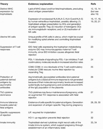
Since Medawar's hypothesis, a variety of mechanisms have been proposed to explain the survival of the semiallogeneic fetus in pregnancy (Table 1 and references therein), particularly with regard to the active generation of tolerance. These mechanisms may act through distinct or overlapping pathways, and some of them are associated with the generation or action of regulatory T cells (Treg). Redundancy in the systems allowing pregnancy success is suggested by the fact that normal pregnancies are observed in mice lacking the T-cell suppressor IDO (Ref. Reference Baban35) or T helper 2 (Th2) cells (Refs Reference Svensson36, Reference Fallon37). The Th1–Th2 cytokine balance and Treg have been seen as particularly important in pregnancy success and are discussed further below. This review also summarises recent work on the enzyme HO-1, which may act partly by modulating immune responses to the fetus.
Some of the current theories explaining the survival of the semiallogeneic fetus within the maternal uterus

Abbreviations: DC, dendritic cell; HO-1, heme oxygenase 1; IDO, indoleamine 2,3-dioxygenase; LIF, leukaemia inhibitory factor; MHC, major histocompatibility complex; NK, natural killer; PDL-1, programmed death ligand 1; Treg, regulatory T cells.
Th1–Th2 cytokine balance in pregnancy
Over the past decade, the Th1–Th2 cytokine balance has been seen as a very important mechanism determining the survival of the fetus in the maternal uterus (Refs Reference Lin27, Reference Raghupathy38, Reference Saito39, Reference Piccinni40). Production of Th2-type cytokines such as the interleukins IL-4 and IL-10 locally at the fetal–maternal interface was thought to favour the maintenance of mammalian pregnancy (Refs Reference Lin27, Reference Piccinni40), whereas a Th1-dominant pattern [up-regulation of IL-2, IL-6, interferon γ (IFN-γ) and tumour necrosis factor α (TNF-α)], as seen during miscarriage, was thought to mediate fetal rejection (Refs Reference Lin27, Reference Raghupathy38, Reference Zenclussen41) by provoking an inflammatory milieu incompatible with pregnancy success. Other immunosuppressive molecules, especially transforming growth factor β (TGF-β), a Th3 cytokine, have also been proposed to play an important role during pregnancy (Refs Reference Gorivodsky42, Reference Dünker and Krieglstein43).
This importance of the Th1–Th2 balance for pregnancy outcome was questioned recently because normal pregnancies were observed in mice that lacked Th2 cytokines, as in IL-10/IL-4 knock-out mice (Ref. Reference Svensson36) and even in mice with quadruple gene deletions leading to lack of IL-4, IL-5, IL-9 and IL-13 (Ref. Reference Fallon37). This suggests that Th2 cells are not essential for normal pregnancy, and other mechanisms may prevent alloreactive Th1 attack on the fetus. Furthermore, up-regulated levels of Th1 cytokines observed during miscarriage might not be the main cause of abortion but rather a result of immunological rejection and inflammation. Nevertheless, IL-4 and IL-10 are related to successful pregnancy outcome in humans (reviewed in Ref. Reference Dealtry, O'Farrell and Fernandez44), and the administration of IL-10 significantly decreased the abortion rate in the CBA/J x DBA/2J murine model (which has a high rate of abortion) (Ref. Reference Chaouat45). In addition, although IL-10 is not essential for maternal immune tolerance, maternal IL-10 is a determinant of the growth trajectory of progeny in the uterus and after birth (Ref. Reference White46). Recent evidence outlined below has shed more light on a role for Th1 cells in pregnancy failure.
Th1 cells and miscarriage: evidence from murine models
Complex selective mechanisms are thought to regulate leukocyte access to the fetal–maternal interface (Ref. Reference Kruse47), and, unlike Th2 cells, Th1 cells are able to bind to the cellular adhesion molecules P- and E-selectins on endothelial cells (Ref. Reference Austrup48). Interestingly, blockage of P-selectin (using a monoclonal antibody) positively influences pregnancy outcome by significantly diminishing the abortion rate in a murine model (Ref. Reference Bertoja49), consistent with a role for Th1 in miscarriage. Furthermore, P-selectin is augmented in decidua of women suffering from spontaneous abortion (Ref. Reference Zenclussen50).
Although the number of Th1 cells at the fetal–maternal interface in mice and humans during miscarriage is known to be augmented (Refs Reference Raghupathy38, Reference Zenclussen41), it has been less clear whether these cells are reactive against paternal antigens. The first evidence of an increased Th1 response towards male antigens was provided by Tangri et al. (Ref. Reference Tangri and Raghupathy28). In a more recent study, the ex vivo ability of spleen and decidual lymphocytes from abortion-prone (CBA/J x DBA/2J) and normal (CBA/J x BALB/c) mice on day 14 of pregnancy to produce Th1 and Th2 cytokines after both nonspecific stimulation and specific stimulation by male antigens was analysed. An enhanced frequency of paternal-alloantigen-specific Th1 memory/effector lymphocytes in decidual tissue from abortion-prone mice was found (Ref. Reference Zenclussen30), although spleen cells showed comparable polyclonal and paternal-alloantigen-specific Th1 frequencies in both combinations. These data suggest that systemic generation of paternal-alloantigen-specific Th1 memory/effector cells is not inhibited during normal pregnancy but their activity is normally down-regulated at the fetal–maternal interface.
Th1 cells and preeclampsia
Transfer of activated Th1 cells into pregnant, but not nonpregnant, mice leads to signs of preeclampsia (Ref. Reference Zenclussen51). In this model, uterine lymphocytes from preeclamptic mice produced significantly more Th1 cytokines (TNF-α and IL-12) compared with normal pregnant mice, and transfer of labelled cells showed that most of the Th1-infiltrating cells at the fetal–maternal interface were host and not transferred cells. It is possible that Th1-type cytokines produced by the transferred cells expand and stimulate host cells primed to paternal antigens to secrete inflammatory cytokines. This would be consistent with the notion that a key cause of preeclampsia is an inflammatory host response (Refs Reference Pijnenborg52, Reference Faas53). It is also possible that the Th1 cytokines secreted by transferred immune cells induce infiltration of macrophages or other antigen-presenting cells, which prime cytotoxic T cells to paternal antigens expressed by fetal cells.
IL-6 and asymmetric antibodies
While the negative effects of traditional Th1 cytokines on pregnancy were clear (Refs Reference Lin27, Reference Raghupathy38, Reference Saito39), the role of the pleiotropic cytokine IL-6 (Ref. Reference Akira, Taga and Kishimoto54) has been controversial. IL-6 contributes to normal trophoblast growth and placental development in humans (Ref. Reference Nishino55), but may have negative effects in the mouse. IL-6 production by trophoblasts and immune cells at the fetal–maternal interface is increased in abortion-prone mice (Ref. Reference Zenclussen41), IL-6 is toxic to mouse embryos (Ref. Reference Wu56), and IL-6 has been shown to be one of the mediators in the lipolysaccharide-injection induced-abortion model (Refs Reference Evans57, Reference Wang, Tawfik and Wood58).
It is known that IL-6 regulates the synthesis of various molecules at the fetal–maternal interface in a dose-dependent fashion, such as gelatinases (Ref. Reference Meisser59) and asymmetric antibodies (Ref. Reference Margni and Zenclussen60). Asymmetric antibodies are elevated in sera and the placenta during pregnancy, where they may have a protective role by blocking paternal antigens and avoiding the development of a classical immune reaction characterised by complement fixation, clearance of antigens and phagocytosis (Refs Reference Malan Borel26, Reference Margni and Malan Borel61, Reference Zenclussen62). High IL-6 levels are accompanied by low levels of asymmetric antibodies, which may result in miscarriage (Refs Reference Zenclussen41, Reference Zenclussen62, Reference Zenclussen63). IL-6 might also act negatively in pregnancy by inhibiting the generation of TGF-β-induced Treg (Ref. Reference Bettelli64).
Regulatory T cells in pregnancy
CD4 +CD25 + Treg are a unique subpopulation of T cells (reviewed in Ref. Reference Sakaguchi65) known to play a major role in preventing autoimmunity and tolerating allogeneic organ grafts (Refs Reference Waldmann66, Reference Sakaguchi67), acting either by cell–cell contact or by secreting immunomodulatory factors such as IL-10 and TGF-β. The acceptance of paternally derived tumour cells during pregnancy (Ref. Reference Tafuri10) supports the involvement of systemic regulatory processes in pregnancy. The existence of a regulatory T-cell population was first proposed in the early 1980s by Chaouat and Voisin (Ref. Reference Chaouat and Voisin68), although the identification of this population was hampered by the techniques available at that time.
Recent studies have shed more light on the role of Treg in pregnancy. During normal pregnancy in mice, there is an expansion of Treg (Ref. Reference Aluvihare, Kallikourdis and Betz29) and, interestingly, mice undergoing abortion have a diminished number of Treg as compared with normal pregnant mice (Ref. Reference Zenclussen30). Additionally, human studies support the important role of Treg in pregnancy. Normal human pregnancy is associated with an increase in the circulating Treg (Refs Reference Somerset69, Reference Sasaki70). Furthermore, the proportion of decidual CD4 +CD25 + T cells was significantly lower in specimens from spontaneous abortion compared with specimens from induced abortions (Ref. Reference Sasaki70).
The importance of Treg
In vitro analysis showed an inhibitory capacity of CD4 +CD25 + T cells on maternal alloresponses; for example, Treg inhibit the proliferation of decidual effector cells as well as their IFN-γ production in vitro (Refs Reference Aluvihare, Kallikourdis and Betz29, Reference Zenclussen30). Accordingly, depletion of CD25 + cells by using a monoclonal neutralising antibody led to impaired pregnancy outcome (Ref. Reference Zenclussen30). Furthermore, in a model system in which lymphocytes (from BALB/c mice pregnant following C57BL/6 matings) were transferred into T-cell-deficient nude (nu/nu) BALB/c females (subsequently paired with C57BL/6 males), no normal pregnancies were observed when CD25 + cells were depleted from the lymphocyte preparation. Although this system is a very artificial one and nu/nu mice do not mate successfully, these data confirm that the absence of Treg can lead to pregnancy failure (Ref. Reference Aluvihare, Kallikourdis and Betz29).
Further work has shown that miscarriage can be prevented by transfer of Treg from normal pregnant mice. Adoptive transfer of CD4 +CD25 + Treg from normal pregnant (CBA/J x BALB/c) but not from nonpregnant CBA/J mice completely prevented spontaneous abortion in abortion-prone (CBA/J x DBA/2) mice (Ref. Reference Zenclussen30) when applied at 0–2 days of pregnancy. Work not yet published indicates that the Treg are acting in a paternal-antigen-specific manner (Zenclussen, A.C. et al. unpublished). Transfer of Treg from normal pregnant mice failed to prevent abortion if carried out on days 4–5 of pregnancy and anti-CD25 applied during days 0–2 of pregnancy led to implantation failure (Ref. Reference Zenclussen30), suggesting that Treg are necessary locally before implantation. This is an interesting point as the situation is very similar to the one observed in transplantation, where Treg are needed before grafting to prevent tolerance (Ref. Reference Karim71).
The administration of a monoclonal antibody against CD86 at an early gestational stage to abortion-prone DBA/2J-mated CBA/J females could suppress maternal immune attack on the fetus. The fact that the treatment with anti-CD86 expanded the peripheral CD4+CD25+ population indicates that the lack of Treg in this particular combination can be corrected by blocking the costimulatory signal of CD86 while keeping the CD80 signal unimpaired (Ref. Reference Zhu25).
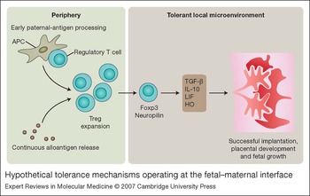
We propose a hypothetical scenario in which Treg are activated in early pregnancy by paternal antigens, possibly even as a result of encountering paternal antigens in the vaginal lumen. Thereafter, Treg migrate to the lymph nodes and after implantation to the fetal–maternal interface. Continuous release of placental antigens into the maternal circulation would allow the maintenance of a Treg population specific for paternal antigens, to allow tolerance towards the semiallogeneic fetus until the time of birth.
The action of Treg: generation of a tolerant microenvironment?
There is no clear evidence that Treg actions in pregnancy include inhibition of Th1 responses (Ref. Reference Zenclussen72). An interesting theory presented by Waldmann and collaborators postulates that Treg might affect several cellular pathways to establish a state of privilege in the tissue microenvironment (Ref. Reference Waldmann66). Some of the putative protective pathways induced after Treg action are IL-10 and TGF-β production, enhanced IDO expression, or HO-1 induction (Ref. Reference Waldmann66).
Interestingly, increased TGF-β protein is found in mice rescued from abortion by Treg transfer (Ref. Reference Zenclussen72); thus augmented local TGF-β levels might be related to Treg-induced tolerance during pregnancy, at least in this particular model of spontaneous abortion. HO-1 mRNA levels were also increased in placenta and decidua in animals that received Treg, suggesting a critical role of HO-1 in Treg-induced tolerance. Interestingly, HO-1 up-regulation was related to an augmentation in the Treg marker neuropilin-1 (Ref. Reference Sollwedel73), which might indicate cross-talk between both systems during pregnancy (see later). Dramatically augmented local levels of LIF after Treg application suggest an important role of LIF in Treg-induced tolerance at the fetal–maternal interface (Ref. Reference Zenclussen72). LIF is essential for implantation success: animals lacking the LIF gene produce normal blastocysts that fail to implant into a LIF-deficient uterus but are able to implant in a wild-type uterus (Ref. Reference Stewart31). Interestingly, LIF was also found to be up-regulated in CBA/J mice that naturally accepted skin allografts from BALB/c mice (Ref. Reference Metcalfe, De and Muthukumarana74), indicating that it may also be involved in tolerance of allografts. Thus, Treg at the fetal–maternal interface may be effective in avoiding allorejection by interacting with tissues and creating a ‘tolerant’ privileged microenvironment characterised by high expression of TGF-β, HO-1 and LIF (Fig. 1).
Hypothetical tolerance mechanisms operating at the fetal–maternal interface. Briefly, we propose that during pregnancy, regulatory T cells (Treg; characterised by Foxp3 and neuropilin-1 expression) are generated after contact with male antigens. This population expands in the periphery as a result of continuous alloantigen release from the placenta. At the fetal–maternal interface, these cells are thought inhibit maternal effector cells and further up-regulate tolerant molecules, such as heme oxygenase 1 (HO-1), leukaemia inhibitory factor (LIF), transforming growth factor β (TGF-β) and interleukin 10 (IL-10). HO-1 and LIF are expressed mainly by trophoblasts, whereas TGF-β can also be released by immune cells, including Treg. Together, these mechanisms may contribute to the tolerance towards the fetal antigens. APC, antigen-presenting cell.

Heme oxygenase 1 during pregnancy
The enzyme HO degrades heme, a protein involved in several vital cellular processes including cell differentiation and proliferation (Refs Reference Abraham, Friedland, Levere and Brown75, Reference Abraham76, Reference Beri and Chandra77, Reference Sassa and Nagai78, Reference Ponka79). It is synthesised in all human nucleated cells, although its importance varies between tissues. Senescent red blood cells, a major source of excess heme, have to be removed from the circulation and are then degraded by the reticulo-endothelial system in the spleen, liver and bone marrow (Ref. Reference Ponka79). Excess free heme can cause cell damage and tissue injury as heme catalyses the formation of reactive oxygen species, resulting in oxidative stress (Refs Reference Vercellotti80, Reference Jeney81); in addition, it can easily intercalate in cellular and intracellular membranes, further destabilising the cytoskeleton (Refs Reference Balla82, Reference Ryter and Tyrrell83). It is thus of vital importance that the amount of free heme is tightly controlled to maintain cellular homeostasis and avoid pathological conditions. HO breaks down the porphyrin ring to yield equimolar amounts of biliverdin, free iron (Fe2 +), and carbon monoxide (CO) (Ref. Reference Beri and Chandra77). The beneficial effects of HO are related to the avoidance of the toxic accumulation of free heme as well as with the antioxidant and protective functions of bilirubin and biliverdin (Refs Reference Otterbein and Choi84, Reference Yesilkaya, Altinayak and Korgun85). Furthermore, the intermediate metabolite CO has been postulated to have protective functions since it promotes reactions that tend to protect the tissues by stimulating blood vessel relaxation (Refs Reference Vedernikov, Graser and Vanin86, Reference Ryter and Choi87).
To date, three isoforms of heme oxygenase have been identified: HO-1, HO-2 and HO-3 (Refs Reference Ryter and Tyrrell83, Reference Maines88). HO-1 and HO-2 share little similarity in amino acid sequence (40%), whereas the HO-2 and HO-3 isoform are far more homologous (90%). All HO isoforms are highly conserved among species. HO is expressed in prokaryotic bacteria as well as in fungi, plants and humans. HO proteins are microsomal and are anchored to the endoplasmic reticulum.
HO-1, -2 and -3 are products of different genes, and show distinct expression patterns and regulation (Ref. Reference Montellano89). Under normal physiological conditions, most cells express low or undetectable levels of HO-1 protein, while HO-2 is constitutively expressed. Very little is known about HO-3. HO-1 gene expression is highly inducible by several factors including heavy metals, bacterial lipopolysaccharides, hypoxia, hyperoxia, heat shock, ischaemia, UV radiation, H2O2, cytokines, nitric oxide, stimuli that deplete cellular glutathione stores, and its substrate heme. HO-1 gene expression is often cell-type- and species-specific (Ref. Reference Morse and Choi90). HO-2 transcription is up-regulated by only a few agents such as opiates and adrenal glucocorticoids (Refs Reference Li and Clark91, Reference Liu92). HO-1 is active even at low substrate concentrations. By contrast, HO-2 has low activity at low substrate concentrations, and even when it is at its maximal activity, it is limited to less than 10% that of HO-1 (Ref. Reference Maines and Panahian93). Specific activity of HO in different organs greatly varies. The highest HO activity is found in spleen, testis, and brain. HO-2 is abundant in brain, testis, placenta and liver. HO-3 has been identified in brain, kidney, liver, heart, testis, and spleen and has been reported to have only a very poor heme-degrading capacity (Ref. Reference McCoubrey, Huang and Maines94).
Up-regulation of HO-1 allows the acceptance of mouse and rat allografts (Refs Reference Woo95, Reference Chauveau96), while its down-regulation or absence is directly related to acute graft rejection (Ref. Reference Soares97). With regard to pregnancy, immunohistochemical staining shows that HO-1 and HO-2 are expressed in different trophoblast types of the placenta of rodents and humans (Refs Reference Zenclussen98, Reference Ihara99, Reference Zenclussen100). The expression of HO-1 protein and mRNA peaks during implantation [Ritschel, S. (2006) Diploma Thesis. Technische Fachhochschule, Berlin, Germany].
Heme protein catabolism by trophoblast-derived HO would be expected to prevent accumulation or recirculation of free heme, which could be extremely toxic to the mother and fetus. Indeed, down-regulated levels of HO-1 and HO-2 have been observed at the fetal–maternal interface in mice undergoing abortion (Ref. Reference Zenclussen98). Furthermore, human miscarriage and preeclampsia have been associated with diminished placental HO levels (Refs Reference Barber101, Reference Zenclussen102). A microsatellite polymorphism of the HO-1 gene (leading to low levels of HO-1 expression) is also associated with idiopathic recurrent miscarriage in humans (Ref. Reference Denschlag103). Interestingly, HO-1 knock-out mice are not able to reproduce (Ref. Reference Poss and Tonegawa104), suggesting a crucial role for HO-1.
HO-1 manipulation and its impact on pregnancy outcome
The finding of diminished levels of HO-1 in animals undergoing abortion led to the assumption that an augmentation in HO-1 levels during implantation would improve pregnancy outcome. In fact, a specific increase in systemic HO-1 as a result of expression of HO-1 following adenoviral gene transfer improved pregnancy outcome in a murine model of abortion by diminishing the fetal rejection rate (Ref. Reference Zenclussen32). A more clinically relevant approach, the up-regulation of HO-1 by means of cobalt protoporphyrin (CoPPIX), also significantly diminished abortion rates (Ref. Reference Metcalfe, De and Muthukumarana74). This is consistent with earlier work showing that increased levels of HO-1 following CoPPIX treatment inhibited early inflammatory events following allograft transplantation and therefore prevented chronic graft rejection (Refs Reference Woo95, Reference Soares97). Conversely, animals that received a single dose of zinc protoporphyrin (ZnPPIX), which is known to inhibit not only HO-1 but also HO-2 (Ref. Reference Sollwedel73), showed increased abortion rates, confirming that a down-regulation of HOs during implantation endangers pregnancy.
In the adenoviral HO-1 gene transfer model, secretion of Th2 cytokines was increased, while Th1 cytokine production was decreased, as shown by the ratio of IL-4 to IFN-γ and IL-10 to TNF-α in decidua or spleen (Ref. Reference Zenclussen32). Thus, the up-regulation of HO-1 may protect fetal tissues by stimulating maternal immune cells to produce less Th1 and more Th2 cytokines. However, contrary to expectations, CoPPIX treatment did not shift the cytokine profile to a Th2 one as the gene therapy approach did (Ref. Reference Sollwedel73). Regarding the possible influence of HO-1 in regulation of apoptosis-related events at the fetal–maternal interface, the adenoviral HO-1 treatment down-regulated placental caspase-3 activity and the number of apoptotic cells as compared with abortion-prone control mice (Ref. Reference Zenclussen32). Augmented mRNA levels of the anti-apoptotic factor Bag-1 (Ref. Reference Townsend105) reinforce the anti-apoptotic hypothesis (Ref. Reference Zenclussen32). Induction of HO-1 by CoPPIX also diminished caspase-3 activity and slightly diminished the number of apoptotic cells, while up-regulating Bag-1, suggesting that HO-1 might protect the fetal tissue from apoptosis/necrosis (Ref. Reference Sollwedel73). Bag-1 overexpression following HO-1 up-regulation may be directly involved in the prevention of abortion (Refs Reference Zenclussen32, Reference Sollwedel73).
Choi et al. recently proposed that Foxp3, a transcription factor essential for Treg development, could induce HO-1 expression (Ref. Reference Choi106), suggesting a connection between both pathways. In addition, neuropilin-1 mRNA levels were significantly augmented in placenta after CoPPIX treatment on day 14 of pregnancy. This suggests an augmentation in Treg number or activity at the fetal–maternal interface following HO-1 up-regulation.
HO in preeclampsia
Preeclampsia, which is characterised by placental hypoxia and abnormal utero-placental blood flow (Refs Reference Jaffe, Dorgan and Abramowicz107, Reference Trudinger, Giles and Cook108, Reference Appleton109, Reference Zhou, Damsky and Fisher110, Reference Marks111), is associated with reduced HO activity: diminished HO-1 and HO-2 levels were found in invasive and syncytiotrophoblasts in samples from preeclamptic patients as compared with age-matched controls (Ref. Reference Zenclussen102). This may result from the hypoxia, since reduced O2 availability in microsomal preparation from chorionic villi of noninfarcted areas of placentas from preeclampsia or normal pregnancies decreased HO activity (Ref. Reference Appleton109).
Decreased HO activity may lead to platelet aggregation, reduced blood flow and the inadequate secondary cytotrophoblast invasion seen in preeclampia (Refs Reference Pijnenborg52, Reference Zhou, Damsky and Fisher110) through lower levels of CO, a product of HO activity. CO inhibits the transcription of hypoxia-inducible genes (Ref. Reference Marks111) and has a key role in cell–cell communication and in blood vessel relaxation (Ref. Reference Verma112). Indeed, a significant dose-dependent constriction within the placental vasculature in the presence of the HO inhibitor ZnPPIX was observed in in vitro studies, consistent with a role for CO in placental function (Ref. Reference Lyall113). Interestingly, smokers have a lower risk of preeclampsia, probably due to high CO levels in placental tissue (Ref. Reference Khoury114).
Conclusions and clinical implications
Although many mechanisms have been proposed to explain fetal tolerance, further research is necessary to elucidate fully the complex net of interactions guaranteeing implantation of a ‘foreign’ fetus, its growth and normal placental development. Nevertheless, there is enough experimental and clinical evidence to conclude that both Treg and HO-1 are very important for pregnancy outcome (Fig. 1). Future work is likely to focus on investigating their mechanisms of action and their interaction with other relevant pathways. Novel approaches such as microarray technologies may help in revealing new molecules associated with tolerance. The great advantage of the ongoing animal models of fetal tolerance is that tolerance can be achieved by manipulating early events.
Understanding of the interactions of components implicated in prevention of rejection of the fetus is essential for the design of therapies to prevent pregnancy complications such as spontaneous abortion and preeclampsia. The concept of redirecting the maternal immune system to recognise and tolerate paternal cells was introduced in the late 1970s by vaccinating mothers with cells from their partners before pregnancy (Refs Reference Taylor and Faulk115, Reference Takeshita116, Reference Hill117); this was considered a promising treatment because blood transfusion given before transplants was reported to decrease the rejection of organ allografts. Although very controversial (Refs Reference Takeshita116, Reference Hill117, Reference Christiansen, Nielsen and Kolte118), this treatment has been shown to be effective in cases of immunological spontaneous abortions (Ref. Reference Mowbray119). It was recently reported at a scientific meeting (Ref. Reference Marget120) that the treatment of patients with lymphocytes from their partners led to augmented levels of Foxp3, suggesting generation of Treg. Although speculative, the idea of using maternal Treg specific for paternal antigens as a therapy is tempting. Murine data show that it is possible to generate ex vivo antigen-specific cells with suppressor activity. Whether this therapy is an applicable one remains to be discussed in the light of ethical and safety issues.
Acknowledgements and funding
We thank the peer reviewers for their helpful comments, and Juliane Rau for her help in the graphic design. Part of the data discussed in the present review was funded by the Charite and by the Deutsche Forschungsgemeinschaft (DFG 526/4-1 and 526/5-1) to A.C.Z. P.W. is a DAAD fellow.