Introduction
Glucocorticoids (mainly cortisol in humans, corticosterone in rodents) have profound effects on brain functions. Chronic glucocorticoid excess has been suggested to cause or accelerate some aspects of degenerative brain aging, notably hippocampus-associated cognitive disorders (Seckl and Olsson, Reference Seckl and Olsson1995; Lupien et al., Reference Lupien2005).
Morning serum levels of adrenal and gonadal hormones have been reported to be elevated in mild to moderate Alzheimer's disease (AD), linked to abnormalities at several levels of the hypothalamus–pituitary–adrenal (HPA) axis (Näsman et al., Reference Näsman, Olsson, Fagerlund, Eriksson, Viitanen and Carlström1996; Rasmuson et al., Reference Rasmuson, Näsman, Eriksson, Carlström and Olsson1998; Reference Rasmuson, Näsman, Carlström and Olsson2002). Longitudinal studies on basal cortisol levels in AD have, however, given conflicting results. No association between longitudinal and cross-sectional findings could be demonstrated in one study (Swanwick et al., Reference Swanwick1998), but other studies have reported that increasing levels of glucocorticoids correlate to disease stage (Umegaki et al., Reference Umegaki2000; Csernansky et al., Reference Csernansky2006). Studies on diurnal rhythmicity of glucocorticoids in AD have also given divergent results. A flattened diurnal curve of cortisol in AD was reported in one study (Ferrari et al., Reference Ferrari2001) while another study showed no differences between a population of AD patients and a population of healthy elderly (Hartmann et al., Reference Hartmann, Veldhuis, Deuschle, Standhardt and Heuser1997).
Of importance, we have shown an increased glucocorticoid production in women with AD that may result from a primary increase in HPA axis activity and/or secondary to increased cortisol clearance (Rasmuson et al., Reference Rasmuson, Andrew, Näsman, Seckl, Walker and Olsson2001). In line with these findings, sex differences in the regulation of the HPA axis were evident in a meta-analysis of cortisol response after either pharmacological or physiological challenge in healthy participants with a three-fold increase in cortisol response in women compared with men (Otte et al., Reference Otte, Hart, Neylan, Marmar, Yaffe and Mohr2005).
Dehydroepiandrosterone (DHEA) is the most abundant steroid in human blood and has been linked to a broad range of physiological effects, including neuronal excitability and neuroprotection, and also has been suggested to counteract the adverse effects of glucocorticoids (Svec and Lopez, Reference Svec, Lopez and 1989). A causal link between decreased levels of dehydroepiandrosterone sulfate (DHEAS) and age-related cognitive decline has been suggested, supposed to be mediated at least in part through loss of an antiglucocorticoid effect (Näsman et al., Reference Näsman1991).
Inflammation mediated by activated microglia is an important factor in AD pathophysiology (Akiyama et al., Reference Akiyama2000; McGeer and McGeer, Reference McGeer and McGeer2003). Of note, immune-mediated cytokine release may affect HPA axis regulation at several levels (Bornstein and Chrousos, Reference Bornstein and Chrousos1999). There may thus be a link between an increased drive of the HPA axis and an inflammatory component in AD. In this exploratory study, we hypothesized that there is a flattened diurnal curve of cortisol and DHEA in mild to moderate AD and that proinflammatory cytokine levels are linked to steroid hormone levels in AD.
Methods
Study population
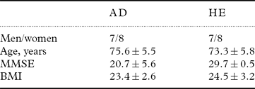
Fifteen patients with mild to moderate AD were included in the study (Table 1). They were selected from patients referred to the Department of Geriatric Medicine, Umeå University Hospital, Sweden. All patients were living at home and investigated as outpatients. A medical history was obtained from interviews with the patients and their family or caregivers. A full physical examination and a routine laboratory screening including plasma glucose, serum albumin, vitamin B12, folate, and thyroid hormone determination were done to rule out various infections, metabolic disorders, and other causes of dementia. Body mass index (BMI) was calculated by dividing body weight (kilograms) by height squared (meters). None of the patients had abnormal albumin levels or showed any signs of liver disease. Serum creatinine levels did not exceed 120 μmol/l. Computed tomography (CT) scans were performed to exclude focal processes in the central nervous system (CNS). The patients underwent neuropsychological assessment, and cognitive status was investigated using the Mini-Mental State Examination (MMSE) (Folstein et al., Reference Folstein, Folstein and McHugh1975). The Montgomery-Åsberg's Depression Scale was used to rate depressive symptoms (Montgomery and Åsberg, Reference Montgomery and Åsberg1979). All patients met the (NINCDS-ADRDA) criteria for probable AD. The Clinical Dementia Rating (CDR) (Hughes et al., Reference Hughes, Berg, Danziger, Coben and Martin1982) was used as a global rating scale; one patient had very early dementia (CDR = 0.5), four patients had mild dementia (CDR = 1), and ten had moderate dementia (CDR = 2).
Demographic and clinical characteristics of the patients: Alzheimer's disease (AD) and healthy elderly controls (HE)

All values are mean ± SD.
MMSE = Mini-Mental State Examination; BMI = Body Mass Index
The patients were not screened systematically for behavioral abnormalities. No patients with sleep disturbances (alterations of the sleep–wake cycle) were included, and none was taking any medication.
Fifteen healthy elderly controls were recruited and evaluated by physical examination, MMSE, and routine laboratory screening. None was taking any medication. The control group participants underwent a structured interview for detection of concomitant depression. Exclusion criteria for patients and controls were acute medical illness, endocrine disorders, depression, infectious disease, renal dysfunction, prostatic hyperplasia, smoking, and excessive alcohol intake. None of the women was on steroid therapy, including estrogen.
The study was approved by the Ethics Committee at Umeå University. A complete description of the study was provided to each participant and a close relative, and written informed consent was obtained. When patients with AD could not give written consent to participate in the study, a close relative did so.
Measurements
An intravenous catheter was inserted in an antecubital vein at 0700 h; after one hour of rest, i.e. at 0800 h, the first blood sample was drawn. Blood samples were then obtained from 0900 h (1200, 1500, 1600, 1800, 2000, 2200, 2300, 0300, 0630) to 0730 h. Between 1500 h and 1600 h, 2200 h and 2300 h, and 0630 h and 0730 h, respectively, samples were taken every 20 minutes for adrenocorticotropic hormone (ACTH) analyses. For practical reasons, samples for ACTH analysis were not taken during the night. Samples for cytokines were collected at 0800 h (within one hour after insertion of the catheter to minimize inflammatory reactions in the vessel wall that might increase cytokine levels). Blood for determinations of plasma ACTH was immediately placed in pre-chilled tubes, kept on ice, and then centrifuged for 10 min at 2000 ×g. Blood for determination of serum cortisol, serum DHEA, and serum cytokines/soluble receptors interleukin-6 (IL-6) and tumor necrosis factor alpha (TNF-α) and soluble TNF receptors (TNFRs) I and II was centrifuged within an hour of collection.
Samples were stored at –80°C until assayed. Meals were standardized and served at regular time-points, and participants were allowed to move freely in the room. Lights were switched off at 2300 h.
Hormone assays
Serum levels of cortisol were determined by radioimmunoassay using commercial kits from Diagnostic Products Corp. (Los Angeles, CA, USA). The detection limit was 11 nmol/l. The intra- and inter-assay variation coefficients were 4.5% and 7%, respectively. Serum levels of DHEA were analyzed by radioimmunoassay after extraction with diethyl ether. The detection limit and intra- and inter-assay coefficients of variation were 0.8 nmol/l, 5%, and 7%, respectively. Serum levels of IL-6, TNF-α, sTNFRI, and sTNFRII were analyzed by immunoassay using commercial kits from R&D Systems (McKinley Place, Minneapolis, MN, USA). The detection limits and intra- and inter-assay variation coefficients were respectively as follows: IL-6, 0.039 pg/ml, 7.4%, and 7.7%; TNF-α, 0.12 pg/ml, 6.6%, and 13.4%; sTNFRI 0.77 pg/ml, 4.4%, and 6.1%; and sTNFRII 0.6 pg/ml, 3.5%, and 4.1%. Plasma levels of ACTH were analyzed by immunoradiometric assay using commercial kits from Nichols Institute Diagnostics (San Juan Capistrano, CA, USA). The detection limit and intra- and inter-assay coefficients of variation were 1.0 ng/l, 3.0%, and 7.8%, respectively.
Statistical analyses
The data were analyzed using a computerized statistical program, SPSS 17.0 (SPSS Inc). Normal distribution of data could not be confirmed, and non-parametric tests were thus used. Because of a few missing values, cortisol and DHEA levels at 1500 h, 2000 h, and 2200 h were excluded from the calculations for men. Spearman correlation tests were used to test correlations between variables. The area under the curve (AUC) levels were calculated for cortisol and DHEA, according to the trapezoid rule. P values are presented without corrections for multiple comparisons. In all calculations on DHEA, adjustments for age were made. After log transformation of the data, the linear regression model in SPSS was used to estimate the independent influence of AD per se on hormonal variables, presented as the standardized coefficient (std. coef.). P < 0.05 was considered to be statistically significant.
Results
There were no differences in IL-6, ACTH, cortisol, or DHEA levels between healthy men and women. Within the AD group, there were no sex differences in age and MMSE. Plasma levels of ACTH, serum levels of DHEA, and AUC levels for cortisol and DHEA, respectively, did not differ between patients with AD and healthy elderly participants (both men and women).
Diurnal hormone levels in men with AD versus healthy elderly men
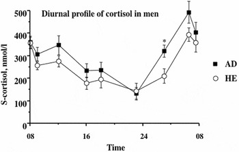
AUC levels for cortisol and DHEA did not differ, respectively, between men with AD and healthy elderly men. Men with AD had lower plasma ACTH levels compared with healthy elderly men throughout the whole 24-hour period and significantly so at 2300 h (p < 0.05) (data not shown). In contrast, men with AD had higher cortisol levels at all time-points and significantly so at 0300 h (p < 0.05) (Figure 1). After inclusion of age and BMI in the linear regression models, disease per se had an independent influence on increased serum cortisol levels at 0300 h (std. coef. = –0.66; p < 0.05) and decreased plasma ACTH levels at 2300 h (std. coef. = 0.66; p < 0.05) in men with AD.
Diurnal profile of serum cortisol nmol/l in men with Alzheimer's disease (AD; n = 7) and healthy elderly men (HE; n = 7). *p < 0.05, AD vs. HE.

Diurnal hormone levels in women with AD versus healthy elderly women
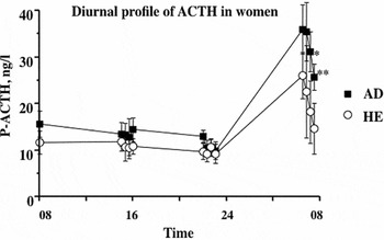
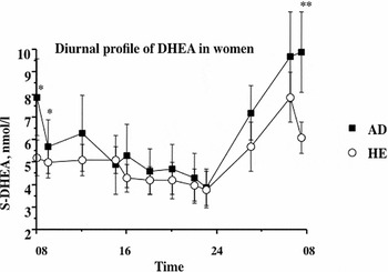
AUC levels for cortisol did not differ between women with AD and healthy elderly women. AUC levels for DHEA were increased in the AD group, but the difference was not statistically significant (p = 0.06). Women with AD had significantly increased plasma ACTH levels in the morning at 0710 h and 0730 h (p < 0.05 and p < 0.01, respectively) (Figure 2). Serum DHEA levels, adjusted for age, were significantly elevated in women with AD at 0730, 0800, and 0900 h (p < 0.01, p < 0.05, and p < 0.05, respectively) (Figure 3). After inclusion of age and BMI in linear regression models, disease per se had an independent influence on increased plasma ACTH levels in women at 0710 and 0730 h (std. coef. = –0.55; p < 0.05 and std. coef. = –0.72; p < 0.01, respectively) and on increased serum DHEA levels at 0730, 0800, and 0900 h (std. coef. = –0.81, p < 0.01; std. coef. = –0.71, p<0.05; and std. coef. = –0.58, p < 0.05, respectively).
Diurnal profile of plasma adrenocorticotropin (ACTH) ng/l in women with Alzheimer's disease (AD; n = 8) and healthy elderly women (HE; n = 8).*p < 0.05, AD vs. HE; **p < 0.01, AD vs. HE.

Diurnal profile of serum dehydroepiandrosterone (DHEA), nmol/l in women with Alzheimer's disease (AD; n = 8) and healthy elderly women (HE; n = 8). Serum DHEA levels adjusted for age. *p < 0.05, AD vs. HE; **p < 0.01, AD vs. HE.

Cytokines and soluble cytokine receptors
Women with AD had significantly increased serum levels of IL-6 at 0800 h compared to healthy elderly women (p < 0.05). No other statistically significant differences between groups regarding serum levels of proinflammatory cytokines or soluble cytokine receptors could be demonstrated (data not shown). No correlations between IL-6 levels and cortisol, DHEA, and ACTH levels, respectively, could be demonstrated.
Discussion
The main findings in this exploratory study are: (i) morning levels of DHEA in women with AD were increased compared to healthy elderly women, preceded by significantly increased morning ACTH levels; (ii) levels of IL-6 were significantly increased in women with AD; and (iii) early morning serum cortisol levels in men with AD were increased, although plasma levels of ACTH were lower. These findings suggest that there are sex-associated differences in the regulation of the HPA axis in AD, with an increased ACTH drive in women, resulting in elevated DHEA levels.
To the best of our knowledge, this study is the first to assess circadian profiles of DHEA in AD. Lower circulating serum DHEAS levels have been reported in both vascular dementia and AD (Ferrari et al., Reference Ferrari2001), and a causal link between decreased levels of DHEAS and an age-related cognitive decline has been discussed (Näsman et al., Reference Näsman1991). The results from this study are in line with an earlier report showing that medication-free AD patients with mild to moderate dementia have significantly increased levels of DHEA (Rasmuson et al., Reference Rasmuson, Näsman, Carlström and Olsson2002). Further strengthening this concept, a recent study on cerebrospinal fluid reported increased levels of DHEA in AD, linked to increased levels of DHEA in temporal cortex specimens from autopsies (Naylor et al., Reference Naylor2008). Importantly, the steroidogenic cytochrome P450 enzyme CYP7B1 is mainly expressed in the hippocampus and mediates the conversion of DHEA to 7α-hydroxy-DHEA (Rose et al., Reference Rose1997), which may have antiglucocorticoid and immunomodulatory properties and thus positive effects on neural functions (Pringle et al., Reference Pringle, Schmidt, Deans, Wulfert, Reymann and Sundstrom2003). In vitro studies of CYP7B1 mRNA suggest that the activity of this enzyme is decreased in AD (Yau et al., Reference Yau2003), and a recent experimental study showed that CYP7B1 bioactivity in hippocampal tissue in memory-impaired aged rats is decreased compared with both young and aged memory-intact rats (Yau et al., Reference Yau, Noble, Graham and Seckl2006). Furthermore, supplementation with 7α-hydroxy pregnenolone improved memory (Yau et al., Reference Yau, Noble, Graham and Seckl2006). We have not explored putative links between gonadal hormones and DHEA/cortisol levels in this study, but in an earlier study, we found no association between estradiol and sex differences in circulating levels of androgens (Rasmuson et al., Reference Rasmuson, Näsman, Carlström and Olsson2002).
Here, we also found no major differences in 24-hour cortisol levels between AD patients and controls. However, early morning cortisol levels in men with AD were significantly increased with concomitantly lower ACTH levels, while women with AD had significantly increased morning ACTH levels. Earlier data on the 24-hour profiles of cortisol in AD are conflicting, which may at least partly relate to heterogeneous study populations including concomitant medications (Hartmann et al., Reference Hartmann, Veldhuis, Deuschle, Standhardt and Heuser1997; Ferrari et al., Reference Ferrari2001). An increased glucocorticoid production rate with an increased metabolic clearance has been reported in women with AD (Rasmuson et al., Reference Rasmuson, Andrew, Näsman, Seckl, Walker and Olsson2001). The ACTH increase in women with AD versus healthy elderly women in the present study may therefore relate to an increased clearance of A-ring reduced metabolites in AD with a compensatory increase in plasma ACTH levels to keep circulating hormone levels constant. No similar studies have been made in males, and further studies on putative sex differences in glucocorticoid metabolism are thus needed in this patient group (Rasmuson et al., Reference Rasmuson, Andrew, Näsman, Seckl, Walker and Olsson2001).
We have previously shown a decreased suppressibility by dexamethasone of the HPA axis in men with mild to moderate AD (Näsman et al., Reference Näsman, Olsson, Viitanen and Carlström1995). A reason for this decrease may be an increased central drive of the HPA axis. However, ACTH levels were decreased in men with AD in this study, indicating that there may be other important regulators of the HPA axis in AD contributing to abnormal negative feedback. Several other factors (in addition to the corticotropin releasing hormone–ACTH system) are involved in HPA axis regulation, including neurotransmitters, neuropeptides, nitric oxide, and proinflammatory cytokines (Bornstein and Chrousos, Reference Bornstein and Chrousos1999). Of note, IL-6 stimulates HPA axis activity at several levels in humans (Mastorakos et al., Reference Mastorakos, Chrousos and Weber1993; Späth-Schwalbe et al., Reference Späth-Schwalbe1994), which opens up the possibility of a putative link between an increased drive of the HPA axis and the inflammatory component in AD. The neuritic plaques (a hallmark of AD) are foci for local inflammatory responses as seen by the presence of cytokines and acute phase proteins. Of note, results from analyses of two prospective longitudinal cohorts from the Rotterdam Study and the Framingham Study, respectively, have suggested a link between low-grade inflammation and risk for AD (Engelhart et al., Reference Engelhart2004; Tan et al., Reference Tan2007). We found increased plasma levels of IL-6 in women with AD compared with healthy elderly women. However, we found no statistically significant association between morning levels of IL-6 and ACTH or cortisol and DHEA, respectively, but the exploration of more time-points would obviously be of interest.
This study was carried out among well-characterized and medication-free participants. However, a drawback of the study is the limited number of patients, which may mean that some of the findings could have arisen only by chance. Further, we have presented our p-values without corrections for multiple comparisons. There is thus a need for prospective studies with larger sample sizes, allowing for more detailed subgroup analyses.
In summary, the women with AD in our study had clearly increased morning levels of DHEA versus healthy elderly women, preceded by higher ACTH levels. An increased ACTH drive may thus stimulate androgen synthesis in women. Whether the increased ACTH levels are secondary to increased clearance and/or a decreased sensitivity in the HPA axis feedback remains to be established. We also demonstrated increased levels of the proinflammatory cytokine IL-6 in women with AD, which may contribute to increased DHEA levels via stimulated ACTH secretion. Our data suggest important sex differences in the regulation of the HPA axis, mainly regarding androgens. The findings in this and other studies of increased levels of DHEA in mild to moderate AD do not support treatment with DHEA to enhance cognition.
Conflict of interest
None.
Description of authors’ roles
SR, BN and TO designed the study. SR wrote the protocol. SR and BN undertook the statistical analyses, and SR wrote the first draft of the paper. All authors contributed to and approved the final version of the paper.
Acknowledgments
The authors wish to thank Britta Larsson and Else-Britt Lundström for technical support during the study and Professors Kjell Carlström and Kjell Grankvist for hormone and cytokine analyses.