Introduction
Bilateral striopallidodentate calcinosis (BSPDC) is a rarely occurring neurodegenerative disorder. Bilateral and symmetrical deposition of calcium and other minerals in the basal ganglia, thalamus, cerebellar dentate nucleus and centrum semiovale are the most common indicators of BSPDC. Different terms, including Fahr's disease, are used instead of BSPDC. However, the latter term is more descriptive and appropriate (Reference Manyam1). As a matter of fact, BSPDC should be defined as a syndrome rather than a single-disease entity (Reference Klein and Vieregge2).
Familial forms in addition to sporadic cases were specified in the etiology. Most of the genetic transition is autosomal dominant (Reference Kobari, Nogawa, Sugimoto and Fukuuchi3,Reference Geschwind, Loginov and Stern4). Secondary causes include metabolic disorders of calcium and other minerals, congenital developmental disorders, toxic and anoxic factors and links with systemic and inflammatory diseases (Reference Harrington, Macpherson, McIntosh, Allam and Bone5). Differential diagnoses of the secondary causes should be determined for patients with BSPDC.
Clinical symptoms of BSPDC are varied. There can be asymptomatic individuals, movement disorders depending on the location of calcification (parkinsonism, chorea, tremor, dystonia, athetosis and orofacial dyskinesia), cerebellar symptoms, cognitive impairment, speech disturbance, psychiatric manifestations, pyramidal signs and gait disturbance (Reference Manyam1). Among psychiatric disorders, obsessive compulsive disorders (OCD), anxiety disorders, mood disorders, bipolar disorders, psychosis and changes in personality are observed (6–9). Neurologic symptoms are generally observed after the third decade and show little progress over the years (Reference Manyam1).
Computerised tomography (CT) is the first and most effective way to observe intracerebral calcifications. Intracranial calcifications are also observed in cranial magnetic resonance imaging (MRI), which has recently grown in popularity. However, CT – sensitive, cheap and easy to apply in a short time – is advised for scanning calcifications (Reference Osborn10,Reference Ogi, Fukumitsu, Tsuchida, Uchiyama, Mori and Matsui11). Herein, we present five patients from two families with BSPDC who were diagnosed by cranial CT.
Case reports
Patient 1
This 43-year-old woman exhibited symptoms including shyness and behavioural inhibition, involuntary facial movements in social environments, a slowing of mental activities and a deterioration of speech. She was evaluated at a health centre and referred to our clinic with a pre-diagnosis of metabolic disease. She exhibited no hereditary disposition to the disorder and her physical examination was normal. No symptoms except for occasional stuttering in her speech and slight hypophonia and facial myokymia around her mouth and eyes when she got excited were determined in a neurologic examination. On the other hand, severe anxiety, somatic symptoms of anxiety (tachycardia, nausea, perspiration and dyspnea), typical obsessions and compulsions (repeated hand washing and controlling) were found in a psychiatric examination. Her score on the Mini Mental State Examination (MMSE) was 30/30 and a neuropsychological test found as normal.
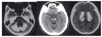
When the patient's cranial CT imaging was examined, calcifications were observed in the bilateral basal ganglia, thalamus, periventricular white matter, centrum semiovale, brain stem and cerebellum (Fig. 1). There were no abnormalities in electroencephalography. Calcium, phosphorus, parathormone levels in her serum were normal, eliminating secondary causes of BSPDC. Other blood test results were normal. In fact, her blood count, sedimentation rate, kidney and liver function tests, vitamin D level, thyroid and growth hormone values, lactate, pyruvate, copper, ceruloplasmin and ferritin levels were all normal. When the fact that no secondary etiology was specified and existing clinical findings were evaluated together, the patient was diagnosed by idiopathic BSPDC.
Cranial computerised tomography scans showing bilateral calcifications of the cerebellum, midbrain, basal ganglia, thalamus and centrum semiovale.

While the diagnosis of BSPDC was explained to the patient and her husband, it was found out that her brother had a neurologic disease with similar symptoms. Her family was invited to our clinic for screening, since it was clear that there was a family history.
Patient 2
This 50-year-old man, the brother of patient 1, complained of occasional stuttering of speech in crowds. He also had to change jobs voluntarily or involuntarily. Psychiatry departments had diagnosed him with social phobia, sexual dysfunction, impulse control disorder and personality disorders. During our interview and evaluation, social phobia, depression and impulse control disorder were confirmed based on Diagnostic and Statistical Manual of Mental Disorders, 4th Edition (DSM-IV) criteria. Despite having occasional stuttering and hypophonia in speech, neurological results were normal. Bilateral calcification areas, similar to those exhibited in patient 1, were detected in his cranial CT. As a result, he was diagnosed with familial BSPDC.
Patient 3
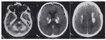
The elder brother of patients 1 and 2, (53-year-old) had been diagnosed with a neurological disease with an unknown etiology. There were no pathologies in previous laboratory tests performed on the patient. Over the years his condition progressively deteriorated rendering him unable to take care of himself. He was unable to walk and in constant need of care. Our physical examination showed that he could not stand up and his general condition was poor. Despite this, he did not cooperate well during examinations. Symmetrical pyramidal signs were present in his bilateral upper and lower extremities. Spastic ataxia with cerebellar dysarthria, bradykinesia and rigidity were also observed. He was crying and laughing at innapropriate times. He did not cooperate with MMSE testing. Bilateral calcification areas were detected in cranial CT imaging (Fig. 2).
Cranial computerised tomography scans showing bilateral calcifications of the cerebellum, pons, thalamus, basal ganglia and centrum semiovale.

Patient 4
This 45-year-old male was admitted to our clinic complaining of inanimation, adduction, forgetfulness and speech problems. He had nothing remarkable in his medical and familial history and had a normal neurological examination despite having occasional stuttering and hypophonia in speech. Depression and social phobia were detected in his psychiatric evaluation based on DSM-IV criterias, His neuropsychological tests yielded no pathology and his MMSE score was 30/30. His cranial CT showed calcifications in bilateral basal ganglia, thalamus, periventricular white matter, centrum semiovale and cerebellum. Blood tests were performed in order to rule out secondary causes but no pathology was detected. The patient was diagnosed with idiopathic BSPDC.
Patient 5
This 21-year-old male is patient 4's elder son. He consulted our clinic upon his father's request. He described adduction, shyness, inability to get into unfamiliar environments and occasional stuttering. His neurological examination was normal except for occasional stuttering and hypophonia in speech. A psychiatric evaluation yielded social phobia and anxiety disorder, and his cranial CT showed bilateral calcification areas.
Discussion
Although BSPDC was identified several years ago and is a very well-known syndrome, its low occurrence rate and broad range of symptoms cause it to be overlooked. Primarily found in sporadic, autosomal dominant or familial forms, BSPDC may develop in secondary forms for various reasons. Even though movement disorders, neuropsychiatric symptoms and cerebellar symptoms stand out in clinical findings, there can be asymptomatic individuals (Reference Manyam1). The five cases we present include findings that belong to different segments of the neuro-axis. In some cases the diagnoses could only be reached by consulting the patients' family histories. Unfortunately, many patients – like patient 1 – conceal family history during anamnesis. Possible secondary causes were ruled out with detailed investigations.
In general, BSPDC is not considered in patients who only address their psychiatric complaints, but approximately 40% of BSPDC patients consult a psychiatrist (Reference Harrington, Macpherson, McIntosh, Allam and Bone5,Reference Ogi, Fukumitsu, Tsuchida, Uchiyama, Mori and Matsui11). A correlation has been reported between symmetrical pathological intracerebral calcifications and socially inappropriate behaviours, personality disorders, OCD, mood disorders, depression and even psychosis (3,7,12–15). Therefore, in order to detect undetermined cases, patients consulting with isolated psychiatric symptoms should be subjected to a thorough familial and medical history and a careful examination. Cummings et al. stated that psychiatric symptoms could begin to occur as early as 30 years of age and as late as 50, while motor and cognitive symptoms could occur mostly in patients over 50 (Reference Cummings, Gosenfeld, Houlihan and McCaffrey16). There is a correlation between the extensity of calcifications and symptoms and clinical findings. It has been noted that, in general, symptomatic patients had more intense calcifications (Reference Shibayama, Kobayashi and Nakagawa17). While pallidal lesions cause disorders related to motivation, decision making and self-awareness, mental disorders are more related to cortical atrophia level. Speech disorders found in BSPDC are in forms of hypophonia, stuttering or cerebellar dysarthria. On the other hand there might be no correlation between brain CT and clinical findings (e.g. asymptomatic individuals).
Literature points to higher occurrence rates of pathological calcifications in males (Reference Manyam1), in line with our case series.
Cranial CT imaging is the most frequently used method in BSPDC (Reference Kazis18). Calcification findings are seen clearly as hyperdense lesions in CT imaging. In contrast with an MRI, a CT scan is a far more sensitive and thorough diagnostic tool. It is also cheaper and saves time (Reference Ogi, Fukumitsu, Tsuchida, Uchiyama, Mori and Matsui11).
BSPDC is a neurodegenerative disorder that can occur because of primary or secondary causes, can have an asymptomatic prognosis and offers different neuropsychiatric findings. We believe that BSPDC should be kept in mind during differential diagnoses in those afflicted with neuropsychiatric and speech disorders. It is important to at least run a CT scan on young adults with speech disorders and neuropsychiatric histories. Then, if intracranial calcifications are detected in appropriate areas, all accessible family members should be examined for familial influences and possible secondary causes should be investigated.