Introduction
Gradenigo's syndrome is an extremely rare clinical symptom triad of deep retro-orbital pain, ipsilateral abducens palsy and purulent otorrhoea in the setting of petrous apicitis.Reference Kitsko, Dedhia, Myers and Snyderman1 Gradenigo's syndrome is not synonymous with petrous apicitis because only 13.6 to 42.1 per cent of cases develop the classic symptom triad of Gradenigo's syndrome.Reference Chole, Flint, Haughey, Lund, Niparko, Robbins, Thomas and Lesperance2,Reference Gadre and Chole3
In the pre-antibiotic era, petrous apicitis occurred once in every 300 cases of acute otitis media.Reference Gadre and Chole3 However, in the modern era of antibiotic use, the incidence has decreased to only 2 cases per 100 000 children with acute otitis media.Reference Goldstein, Casselbrant, Bluestone and Kurs-Lasky4 The largest retrospective study of petrous apicitis showed a mortality rate of 2.3 per cent.Reference Gadre and Chole3 Treatment for Gradenigo's syndrome remains highly controversial; however, antibiotic therapy should be initiated early. Although the recent trend has been towards a more conservative approach to management, many still advocate for aggressive surgical intervention because of the potential for mortality.
Case presentation
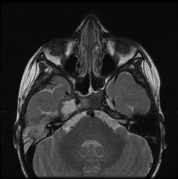
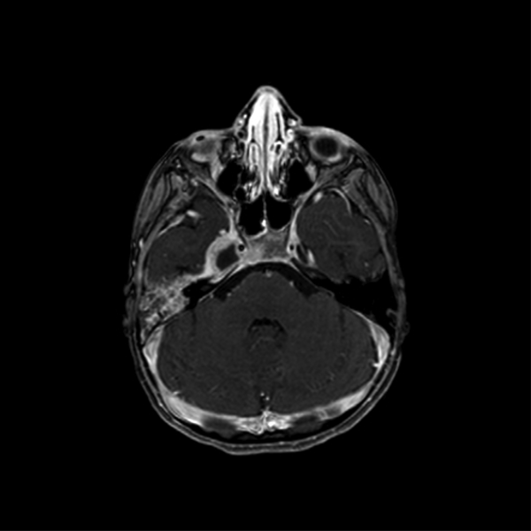
A four-year-old female was admitted to the paediatric in-patient ward for failure to thrive following recent sinusitis. Past medical history was significant for developmental delay and suspected autism spectrum disorder. Physical examination was negative for acute upper respiratory infection or acute otitis media but positive for right sided facial pain, photophobia and right abducens nerve palsy. Subsequent magnetic resonance imaging (MRI) showed a 1.3 × 1.7 × 1.4 cm abscess encompassing the right Meckel's cave (Figures 1 and 2). A contrast-enhanced computed tomography (CT) scan showed petrous apicitis and otomastoiditis, confirming Gradenigo's syndrome. An audiogram showed a mild conductive hearing loss.
Axial contrast-enhanced T2-weighted magnetic resonance image showing a rim-enhancing abscess measuring 1.3 × 1.7 × 1.4 cm within the right Meckel's cave.

Axial contrast-enhanced T1-weighted magnetic resonance image showing the abscess expanding into the region of the right Dorello's canal.

The patient was started on intravenous vancomycin and ceftriaxone. She was taken to the operating theatre for right intact canal wall mastoidectomy with myringotomy and tube placement with findings of a non-purulent middle-ear effusion and granulation tissue in the mastoid cavity and middle ear. Surgical cultures grew coagulase-negative staphylococcus. The patient was discharged on six weeks of ceftriaxone administered by a peripherally inserted central catheter line. At a two-week post-operative visit, she showed notable improvement in neuropathic symptoms. Post-operative audiogram showed normal hearing with a resolution of conductive hearing loss.
Discussion
Up to 85 per cent of children will have at least one episode of acute otitis media in their lives.Reference Goldenberg and Goldstein5 Complications of acute otitis media can be defined as the spread of infection beyond the confines of temporal bone air space and residing mucosa.Reference Neely, Brackmann, Shelton and Arriaga6 One of the most feared complications is petrous apicitis, which occurs in the presence of a pneumatised petrous apex. The prevalence of pneumatisation in the general population is 30 per cent.Reference Goldenberg and Goldstein5,Reference Kazemi7 Because of its rarity, the incidence of petrous apicitis is unknown in the adult population, but the incidence in children is 2 per 100 000 cases of acute otitis media.Reference Goldstein, Casselbrant, Bluestone and Kurs-Lasky4 One study showed a mortality rate of 2.3 per cent of cases, but older reports have estimated rates as high as 18 per cent.Reference Gadre and Chole3,Reference Sears8 Contemporary guidelines for management of acute otitis media have recommended a period of watchful waiting rather than aggressive initial antibiotic therapy, which has raised concern for an increased risk of complications such as petrous apicitis. Despite decreasing rates of antibiotic use in children with acute otitis media, there is a lack of evidence in the literature of a concordant rise in the incidence of complications.Reference Meissner9
Although petrous apicitis can present with many combinations of symptoms, the most well-known presentation is Gradenigo's syndrome consisting of deep retro-orbital pain, ipsilateral abducens palsy and purulent otorrhoea.Reference Kitsko, Dedhia, Myers and Snyderman1 In addition to the classic triad, other symptoms include otalgia (79 per cent), otorrhoea (59 per cent), fever (22 per cent), coma, and paralysis of cranial nerves V, VII, VIII and X.Reference Chole, Flint, Haughey, Lund, Niparko, Robbins, Thomas and Lesperance2 We present a classic case of Gradenigo's syndrome, but one must be familiar with the variety of manifestations of petrous apicitis to not overlook this diagnosis.
The pathophysiology of neuropraxia associated with Gradenigo's syndrome can be explained by inflammation of Dorello's canal and Meckel's cave combined with neural oedema compressing the abducens and trigeminal nerves.Reference Janjua, Bajalan, Potter, Whitney and Sipaul10 The abducens nerve contacts the apex of the petrous part of the temporal bone as it runs under the petroclinoid ligament and through Dorello's canal. The trigeminal nerve and ganglion lie within the petrous apex. In addition, the facial and vestibulocochlear nerves pass through the temporal bone and may be damaged from local inflammatory factors.
Differential diagnosis of petrous apicitis includes inflammatory pseudotumor, sarcoidosis, tuberculosis, intracranial abscess, lateral sinus thrombosis, Tolosa–Hunt syndrome and malignancy.Reference Gadre and Chole3,Reference Janjua, Bajalan, Potter, Whitney and Sipaul10 Malignancy is less likely in the presence of an elevated erythrocyte sedimentation rate or C-reactive protein. Tolosa–Hunt syndrome is an extremely rare condition defined by chronic granulation tissue of the cavernous sinus, and it may present as Gradenigo's syndrome with ocular neuropraxia without petrositis or mastoiditis.Reference Neely, Brackmann, Shelton and Arriaga6 Diagnosis of petrous apicitis is typically confirmed by high-resolution CT scan or MRI.Reference Gadre and Chole3,Reference Goldenberg and Goldstein5
Management of petrous apicitis depends on control of the infection.Reference Chole, Flint, Haughey, Lund, Niparko, Robbins, Thomas and Lesperance2,Reference Gadre and Chole3 Antibiotics should cover the most commonly seen organisms, including Streptococcus pneumoniae, staphylococcus spp., beta-haemolytic streptococci, Haemophilus influenza, pseudomonas spp., Moraxella catarrhalis and various anaerobes.Reference Gadre and Chole3 Cases that do not respond to traditional therapy should raise the suspicion for uncommon organisms such as aspergillus spp. and non-tuberculosis mycobacteria.Reference Kazemi7 Most authors advocate for the use of a cephalosporin antibiotic along with metronidazole with or without the addition of dexamethasone and vancomycin.Reference Gadre and Chole3,Reference Janjua, Bajalan, Potter, Whitney and Sipaul10 Although controversial, the anti-inflammatory and analgesic properties of corticosteroids can alleviate neural pathology such as ocular palsies and headache. In our case, ceftriaxone and vancomycin were chosen pre-operatively for empiric broad-spectrum coverage. The patient was discharged on six weeks of ceftriaxone administered by peripherally inserted central catheter line based on culture results.
The ideal treatment for petrous apicitis is controversial and typically depends on the severity of the disease. Some authors agree that antibiotic therapy combined with modified or radical mastoidectomy and tympanostomy tube placement may be sufficient to allow drainage of the petrous apex air cell tracts.Reference Goldstein, Casselbrant, Bluestone and Kurs-Lasky4 However, more recently authors have advocated for non-surgical intervention with intravenous antibiotic therapy. Petrous apicitis management is challenging because the petrous apex is the most surgically inaccessible portion of the temporal bone.Reference Chole, Flint, Haughey, Lund, Niparko, Robbins, Thomas and Lesperance2 Indications for surgical intervention include abscess formation, osteonecrosis and failure of medical therapy. The choice of surgical approach depends on the patient's pre-operative hearing status, individual temporal bone anatomy and the surgeon's experience. In non-hearing ears, translabyrinthine and transcochlear approaches provide the most direct access but result in destruction of the cochlea. Approaches that preserve hearing include infracochlear, infralabyrinthine, retrolabyrinthine, middle fossa and subarcuate approaches. In the case presented, our neurosurgical colleagues were available if a middle cranial fossa approach was deemed necessary or if the patient failed treatment with mastoidectomy, myringotomy and antibiotics.
Risks of surgical intervention on the petrous apex cannot be overlooked and include facial nerve damage, intracranial infections and thrombosis.Reference Janjua, Bajalan, Potter, Whitney and Sipaul10 Although there may not be one ideal therapy for all cases of petrous apicitis, favourable outcomes are associated with a high index of suspicion, early diagnosis with appropriate imaging studies and prompt intervention (either medical or surgical).Reference Gadre and Chole3 Post-operative management may include intensive care unit monitoring.Reference Kitsko, Dedhia, Myers and Snyderman1 In this period, it is critical to monitor the patient for signs of cerebrospinal fluid leak, cerebral oedema, increased intracranial pressure, pneumocephalus and seizures. Infectious disease and neurosurgery consultations are recommended, and many patients require long-term antibiotic therapy.
• Gradenigo's syndrome describes the rare symptom triad of otorrhoea, ipsilateral abducens palsy and ipsilateral retro-orbital pain classically associated with petrous apicitis
• Without prompt recognition and early intervention, petrous apicitis can be deadly with mortality rates of between 2.3 and 18 per cent
• Only some patients with petrous apicitis develop Gradenigo's syndrome, and diagnosis can typically be made with high-resolution computed tomography or magnetic resonance imaging
• Treatment is controversial but should always include antibiotics and may or may not include surgical intervention
• Surgical approach depends on patient hearing status, individual anatomy, and disease severity, and modified or radical mastoidectomy and tympanostomy tube placement may achieve full resolution despite recent trends towards non-surgical therapy
• Thorough knowledge of temporal bone anatomy and pathophysiology is required to determine the safest treatment approach
Conclusion
Gradenigo's syndrome is seldom seen in modern medicine thanks to widespread antibiotic use for the treatment of acute otitis media. Many otolaryngologists are unfamiliar with its presentation, which can lead to a delayed diagnosis and complicated management. Physicians should keep a high index of suspicion and not rule out petrous apicitis due to an insidious onset or the lack of one or more of the classic symptoms. In paediatric patients presenting with Gradenigo's syndrome, a low threshold must exist for the initiation of antibiotic therapy, and the necessity of surgical intervention should be assessed because numerous surgical approaches exist. Recognising the clinical features of Gradenigo's syndrome while ensuring timely management is vital to preventing severe destruction and mortality.
Acknowledgements
The authors acknowledge the American Academy of Otolaryngology – Head and Neck Surgery for accepting our project for poster presentation at the 2018 American Academy of Otolaryngology – Head and Neck Surgery Foundation Annual Meeting and Otolaryngology Experience, Atlanta, Georgia, October 7–10, 2018. This project was supported by the Department of Otolaryngology at the University of Texas Medical Branch in Galveston, Texas, USA.
Competing interests
None declared