In corrected transposition of great arteries, a suboptimal non-crescentic right ventricle and structural abnormalities cause tricuspid regurgitation. Reference Prieto, Hordof, Secic, Rosenbaum and Gersony1 Right ventricle is preserved after tricuspid interventions in even asymptomatic patients. Reference Baumgartner, De Backer and Babu-Narayan2 Unlike mitral valve, complex tricuspid abnormalities necessitate replacement often with a bioprosthesis rather than repair. Reference Koolbergen, Ahmed and Bouma3 Tricuspid replacement provides a 5-year freedom from death and reintervention only in 65% of patients. Reference Koolbergen, Ahmed and Bouma3 Surgical replacement of degenerating bioprosthesis under ischaemic arrest further complicates right ventricular function. Reference Koolbergen, Ahmed and Bouma3 Parasternal location of right ventricle in corrected transposition with a vertical trajectory from the aneurysmal left atrium pose anatomical challenges for a transcatheter valve-in-valve implantation within a degenerated tricuspid bioprosthesis in systemic right ventricle, reported for the first time.
Case report
A 31-year-old lady with corrected transposition became symptomatic after second decade from progressive tricuspid regurgitation. Left atrium measured 9 cm, and redundant tricuspid leaflets from a 43 mm annulus were non-coapting. The right ventricular ejection fraction was 34%. Tricuspid replacement with a 31 mm Perimount (Edwards Lifesciences, Irvine, CA) bioprosthesis was done at 24 years through combined left atrial and septal approach. Paroxysmal atrial fibrillation on follow-up needed electrical cardioversion thrice and antiarrhythmic drugs. Echocardiography after a recent cardioembolic brain infarction revealed a degenerated severely stenotic bioprosthesis with a mean gradient of 13 mmHg and moderate regurgitation. Heart team recommended a valve-in-valve implantation considering the risks of repeat sternotomy, ischaemic arrest, and recent stroke after informed consent from patient.
Trans-septal puncture and vertical guidewire course from aneurysmal left atrium to parasternal right ventricle were planned on CT. The distance from bioprosthesis to the right ventricular apex was short (Supplemental Figure). Bapat’s mobile application predicted an internal diameter of 28.5 mm for this bioprosthesis. Reference Bapat4
Under general anaesthesia and ultrasound guidance, groin and jugular venous access were obtained. A right anterior oblique projection profiling the bioprosthesis was chosen as the working fluoroscopic plane. Posteroinferior transseptal puncture guided by transesophageal echocardiography was confirmed on fluoroscopy to avoid a puncture superior to the level of the bioprosthesis. Prior surgical atrial septal scars did not permit passage of the 8F transseptal sheath even after the needle penetration. After protecting the left atrium from needle injury by a long coronary wire through the needle, force was used to advance the sheath. Left atrium recorded tall v waves measuring 38 mmHg and a mean pressure of 26 mmHg as well as a transtricuspid gradient of 13 mmHg.
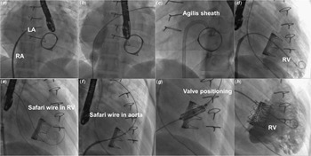
The rigid septal puncture site was predilated using 16 mm Atlas gold balloon (Bard endovascular, Tempe, AZ) before advancing a 8.5F Agilis sheath(Abbott, Chicago, IL) to advance a pigtail catheter into the RV. As the extra-small curve 0.035 Safari wire (Boston Scientific, Marlborough, MA) was not supported in the short right ventricular inflow, it was flipped into the ascending aorta despite losing its coaxiality. The groin venous sheath was upgraded to 14F expandable Python sheath (Meril Lifesciences, Vapi, India) to advance a 30.5 mm MyVal THV(Meril Lifesciences, Vapi, India) into the bioprosthesis. The valve was slowly balloon expanded while adjusting the Navigator delivery system (Meril Lifesciences, Vapi, India) to autocorrect the lack of alignment and recreate its coaxiality in the final stages of deployment. Rapid pacing, slow inflation, and apnoea induced on ventilator allowed control during valve positioning with 20% of the stent protruding on the atrial side (Fig 1).
Septal puncture was made below the level of tissue valve(A) and dilated with 16 mm large balloon(B) before advancing a steerable sheath into the left atrium(LA) towards the valve orifice(C). Angiogram(D) in right ventricle(RV) shows small inflow dimension compared to outflow and severe tricuspid regurgitation. The small RV inflow provided inadequate support for the guidewire loop(E) which was flipped into the aorta(F) thereby losing coaxiality with the valve(G) that got autocorrected during valve expansion(H).

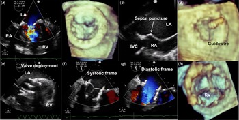
Transoesophageal echocardiogram(Fig 2) and simultaenous haemodynamic pressure traces confirmed normal function of the transcatheter valve. There was no significant interatrial shunt. Uneventful post-procedural course led to discharge after two days while receiving aspirin, rivaroxaban, amiodarone, and metoprolol.
Transesophageal echocardiogram(A) showed severe bioprosthesis stenosis and degeneration on three dimensional volume rendering(B). Echocardiographically guided posteroinferior transseptal puncture(C) assisted passage of guidewire through the tissue valve(D). After deployment of the balloon expandable valve(E), systolic(F) and diastolic(G) frames as well as volume rendered ventricular view(H) of the transcatheter valve demonstrate normal valve function.

Discussion
This is the first reported case of transcatheter valve-in-valve implantation within a tricuspid bioprosthesis in corrected transposition after a detailed literature search. Unlike mitral valve, tricuspid valve in systemic right ventricle warrant modifications during edge-to-edge tricuspid repair. Reference Picard, Tadros and Asgar5,Reference Luedike, Riebisch, Weymann, Ruhparwar, Rassaf and Mahabadi6 High surgical risks of tricuspid replacement in right ventricular dysfunction may force a heart transplantation. Reference Picard, Tadros and Asgar5,Reference Luedike, Riebisch, Weymann, Ruhparwar, Rassaf and Mahabadi6 Unlike mitral valve, the following procedural modifications may be needed. Reference Guerrero, Salinger and Pursnani7
When atrial septal surgical scars hinder transseptal puncture, needle is electrified with a bougie of an electrocautery and a coronary wire advanced through it to avoid inadvertent left atrial injury. Predilating rigid atrial septum with a large 16 mm facilitates to advance transcatheter heart valves larger than 26 mm and reduce the procedural time. But advancing this balloon through the degenerated prosthesis is not advised to prevent embolisation of fractured leaflets. Small left atrial appendage thrombus or clots from degenerating bioprosthesis are excluded by transoesophageal echocardiography in patients with recent cardioembolic strokes. Unfavourable angulated trajectory from left atrium to a parasternally located right ventricle in corrected transposition is further worsened by a superior transseptal puncture that is avoided by fluoroscopic selection of the puncture site below the plane of the prosthetic ring. Unlike equal inflow and outflow dimensions of left ventricle that allow a deep positioning of the Safari wire for support, short right ventricular inflow dimension in corrected transposition denies any wire support. This may necessitate the wire loop to be flipped into the ascending aorta. The generous subaortic conus in corrected transposition prevents any possibility of neo-subaortic narrowing, unlike in mitral procedure. Finally, unlike Sapien S3 valve mounted and crimped proximal to the balloon on the delivery system that needs alignment with the balloon markers within the inferior caval vein, the MyVal is crimped directly on the balloon easing this step. Reference Sivaprakasam, Reddy, Sengottuvelu and Sivakumar8
Supplementary material
For supplementary material accompanying this paper visit https://doi.org/10.1017/S1047951121005059
Acknowledgements
Pramod Sagar compiled computed tomographic and echocardiographic images.
Financial support
None.
Conflicts of interest
None.
Ethical standards
The procedure complied with ethical standards of Madras Medical Mission ethical committee and Indian council of medical research.