Introduction
The number of undescribed fungi found growing on lichens continues to swell, even in Europe, the region most intensively studied for these fungi. This is not only a matter of additional species discovered in already established genera, but of species for which no appropriate genus to accommodate them has been described. One such new fungus is described here.
Oieleku Fagus wood (Oiartzun, NE Gipuzkoa, northern Spain), where this evidently undescribed fungus was discovered, is an old oceanic forest composed of c. 200 year-old trees, whose biodiversity is being studiedwithin the Life Aiako-Harria project. The lichen biota is interesting, with Lobarion well preserved and rare species hardly recorded elsewhere in Spain, such as Bryophagus gloeocapsa, Coenogonium luteum, Japewiella tavaresiana, Lecidea exigua, Lobaria virens, Micarea lutulata, Parmeliella parvula, Parmelinopsisminarum (very abundant), Phyllopsora rosei, and Punctelia jeckeri. Further, the wood is especially interesting for the lichenicolous fungi present, which include some species that are scarcely recorded in the Peninsula, notably Cornutispora triangularis, Diplolaeviopsis ranula and Monodictys fuliginosa.
Material and Methods
Specimens were studied using a Nikon and Meiji zoom stereomicroscope and Olympus CH and Olympus BH–2 research microscope fitted with Nomarski differential interference contrast optics and a drawing tube. Preparations were made in water and lactofuchsin, and measurements were made on material mounted in water. Photographs were taken with a Nikon Coolpix 4500 digital camera attached to the Olympus microscopes.
Taxonomy
Calongia D. Hawksw. & Etayo gen. nov
MycoBank no.: MB 515575
Conidiophora macronemata, mononemata, erecta, non ramosa, rigidiuscula, rubro-brunnea, 0–multiseptata, cum apicibus gibbus. Cellulae conidiogenae terminales, numerosae, subglobosae vel ampulliformes, polyblasticae, in vesiculo hyalino inclusae. Conidia singularia, praecipue angusto-ellipsoidia, (0–)1-septata, laevia, fusca.
Typus: Calongia gibelluloides D. Hawksw. & Etayo 2010.
Conidiophores macronematous, mononematous, originating singly, or more rarely in groups of 2–3, arising vertically from a single red-brown rhomboidal basal cell; unbranched, rigid, non-septate or septate, red-brown, smooth-walled, the apical part swollen; the apical part and the conidiogenous cells and conidia, at first enclosed in a hyaline subspherical membranous enveloping sheath. Conidiogenous cells arising directly from the expanded apex of the conidiophores, several on a single conidiophore, discrete, subglobose to broadly ampulliform, brown, polyblastic with 1–several conidiogenous loci, not proliferating, the conidiogenous loci mainly on the apical part, the conidia leaving minute circular scars; conidiogenesis events closest to type six of Sutton (Reference Sutton, Reynolds and Taylor1993) and Kirk et al. (Reference Kirk, Cannon, Minter and Stalpers2008). Conidia solitary, narrowly ellipsoid to elongate-reniform or elongate-soleiform, (0–)1-septate, smooth-walled, greyish brown, cessation schizolytic.
Etymology. Named in honour of Francisco “Paco” de Diego Calonge (b. 1938), President of the Sociedad Micológica de Madrid and a leading specialist on gasteromycetous fungi, on the occasion of his retirement from the Real Jardín Botánico de Madrid of which he once served as Director.
Calongia gibelluloides D. Hawksw. & Etayo sp. nov
MycoBank no.: MB 515576
Conidiophora 15–25(–45) × 3–3·5(–4·5) μm, cum apicibus 4·5–6·5 μm crassis et in vesiculo 20–40 μm diam. Cellulae conidiogenae (2·5–)4–6 μm diam. Conidia 5–9 × 2–3 μm.
Typus: Spain: Gipuzkoa: Oiartzun, Oieleku, wood near the fountain, on soredia of Flavoparmelia caperata on Fagus sylvatica, 520–550 m, 43º15′10″ N, 1º49′36″ W, 1 July 2006, J. Etayo & A. Etayo (K(M) 164014—holotypus; hb. Etayo 23364—isotypus).
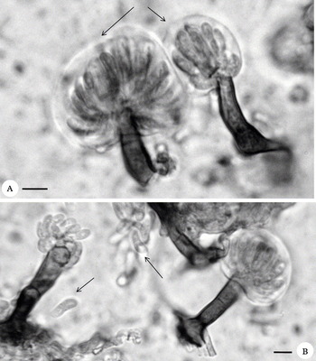
Calongia gibelluloides (hb. Etayo 23364). A, conidiophores and conidia with the enclosing vesicular membranous sheath intact (arrows); B, conidia (arrows), the lower with the median septum just visible. Scales = 10 μm.

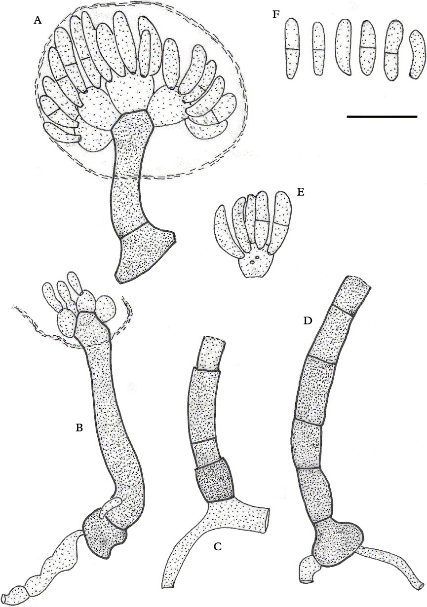
Calongia gibelluloides (K(M) 164014—holotype). A, conidiophore with the enclosing vesicular membranous sheath intact; B, conidiophores with most conidia discharged; C, basal part of a conidiophores showing percurrent proliferation; D, basal portion of a conidiophore with four transverse septa; E, conidiogenous cell with scars where conidia have secessed; F, conidia. Scale = 10 μm.

Mycelium arising on soredia of the host lichen, growing on the surface over one or sometimes more soredia, hyphae discrete, subhyaline to pale fuscous brown, smooth-walled, sparsely septate, even in thickness to almost torulose in parts, 1·5–2·5(–3) μm thick. Conidiophores macronematous, mononematous, originating singly, or more rarely in groups of 2–3, arising vertically from a single red-brown rhomboidal basal cell, 6–7 μm wide; unbranched, rigid, 0–2(–5) septate, not constricted at the septa, red-brown, smooth-walled, wall c. 0·3 μm thick, sometimes proliferating and growing through a broken lower cell, 15–25(–45) μm tall and 3–3·5(–4·5) μm thick, the apical part swollen to 4·5–6·5 μm wide; the apical part and the conidiogenous cells and conidia, at first enclosed in a subspherical enveloping sheath, 20–40 μm diam., the sheath membranous and colourless, 2·5–3 μm thick, at maturity becoming broken and then appearing as irregular collar-like remnants adhering to the conidiophores just below the swollen apical part, and finally disappearing. Conidiogenous cells arising directly from the expanded apex of the conidiophores, 4–6(–8) on a single conidiophore, subglobose to broadly ampulliform, brown, polyblastic with 1–several conidiogenous loci, discrete, not proliferating, individually (2·5–)4–6 μm diam., the conidiogenous loci mainly on the apical part, the positions of released conidia indicated by minute circular scars c. 0·5 μm diam; conidiogenesis events closest to type six of Sutton (Reference Sutton, Reynolds and Taylor1993) and Kirk et al. (Reference Kirk, Cannon, Minter and Stalpers2008). Conidia solitary, not in chains, narrowly ellipsoid to elongate-reniform or elongate-soleiform, rounded at the apex and rounded to slightly truncate at the base, (0–)1-septate, smooth-walled, greyish brown, 5–9 × 2–3 μm, cessation schizolytic.
Hosts. Flavoparmelia caperata soredia and squamules of an unidentified corticolous Cladonia. The sorediate areas on the thallus of F. caperata become discoloured and pinkish brown, and the black pin-like conidogenous structures can just be observed under a dissecting microscope at × 40 – appearing as small black dots. The fungus is probably saprobic on the soredia, which were also infected by Arthonia coronata, a species common on the soralia of Flavoparmelia in northern Spain. The type collection is also infected by another lichenicolous fungus, the anamorph of Abrothallus microspermus (i.e. Vouauxiomyces truncatus) on the thallus. The collection on the Cladonia squamules is also accompanied by an additional lichenicolous fungus, Lichenoconium usneae. The squamules are decolourized, whitish, and in a poor condition as commonly seen on trees in these oceanic forests.
Etymology. The species epithet recalls the similarities of the conidiogenous heads to those found in species of Gibellula Cavara 1894.
Distribution. Spain (Gipuzkoa). Currently known definitively only from the Oieleku collections, but we have observed whitish corticolous Cladonia and Flavoparmelia caperata specimens with similar discolourations of the sorediate areas on the thalli in southern France and northern Spain, although we failed to find any sporulating material of the fungus in them. However, the fungus is so small that it is probably often unnoticed.
Additional specimen examined. Spain: Gipuzkoa: Oiartzun, Oieleku, wood near the fountain, on whitish squamules of Cladonia on Fagus sylvatica, 520–550 m, 43º15′10″ N, 1º49′36″ W, 1 vii 2006, J. Etayo & A. Etayo (hb. Etayo 23372).
Discussion
No fungus similar to Calongia gibelluloides was encountered during the revision of all the then reported lichenicolous hyphomycetes by Hawksworth (Reference Hawksworth1979), and none appears to have been described since. An extensive search amongst the literature on hyphomycetous fungi in general also failed to reveal any genus with the characteristics of Calongia gibelluloides. The fungus appears to be smaller than any dematiaceous hyphomycete previously described, apart from some black yeasts, and we found none with a similar enveloping membranous sheath. Indeed, such a sheath is more characteristic of the sporangial walls of zygomycetous fungi in Mucorales than any ascomycetous hyphomycetes. However, the septa in the mycelium and conidiophores provide evidence that indicates this is not a mucoraceous fungus. The most superficially similar structures we could locate descriptions of were the conidiogenous heads in species of the synnematous genus Gibellula Cavara 1894, a genus with teleomorphs in the hypocrealean Torrubiella Boud. 1885 – a mainly tropical genus of entomogenous fungi in Cordycipitaceae. However, there are significant differences. In Gibellula the conidiophores arise laterally from a thick and conspicuous multicellular rope-like synnema, the conidiophores are ornamented, the conidia are always non-septate, the conidiogenenous cells are phialidic and borne on metulae subtended by a swollen vesicular cell, each conidiogenous cell has only one conidium locus, and there is no enveloping membranous sheath evident even in the earliest stages. Fine photographs of several Gibellula species are included in Samson et al. (Reference Samson, Evans and Latgé1988) and Tzean et al. (Reference Tzean, Hsieh and Wu1997a); detailed treatments of selected species are provided by Samson & Evans (Reference Samson and Evans1992) who also provide a key to the eight known species.
Also meriting comparison with Calongia are Custingiophora Stolk, Hennebert & Klopotek 1968 and Stachybotrys Corda 1837. However, both lack an enveloping sheath and have phialidic conidiogenous cells with a single conidiogenous locus. In addition, Custingiophora does have a vesiculate apical cell to the conidiophores, but the conidia are minute (2–3 × 1–1·5 μm) and broadly ellipsoid, hyaline or subhyaline. Stachybotrys, in contrast, does have the apices of the conidiophores somewhat swollen but the conidiophores are generally less rigid, often hyaline or pale brown, and the conidia broadly ovoid to ellipsoid, non-septate and often ornamented. Illustrations, including the type species of these two genera, are included in Ellis (Reference Ellis1971).
We hesitate to suggest a phylogenetic placement for Calongia at this time, especially in the absence of any linked teleomorph or molecular evidence, although it would not surprise us if it turned out to be allied to Hypocreales in view of the similarities to some species of Gibellula.
This investigation was undertaken as a part of a research grant to DLH from the Ministerio de Educación y Ciencia of Spain (Proyectos I+D CGL 2008-01600) and from the Diputación Foral de Gipuzkoa and Life Aiako Harria project to JE. JE also thanks Iñaki Aizpuru for help within the project. Paul M. Kirk (Egham, UK) and Keith A. Seifert (Ottawa, Canada) kindly commented on and made suggestions for improvement to our first draft.