The mitral–aortic intervalvular fibrosa is the area of fibrous continuity between the mitral valve and the aortic valve. Pseudoaneurysm formation can rarely occur in this region after trauma. We describe the first case of a congenital pseudoaneurysm in this region, detected prenatally as an isolated cardiac defect, which was followed-up conservatively postnatally. The natural history of a congenital pseudoaneurysm is unclear, but we propose a conservative management strategy with indications for intervention.
Case report
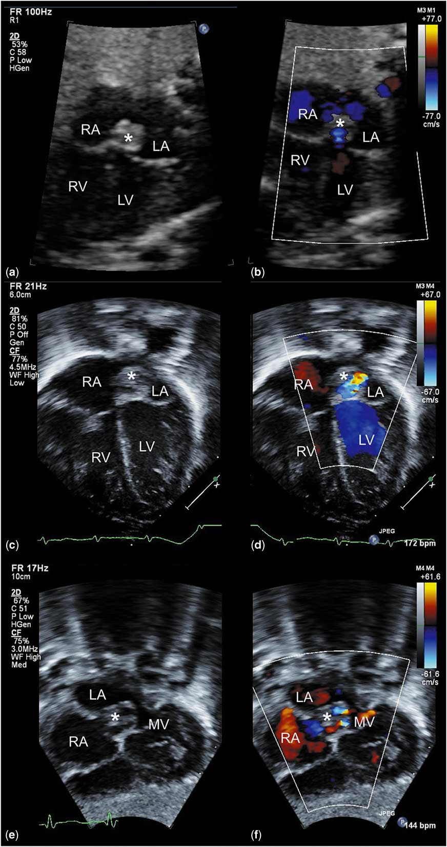
A 27-year-old woman, gravida 2, para 0, was referred for a fetal echocardiogram at 30 weeks and 4 days of gestation after an echogenic focus was seen in the left atrium on fetal ultrasound. The four-chamber view on fetal echocardiogram showed a large, single, slightly mobile mass oriented along the left aspect of the atrial septum in the left atrium (Fig 1a, Supplementary video 1). The mass appeared to have mixed echogenicity, and there was suggestion of flow in the lesion on color Doppler (Fig 1b). The mass was not obstructing mitral, aortic, or foramen ovale flow, and the rest of the cardiac anatomy was normal with normal ventricular function.
( a and b ) Four-chamber view of the fetal heart showing the pseudoaneurysm of the mitral–aortic intervalvular fibrosa (PMAIVF) as a mass of mixed echogenicity oriented along the left side of the atrial septum ( b ) with flow into the lesion on color Doppler ( b ). ( c and d ) The comparative four-chamber view of the heart on postnatal echocardiogram confirming the presence of the mass ( c ) with flow on color Doppler ( d ). Subcostal long-axis view demonstrating the location of the pseudoaneurysm (*) posterior to the root of the aorta ( e ) with color flow swirling inside ( f ). LA=left atrium; LV=left ventricle; MV=mitral valve; RA=right atrium; RV=right ventricle; *=PMAIVF.

A follow-up fetal echocardiogram was performed at 36 weeks of gestation and did not show interval change in appearance, location, or size of the mass. The infant was born at 38 weeks of gestation and was admitted to the neonatal ICU for further evaluation and telemetry monitoring, given the concern that the mass might induce atrial arrhythmias.
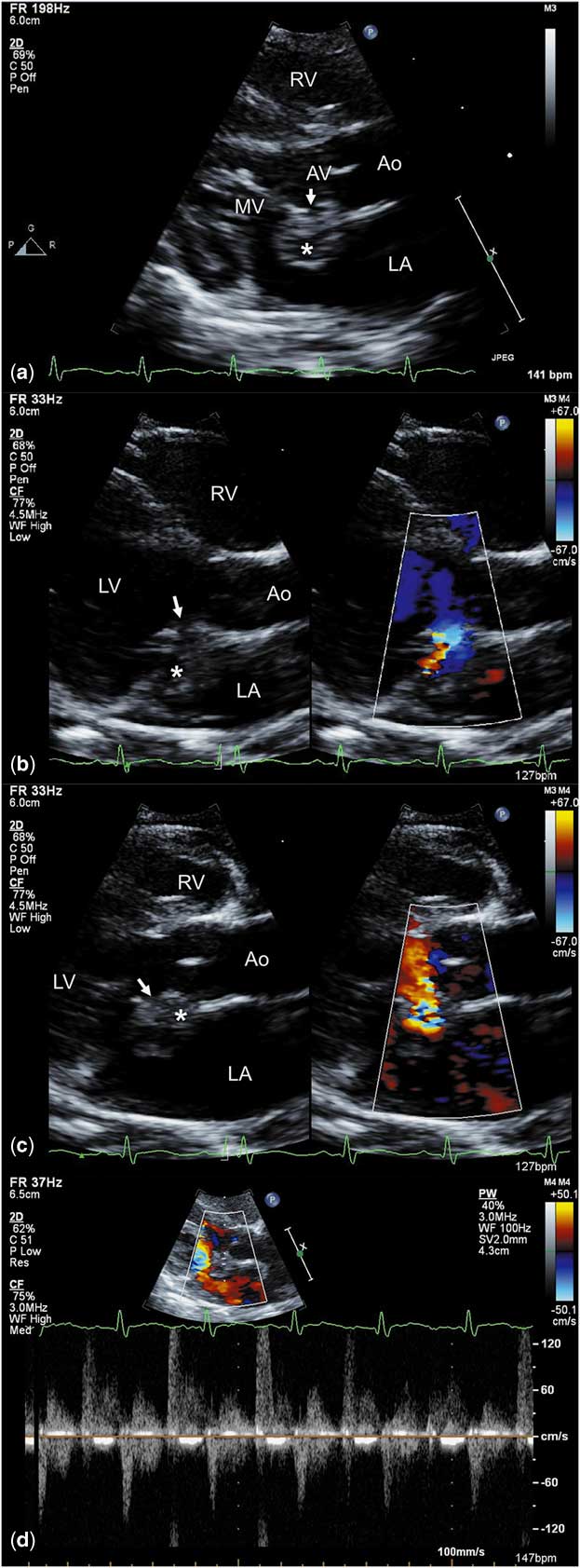
A comparative four-chamber view on the postnatal echocardiogram confirmed the presence of a 9.1×8.2-mm mass with echolucent components in the left atrium (Fig 1c) with flow into the lesion on color Doppler (Fig 1d). The subcostal long axis demonstrated the location of the mass posterior to the aortic root, along the atrial septum (Fig 1e–f). The parasternal long axis demonstrated the presence of an aneurysmal neck arising between the mitral and aortic annuli with a clear lack of fibrous continuity (Fig 2a, Supplementary video 2). Color Doppler imaging demonstrated to-and-fro flow into the echolucent components arising from the distal left ventricular outflow tract, between the aortic annulus and the mitral annulus (Fig 2b–c). The blood flow was antegrade into the pouch during systole and retrograde into the left ventricular outflow tract during diastole (Fig 2b–c). Pulsed-Doppler verified the antegrade and retrograde flow in the neck (Fig 2d). The location of the defect and the pattern of to-and-fro flow in the neck were consistent with a congenital pseudoaneurysm of the mitral–aortic intervalvular fibrosa.
Parasternal long-axis view demonstrating the neck of the aneurysm (arrow) arising between the mitral and aortic annuli ( a ) with blood flow into the pouch during systole ( b ) and into the left ventricular outflow tract during diastole ( c ) on color Doppler. Pulsed-Doppler demonstrating antegrade and retrograde flows into the neck ( d ). Ao=aorta; AV=aortic valve; LA=left atrium; LV=left ventricle; MV=mitral valve; RV=right ventricle; *=pseudoaneurysm of the mitral–aortic intervalvular fibrosa (PMAIVF); ↓=neck of the PMAIVF.

Similar to the prenatal echocardiograms, there was no intracardiac obstruction and otherwise normal cardiac anatomy and function. The infant was observed in the neonatal ICU for 48 hours on telemetry, where he remained in sinus rhythm without evidence of arrhythmias. He was discharged home on day of life 2.
The infant is now 12 months old, and has had serial postnatal echocardiograms showing no significant change in the size or appearance of the lesion. The family was counselled that this is a rare congenital defect, and that the natural history and management are not defined. Given the stable size of the aneurysm, its non-obstructive nature, and the to-and-fro flow in the neck, we are continuing conservative management with close observation with serial echocardiograms. If the pseudoaneurysm begins to increase in size or become obstructive, surgical correction will be discussed. In addition, should the neck of the aneurysm begin to constrict, initiation of aspirin for thrombus prophylaxis will be considered.
Discussion
The mitral–aortic intervalvular fibrosa is an avascular, fibrous structure between the anterior mitral leaflet and the aortic annulus. It is adjacent to the left ventricular outflow tract and is the area where the anterior mitral leaflet becomes continuous with the non-coronary cusp of the aortic valve, providing structural and functional integrity to the aortic and mitral valves. It is bound by the left and right fibrous trigones, the pericardium anteriorly, and the left ventricular outflow tract forms the base.
Pseudoaneurysm formation is uncommon but well described, predominantly in adults, after infective endocarditis or trauma resulting from valve surgery.Reference Şahan, Gül and Şahan 1 – Reference Xie, Li and Cheng 3 Pseudoaneurysm formation has also been reported as a “congenital” diagnosis in cases where preceding infection, trauma, or surgery have not been documented.Reference Gelehrter, Wright and Gless 4 – Reference Zhao, Chen and Yang 8 Gelehrter et alReference Gelehrter, Wright and Gless 4 reported seven patients with pseudoaneurysms originating from the aorto-mitral intervalvular fibrosa. All except one patient had associated CHD, preceding cardiac intervention, infection, or trauma. The only previous prenatal diagnosis was seen in a terminated pregnancy where autopsy confirmed the diagnosis.Reference Zhao, Chen and Yang 8 To our knowledge, our case is the first case of a congenital pseudoaneurysm that exists as an isolated cardiac defect detected prenatally on an echocardiogram and is being followed-up postnatally.
The diagnosis was confirmed postnatally by echocardiography, visualising a pouch communicating with the left ventricular outflow tract with a to-and-fro flow pattern on color Doppler imaging. This feature can help distinguish the lesion from other possible diagnoses such as a dilated coronary sinus, coronary arteriovenous fistula, or other intracardiac masses.
According to previously reported case studies, the lesion can result in fatal complications leading to recommendations to perform surgical correction at diagnosis.Reference Şahan, Gül and Şahan 1 – Reference Xie, Li and Cheng 3 Complications can result from compression or fistula formation with adjacent structures, such as the left atrium and left ventricular outflow tract, rupture, intra-aneurysm clot formation, and systemic embolisation.
Our case demonstrates that pseudoaneurysm of the mitral–aortic intervalvular fibrosa can exist without preceding trauma as a rare, isolated congenital cardiac defect seen on prenatal echocardiography. The natural history of these lesions is unknown, and it is unclear whether a congenital pseudoaneurysm, which exists without preceding trauma, infection, or surgery, in the absence of associated CHD, carries the same risks as a pseudoaneurysm that develops secondary to trauma or in association with other heart defects. Our patient has been followed-up with serial echocardiograms documenting the stability of the lesion, making conservative management with serial echocardiograms of lesions that remain stable in size, without impingement on adjacent structures, and to-and-fro flow in the neck, a reasonable option.
Acknowledgements
None.
Financial Support
This research received no specific grant from any funding agency or from commercial or not-for-profit sectors.
Conflicts of Interest
None.
Ethical Standards
This manuscript does not involve human or animal experimentation.
Supplementary materials
To view supplementary material for this article, please visit https://doi.org/10.1017/S1047951117000890