Information on longevity of digeneans is restricted to a few studies on some medically important species (see review in Sandground, Reference Sandground1936). Thus, fasciolids may persist in various ruminants for 5 years or more and human schistosomes and opisthorchids may live for 20–30 years. However, most digeneans survive in their definitive hosts for several months or 1–2 years (Sandground, Reference Sandground1936). Precise information on the longevity of adult non-human digeneans in laboratory hosts is, for the most part, unavailable. In most studies on digeneans in laboratory hosts, the experiments were terminated prior to the time when all adult worms were eliminated from the hosts (see, for example, Toledo et al., Reference Toledo, Espert, Muñoz-Antoli, Marcilla, Fried and Esteban2005).
Hosier & Fried (Reference Hosier and Fried1991) determined the longevity of the intestinal trematode Echinostoma caproni in ICR mice; the study was not terminated until 29 weeks postinfection (PI) and showed that mice, each infected with 25 encysted metacercariae (cysts), could support a relatively high percentage of worms up to 20 weeks PI. From 20 to 29 weeks PI, there was a diminution in worm growth (degrowth) based on body area measurements. There was also an anteriad shift in the worm population from the ileum–jejunum to the duodenum. The duodenum is an unusual site for this digenean (see review in Fried & Huffman, Reference Fried and Huffman1996).
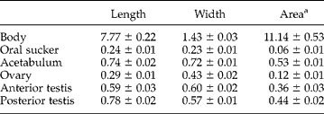
We have recently begun using Balb/c mice in our laboratory because of the availability of this strain from a breeding colony at our institution. Our first study with this strain (Bandstra et al., Reference Bandstra, Murray, Fried and Sherma2007) showed that it was susceptible to E. caproni infection using a cyst inoculum of 75 per mouse. Because of our success with this model, the purpose of this study was to test the longevity of E. caproni in Balb/c mice using five hosts (see details of mouse exposure and maintenance in Bandstra et al., Reference Bandstra, Murray, Fried and Sherma2007), each exposed to 75 cysts. To determine if the exposed mice became infected, faecal samples were taken from each mouse at 2, 4 and 6 weeks postexposure. All mice were positive with E. caproni eggs based on the detection of eggs in the stool. All mice were necropsied at 23 weeks (160 days) PI, following methods described in Stillson & Platt (Reference Stillson and Platt2007); the intestine was divided into five equal zones from the pylorus to ileo-caecal valve to identify worm location in the gut. Worms were removed, pooled and fixed in hot 5% neutral buffered formalin. The worms were left unstained and mounted on glass slides in glycerin jelly. Measurements (n = 10) were made on body length, maximum width, oral sucker, acetabulum, ovary and testes and were converted to areas (table 1).
Mean±SE morphometric values (mm) of ten randomly selected E. caproni worms from two adult Balb/c mice necropsied 23 weeks postinfection.
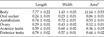
a Values for area are given in mm2.
Two of the five hosts were infected at 23 weeks PI with a total of 33 worms; one host contained 10 and the other 23 worms. All worms were located in sections 3 and 4, the ileum–jejunum region. All worms looked normal, i.e. showed no signs of abnormal testes ovary or uterus. Such abnormalities in the genital system would be indicators of ageing of digeneans in a host (see Bakke, Reference Bakke1988). The worm uteri were distended with eggs, and uterine egg counts of 10 worms showed a mean of 243 ± 6 eggs/worm. The condition of the uterus and the number of eggs/worm suggested that the eggs were viable; however, embryonation experiments were not done. The morphometric data are reported in table 1. These data suggest that body and organ measurements were normal in comparison with similar measurements made on adults of E. caproni (Hosier & Fried, Reference Hosier and Fried1991; Balfour et al., 2001)
Numerous factors affect long-term worm recovery in the mouse–E. caproni model, including strain of mouse, age of cysts, and number of cysts used to infect the mice. The results of this study show that Balb/c mice can sustain normal-appearing worms in the ileum–jejunum region for >5 months; this host will be useful for studies that examine long-term effects of the E. caproni infection on both the parasite and the host.