Introduction
Tuberculosis (TB) is an airborne, infectious disease caused by bacteria belonging to the Mycobacterium tuberculosis family. Annually, there are approximately nine million new cases of TB, with nearly two million deaths worldwide. It is the leading cause of death due to a preventable, curable infectious disease. Approximately 9000 new cases of TB are currently reported each year in the UK. Most cases occur in major cities, particularly London.
Up until the 1980s, TB was thought to be becoming a historical disease, due to the availability of effective antibiotics for treatment and vaccination for prevention. However, the rise in human immunodeficiency virus (HIV) infections, neglect of TB control programmes and emergence of drug-resistant strains have lead to a resurgence in TB. As a result, the World Health Organization declared TB a global emergency in 1993. The incidence of TB in the UK is still on the increase. In 2009, the number of TB cases provisionally reported in the UK was 9153 (14.9 per 100 000 population), representing a 5.5 per cent increase on the 2008 figure.1
It is not uncommon for TB to present to otolaryngologists. Cervical lymphadenopathy is the commonest such presentation. Lymph node TB accounts for nearly half of all non-respiratory sites, and its incidence is rising.2 However, there are a number of more unusual presentations, and a high index of clinical suspicion is always required.
In this article, we present a new case of TB presenting as acute stridor. We also review some of the more unusual ENT manifestations of the disease, and discuss current management strategies.
Case report
A 39-year-old Somalian man presented to the accident and emergency department with a two-day history of noisy and difficult breathing, associated with weight loss and a non-productive cough for several weeks. He had lived in the UK for 18 years, and had never left the country in that time. He had no history of TB contact, but had never received a bacilli Calmette-Guérin (BCG) vaccination. He had no significant past medical history.
On examination, the patient had inspiratory stridor associated with tracheal tug and intercostal recession. His respiratory rate was 22 breaths/minute and his oxygen saturation was 94 per cent on oxygen (10 l/minute). On auscultation, he had reduced air entry bilaterally. Palpation of the neck revealed a symmetrical anterior neck mass. Flexible nasendoscopy showed bilateral vocal fold palsy in the adducted position.
Arterial blood gas analysis (on oxygen 10 l/minute) showed respiratory acidosis (pH 7.28, pCO2 7.5 kPa, pO2 12.1 kPa and HCO3 concentration 23.2 mEq/l).
A chest X-ray showed some left base consolidation only.
In view of the severity of the patient's respiratory distress, due to upper airway obstruction, a decision was made to proceed to tracheostomy under local anaesthesia.
At tracheostomy, a butterfly-shaped swelling separate from the thyroid gland was identified overlying the thyroid cartilage. There was no tracheal displacement. A tracheostomy tube was successfully inserted through the third tracheal ring. The patient made an uneventful post-operative recovery.
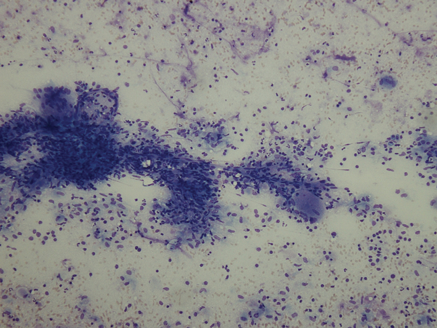
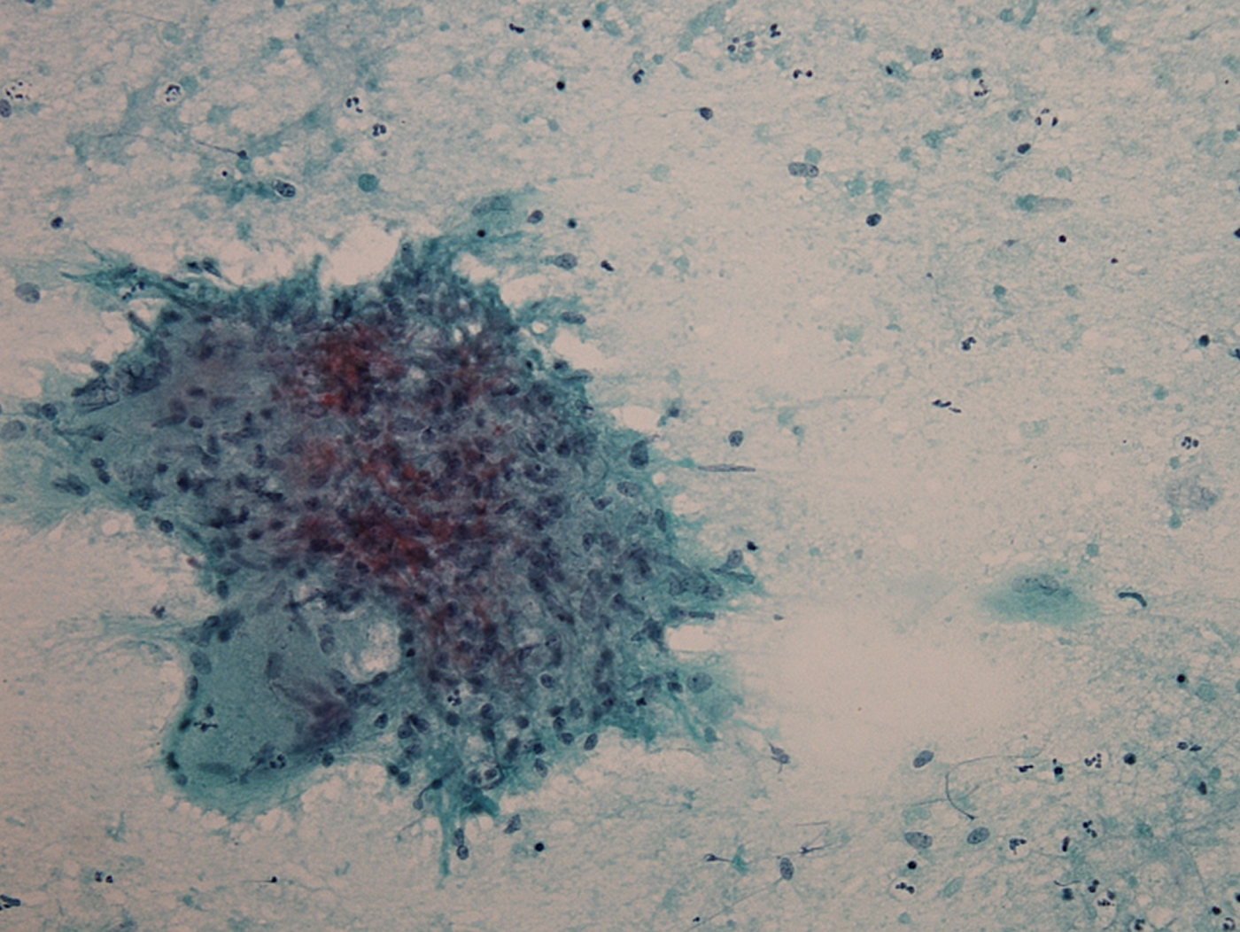
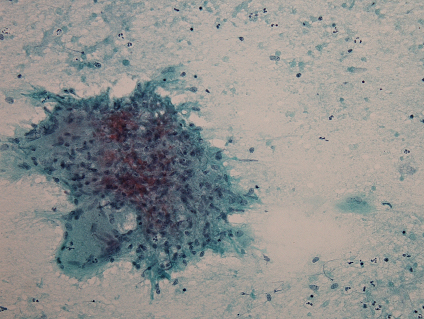
Fine needle aspiration cytology, performed the following day, showed well formed granulomas against a background of polymorphs and lymphocytes, associated with some necrosis but no caseation (Figures 1 and 2). The granulomas were composed mainly of epithelioid histiocytes and occasional multinucleated giant cells. The cytological features were of necrotising granulomatous inflammation, and the possibilities of infection and sarcoid were raised. Ziehl–Nielsen staining for acid-fast bacilli was negative, and microbial culture showed no growth.
Photomicrograph showing granulomas against a background of polymorphs and lymphocytes. (May-Grünwald Giemsa; ×40)
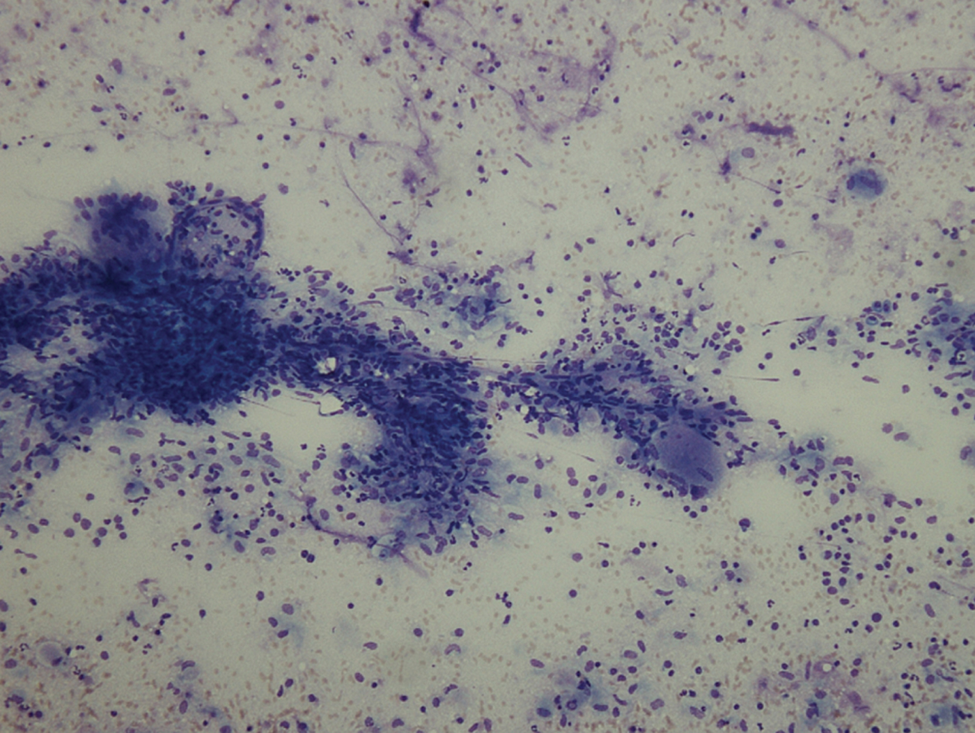
Photomicrograph showing well formed epithelioid granulomas with occasional giant cells and some necrotic background material. (May-Grünwald Giemsa; ×100)
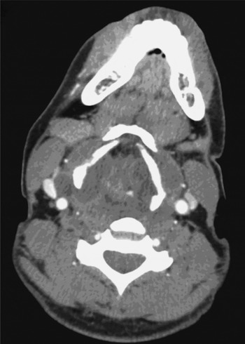
Computed tomography (CT) scanning following tracheostomy showed a mass, separate from the thyroid gland, surrounding the thyroid cartilage and causing airway occlusion (Figure 3). The scan also showed bilateral upper lobe pulmonary infiltrates and coeliac and retrocrural lymphadenopathy, in combination with multiple lesions in the spleen.
Axial computed tomography scan showing complete airway occlusion.

After a review by the communicable diseases team, the patient was commenced on standard TB quadruple therapy, three days after initial presentation. Subsequently, all three sputum specimens obtained from the tracheostomy tube prior to commencement of treatment showed acid-fast bacilli. Tests for HIV and hepatitis B and C serology were negative.
On day 5 of treatment, flexible nasendoscopy was repeated, which showed sluggish but definite movement of both vocal folds. After one week of treatment, the patient was successfully decannulated. At the time of writing, he continued to make a good recovery.
Discussion
Tuberculosis presents most commonly to the otolaryngologist as tuberculous cervical lymph nodes. The typical finding is a chronic, painless mass in the neck. As the disease progresses, the abscess may burst and form a sinus. More unusual otolaryngological manifestations have also been reported, and include endolaryngeal lesions, unilateral vocal fold palsy secondary to mediastinal involvement, laryngitis, otitis media, and masses in the tonsil, parotid, thyroid, nasal cavity and cervical spine.Reference Nalini and Vinayak3
Tuberculosis of the larynx commonly occurs secondary to pulmonary TB, and very rarely occurs in isolation.Reference Soda, Rubio, Salazar, Ganem, Berlanga and Sanchez4 Symptoms include hoarseness, cough, dyspnoea, dysphagia, odynophagia and weight loss, which can make this condition difficult to distinguish from laryngeal carcinoma, even on laryngoscopy. Direct laryngoscopy and biopsy are essential for establishing the diagnosis.Reference Shin, Nam, Yoo and Kim5
Cases of tuberculous tracheitis have also been described, and may present with cough and stridor. Endoscopic examination may show diffuse tracheal narrowing by tuberculous pseudomembranous lesions, which are almost always secondary to pulmonary TB.Reference Tong and Chow6
Tuberculous lesions of the oral cavity are also usually secondary to pulmonary TB. The commonest site for oral lesions is the tongue. The usual presentation is with an ulcer, but macroglossia can also present. Other sites include the gingiva, palate, lips, buccal mucosa and the floor of the mouth. Primary tuberculous lesions in the oral cavity are rare, and usually occur in areas of trauma such as the lateral border of the tongue when exposed to broken teeth, as the mycobacterium favours areas of chronic inflammation.Reference Iype, Ramdas, Pandey, Jayasree, Thomas and Sebastian7 Unlike tuberculous lymph nodes, oral cavity lesions are usually painful and result in poor nutrition. Differential diagnoses include malignancy, traumatic ulcers, aphthous ulcers, and other conditions associated with granuloma formation such as Crohn's disease, cat-scratch disease and syphilis.
Salivary gland TB is also often difficult to distinguish from malignancy. The parotid gland is the most frequently affected. The commonest presentation is a localised mass, resulting from infection of parotid lymph nodes. Other presentations include acute sialadenitis with diffuse glandular enlargement, periauricular fistula or an abscess.Reference Sethi, Sareen, Sabherwal and Malhotra8
Nasal TB is usually secondary to pulmonary TB or lupus vulgaris.Reference Waldman, Levine, Sebek, Parker and Tucker9 Affected individuals may present with nasal obstruction, discharge, pain, crusting, epiphora, recurrent nasal polyps or ulceration. Lesions may be ulcerative, infiltrative or proliferative, and most cases are unilateral.Reference Butt10 The most common sites of involvement are the cartilaginous septum and the inferior turbinate.Reference Hup, Haitjema and de Kuijper11
Tuberculosis of the middle ear clinically resembles chronic otitis media in its early stages, often leading to delayed diagnosis and the risk of serious complications. Symptoms include painless, scanty, thin and odourless discharge, disproportionate hearing loss, and, later, multiple tympanic membrane perforations. Early bacteriological analysis is often negative.Reference Chmielik, Ziolkowski, Koziolek, Kulus and Chmielik12 Diagnosis is made by direct microscopy, culture and polymerase chain reaction.
Cervical spine TB accounts for only 1–3 per cent of spinal TB (also known as Pott's disease). It may initially present to otolaryngologists with dysphagia, stridor, torticollis, hoarseness, pain or neck stiffness.Reference Pageeh and Lamothe13 It should be borne in mind that 40 per cent of patients develop severe neurological symptoms, and approximately 50 per cent develop retropharyngeal cold abscess.Reference Al Soub14
Investigation
The initial investigation of choice for cervical lympadenopathy is fine needle aspiration cytology. Excisional biopsy may occasionally be required, but this should be avoided unless essential for diagnosis, as it may spread the disease and can lead to the development of a chronically discharging sinus. In addition, patients who have TB of the head and neck region must be investigated to exclude pulmonary or systemic TB.
If non-respiratory TB is a possibility, samples should be placed in a dry container and sent for TB microscopy and culture. The Ziehl–Nielsen stain is conventionally used to identify M tuberculosis; this dyes acid-fast bacilli bright red against a blue background. However, fluorescence microscopy (using auramine-rhodamine staining) is now preferred as it has greater sensitivity.Reference Steingart, Henry, Ng, Hopewell, Ramsay and Cunningham15 If acid-fast bacilli are seen, polymerase chain reaction or gene probe testing is conducted to distinguish M tuberculosis from other mycobacteria. Culturing acid-fast bacilli may take four to eight weeks; new, automated systems may be faster and more accurate than the traditionally used Lowenstein–Jensen media.Reference Drobniewski, Caws, Gibson and Young16
The tuberculin sensitivity test, also known as the Mantoux test, can identify individuals who have been exposed to TB, and is cheap and easy to perform. It requires a small dose of TB protein to be injected intradermally. A raised, red area shows a positive reaction. The test response has to be interpreted within a certain timescale to avoid false negative results. Other causes of false negative results include immunosuppression (particularly co-infection with HIV) and medication. False positive results may be due to prior BCG vaccination or exposure to environmental mycobacteria.
Serum interferon gamma tests assess the body's response to specific tuberculosis antigens which are not found in BCG. Serum interferon gamma tests should be used in cases of positive tuberculin sensitivity test, for the diagnosis of latent TB.Reference Soda, Rubio, Salazar, Ganem, Berlanga and Sanchez4, Reference Pai, Riley and Colford17
Typical chest radiograph appearances of TB include upper lobe infiltrates or cavities, mediastinal or hilar lymphadenopathy, and pleural effusions. However, radiographic appearances are suggestive of TB rather than diagnostic. In primary TB cases, as many as 15 per cent of conventional chest radiographs may be normal.Reference Lee, Song, Lim, Kim, Kim and Lee18
In the immunocompromised population, lymphadenopathy may occasionally occur in isolation, and it may not be detected on conventional radiographs. Additional imaging with CT is often required, because this modality is more sensitive in depicting lymphadenopathy.
Treatment
The appropriate treatment regimen should be started without waiting for culture results, if the clinical picture and cytology and/or histology are consistent with a diagnosis of TB.Reference Ormerod, McCarthy, Rudd and Horsfield19
For the last 15 years, the ‘gold standard’ for TB therapy has been six months of daily treatment with rifampicin and isoniazid, supplemented for the first two months with pyrazinamide and ethambutol. Most cases of TB are still curable with this regime.
• The UK incidence of tuberculosis is increasing, particularly in urban areas
• Cervical lymphadenopathy is the commonest ENT tuberculosis presentation, but there are many other ENT manifestations
• A high index of clinical suspicion is required to ensure early diagnosis
• A multidisciplinary team approach is essential for appropriate management
A minority of TB cases show resistance to these antibiotics. In England, 6–8 per cent of cases are resistant to isoniazid, and 1 per cent of cases are resistant to more than one of the standard antituberculous antibiotics.Reference Ormerod, McCarthy, Rudd and Horsfield19 Multidrug-resistant TB is defined as TB caused by organisms that are resistant to isoniazid and rifampicin, two of the first-line drugs in TB treatment. A risk assessment for drug resistance should be made for each patient with TB, based on the following risk factors: (1) a history of prior TB drug treatment, and prior TB treatment failure; (2) contact with a known case of drug-resistant TB; (3) birth in a foreign country, particularly one with a high incidence of TB; (4) HIV infection; (5) residence in London; (6) age between 25 and 44 years; and (7) male gender.2
Resistance to rifampicin is used as a marker for multidrug-resistant TB, as a high proportion of rifampicin resistance (approximately 95 per cent) is associated with concurrent resistance to isoniazid. Polymerase chain reaction based techniques are used to detect mutations in the rpoB gene (a common cause of rifampicin resistance) in patients with risk factors for multidrug-resistant TB. If rifampicin resistance is found, infection control measures and drug treatment for multidrug-resistant TB are commenced, until the results of standard drug susceptibility tests are available.2
Since 2006, extensively drug-resistant TB has been identified in all regions of the world. This is defined as multidrug-resistant TB that is also resistant to any one of the fluoroquinolones and to at least one of three injectable second-line drugs (amikacin, capreomycin or kanamycin).Reference Raviglione and Uplekar20 The emergence of multidrug-resistant TB and extensively drug-resistant TB has threatened the previous progress made in controlling TB. Patients with extensively drug-resistant TB have significantly worse treatment outcomes than those with multidrug-resistant TB. The approach to the management of multidrug-resistant TB is under continuous development. The latest recommendations on the management of drug-resistant TB are published in the emergency update of the World Health Organization's Guidelines for the Programmatic Management of Drug-Resistant Tuberculosis.21
Conclusion
Tuberculosis has a wide variety of possible ENT presentations, and diagnosis requires a high index of clinical suspicion. Clear investigation strategies are important in order to prevent delay in diagnosis and treatment. Close liaison with microbiology and infectious disease teams is essential for appropriate management.
Acknowledgements
The authors would like to acknowledge the support and advice of Mr J Lim and Dr J Seet.