Introduction
Functional endoscopic sinus surgery (FESS) is now widely accepted to be the primary surgical intervention for sinonasal inflammatory disease.Reference Fokkens, Lund, Mullol, Bachert, Alobid and Baroody 1 One of the key prerequisites for performing successful and safe surgery is a thorough knowledge of the paranasal sinus anatomy. Despite this knowledge, unusual anatomical variants, previous surgery and severe disease may lead to more hazardous conditions, putting the patient at risk of potentially catastrophic complications.
Computed tomography (CT) scans of the paranasal sinuses are now regarded as mandatory prior to FESS, as they provide accurate information regarding disease extent and detailed anatomy.Reference Lund, Savvy and Lloyd 2 Anecdotal evidence suggests that in the UK, the majority of surgeons who request CT scans of the paranasal sinuses interpret the images themselves prior to surgery. However, no formal study has confirmed this practice. Publicised radiological standards make reference to the importance of expert reporting of all radiological images. 3 Expert reporting may identify disease or important anatomical variants that might otherwise be missed by those without a radiological background or by those who do not perform a high volume of FESS procedures. In rare circumstances, ignoring the findings of a formal radiological report can leave clinicians open to medicolegal litigation in the event of operative complications.
The level of anatomical detail provided in a radiology report has been shown to differ significantly in practice.Reference Deutschmann, Yeung, Bosch, Lysack, Kingstone and Kilty 4 This may in part be related to differences in the expectations of the perceived use of paranasal sinus CT scans between radiologists and otolaryngologists. Otolaryngologists predominantly request paranasal sinus CT as a ‘road map’ to identify surgically relevant anatomy, rather than for assessing disease extent. A lack of understanding of the specific surgical needs by the reporting radiologists may lead to suboptimal reports that are inadequate for operative planning, especially for those less comfortable with interpreting CT images themselves.
Several ‘checklists’ have been published that highlight key anatomical areas within the paranasal sinuses.Reference Hoang, Eastwood, Tebbit and Glastonbury 5 – Reference Zinreich, Kennedy, Rosenbaum, Gayler, Kumar and Stammberger 7 To date, there are no published (radiological or ENT) guidelines for the interpretation of paranasal sinus CT scans performed specifically for peri-operative planning. This study therefore aimed to ascertain the opinions of practising UK otolaryngologists regarding identifying important surgically relevant areas to be described within a radiological report, which could be used to improve pre-operative planning and safety.
Materials and methods
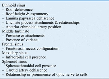
A review of the literature was performed by searching databases that included Medline, PubMed and Embase for the following keywords: endoscopic sinus surgery, computed tomography (CT), anatomy and paranasal sinuses. Current evidence relating to key areas in surgical and radiological sinonasal anatomyReference Lund, Savvy and Lloyd 2 , Reference Lloyd, Lund and Scadding 8 , Reference Arslan, Aydinhoglu, Bozkurt and Egeli 9 was retrieved and used to develop a questionnaire (Table I).

The survey aimed to address two main questions: (1) how comfortable are UK otolaryngologists at interpreting CT scans of the paranasal sinuses themselves; and (2) what areas are considered important for radiologists to make a formal comment upon within their report.
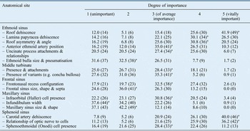
Each question regarding individual anatomical areas requested a response on a five-point scale, which reflected varying degrees of importance, with a score of 1 representing unimportant and 5 reflecting vitally important (Table II).
Importance of individual anatomical areas of paranasal sinus computed tomography to be reported

Data represent the importance of each anatomical area, in terms of percentages (and numbers) of responders. *Indicates the grade (1–5) given by the highest percentage (numbers) of responders
After an initial local pilot study assessing the appropriateness of the questionnaire, the survey was edited and approved by the ENT-UK Survey Guardian prior to distribution. It was circulated via e-mail invitation to the ENT-UK panel, the British Association of Otorhinolaryngology – Head and Neck Surgery, between October 2013 and November 2013. The results for each question were collated and statistically analysed (chi-square analysis) using SPSS® version 17.0. Statistical significance was defined as p < 0.05.
Results
Questionnaire invites were sent to 332 members of the ENT-UK panel. From these, there were 117 (35.2 per cent) fully complete responses. Responders to the survey included: consultants (84 per cent), specialty trainees (7 per cent), associate specialists (6.1 per cent) and others (2.9 per cent). The average experience in otolaryngology was 19.82 years (range, 6–40 years). From the consultant group, 42 per cent were subspecialist rhinologists.
Twenty-nine per cent of the consultant clinicians stated that they were not comfortable analysing all anatomical areas in question on CT imaging. In addition, 39 per cent admitted to feeling uncomfortable in operating within certain anatomical areas during FESS without a formal radiological report. The subgroup analysis showed that the non-rhinologist group were less comfortable in interpreting all areas of the CT scan compared to the rhinologist group (p = 0.015).
Importance of anatomical areas
Detailed responses for the questionnaire are shown in Table II. Anatomical areas graded with the highest importance (5 out of 5) included the carotid canal and ethmoid roof (dehiscence of both), and the optic nerve and its relationship to accessory air cells. Anatomical areas graded 4 out of 5 and labelled ‘very important’ included the lamina papyracea (dehiscence) and the ethmoid roof (asymmetry and anteroposterior angle). Anatomical areas graded 3 out of 5 and labelled of ‘average importance’ included the anterior ethmoidal artery (its position), sphenoethmoidal (Onodi) cells (their presence), the frontonasal recess (configuration), the uncinate process (attachments and relationships), infraorbital (Haller) cells (their presence), the middle turbinate (presence and attachments) and middle turbinate variants. There was agreement between the two subgroups (rhinologists and non-rhinologists) when grading the vast majority of anatomical subsites.
Discussion
Computed tomography imaging is essential in identifying patient-specific paranasal sinus anatomy pre-operatively, to enable safe and efficient surgery. Our study highlights the surgically relevant anatomical areas identified by UK otolaryngologists.
Formal radiological reporting is considered to be the ‘gold standard’ in image interpretation; however, limited evidence suggests that the content can be variableReference Deutschmann, Yeung, Bosch, Lysack, Kingstone and Kilty 4 and is not always used by the operating clinician. This practice may have several important implications relating to possible medicolegal consequences, and may have an impact upon radiological services and inter-departmental relationships.
Medicolegal implications
Medical malpractice cases linked to FESS have become more commonplace. Lynn-Macrae et al. performed an analysis of the US legal database, assessing FESS-related litigation from 1990 to 2003.Reference Lynn-Macrae, Lynn-Macrae, Emani, Kern and Conley 10 They reported that 76 per cent of malpractice cases were allegedly a result of negligent technique, with the highest payouts being awarded to those with disabilities from blindness, diplopia, anosmia, cerebrospinal fluid leak and brain damage.
These specific complications arise from damage to the skull base or orbit. Severe sinonasal disease or variable anatomy may be significant contributory factors in accidental damage to these areas. Therefore, from the medicolegal perspective, inadequate pre-operative planning (e.g. failure to appreciate endogenous risk factors in patients, such as variable anatomy on a CT scan) may be perceived as contributing to negligent technique. Further analysis has shown that these patient factors (e.g. variable anatomy) have a stronger relationship with the risk of such complications compared to surgical factors (e.g. technique and experience).Reference Re, Maquiulo, Romero, Gioacchini and Pasquini 11
Burden for radiologists
Radiological services within the UK are coming under increasing pressure, with a growing annual workload of the order of 2–5 per cent per annum.Reference Re, Maquiulo, Romero, Gioacchini and Pasquini 11 , 12 In 2002, the Royal College of Radiologists' publication Clinical Radiology: a Workforce in Crisis highlighted the increased workload of the consultant radiologist. 13 Plans to include additional commitments such as multidisciplinary team working has meant that there is a greater need for rationalisation of the time committed to image reporting. Guidance published by the Royal College of Radiologists recommends that numbers of CT and magnetic resonance imaging scans to be reported by radiologists should be at three to six per hour for standard scans and at one to two per hour for complex scans, 14 thus giving an estimate of the potential burden for the reporting of sinus CT scans.
The average growth rate of CT image requests has been shown to be in the order of 10.1 per cent per annum since 2003. 12 Therefore, in the context of greater CT scan reporting and an increasing workload, it is more important than ever that radiologists spend their time reporting on images that really make a difference to surgery and patient safety.
What our survey says
The majority of specialists within our study (70.1 per cent) indicated that they are fully confident in interpreting all of the anatomical sites within the CT scan. A notable number of consultant clinicians (39 per cent) stated that they would not feel comfortable operating in certain anatomical areas without a formal radiological report. These findings may reflect varying experience (within consultant clinicians) in the UK in performing FESS. It is likely that more complex cases (e.g. frontal sinus or sphenoid disease) are commonly referred onto subspecialists who deal with larger caseloads. Subspecialist rhinologists and FESS surgeons represented 42 per cent of the study group, which suggests that the findings from our study reflect the opinions of a wider range of ENT clinicians, including the generalist, all of whom commonly perform FESS in the UK.
Despite the anecdotal opinions, our survey of the ENT-UK panel indicates that there is diversity in the peri-operative strategy for those performing FESS in the UK. When surveying a sizeable number of practising UK otolaryngologists (with an average time in practice of 19.82 years; range of 6–40 years), this study identified an ongoing need for a formal radiological report for a significant number of clinicians performing FESS. Unsurprisingly, it is likely that the greatest benefit of formal reporting may come to those who are not subspecialist rhinologists, and are therefore less confident operating in certain areas (e.g. frontal sinus or sphenoid sinus), especially if they are not planning to refer the case onwards.
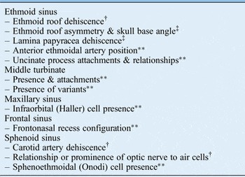
This study identified key anatomical areas that hold greater importance to otolaryngologists (Table III). Unsurprisingly, the sites identified represent areas that harbour risk of catastrophic complications relating to the orbit and skull base.
Checklist of key areas*

* These represent the key areas to be commented upon within a formal radiological report. †Vitally important; ‡very important; **important
The study attempts to ascertain the importance of a potential comment about a specific anatomical subsite within a formal radiological report. This does not reflect the importance of the anatomical area per se. This may explain why a number of participants (n = 9) labelled all areas with ‘no importance’ to be reported by the radiologist. Reasons cited by clinicians for this pattern of response included their own comfort in interpreting all aspects of the CT scan alone and the subsequent lack of need for using the radiologist's report. These clinicians were predominantly subspecialist rhinologists. This finding highlights a limitation of the study design that requires all participants to undertake the main body of the questionnaire despite stating they did not use a radiologist report. Twenty-seven of the participants who stated they were comfortable in analysing all areas of a CT scan continued to rate the importance of individual anatomical areas. Therefore, all responses were included in the final analysis.
The inclusion of primary and revision surgery within the survey may be regarded as a further limitation. Feedback suggests that experts consider radiological reporting to have greater importance in revision cases. However, it could be argued that peri-operative planning for any surgery (primary or revision) should be consistent in order to reduce any undue risk of medical negligence. Production of this particular questionnaire required a concise structure that followed strict production and dissemination guidelines (ENT-UK 15 ). For this study, addressing the limitations already described with regard to survey design and separation of primary and revision surgery would have significantly prolonged the questionnaire, and thus impacted upon participant engagement.
-
• Paranasal sinus computed tomography is essential for pre-operative planning of functional endoscopic sinus surgery
-
• Radiological reports may be variable because of the lack of published guidance
-
• Key anatomical areas identified by ENT-UK panel are described
-
• This study may be used to produce surgically relevant radiological reports
The results from this study may be utilised as an aide memoir to guide radiologists producing reports for paranasal sinus CT; the findings highlight those surgically relevant anatomical areas identified as important by UK otolaryngologists that should be included within such reports (Table III).
Conclusion
To our knowledge, this is the first study to obtain formal guidance from a group of practising otolaryngologists regarding surgically relevant areas considered important for description within a radiology report. The information gleaned from this study can be used to guide radiologists who report paranasal sinus CT findings, to improve peri-operative surgical planning and ultimately safety. In an age of increasing medicolegal litigation, such information may help to reduce the risk of medical negligence claims.
Acknowledgements
The Survey Authors acknowledge the voluntary contribution of ENT-UK Expert Panel Members, who help develop knowledge within our speciality by sharing their Reflections on Practice.