Introduction
The pterygopalatine fossa is a small space the shape of an inverted pyramid, which contains the pterygopalatine ganglion and maxillary nerve together with the terminal part of the maxillary artery and its branches. It is related posteriorly to the greater wing of the sphenoid, anteriorly to the posterior wall of the maxilla, superiorly to the inferior surface of the body of the sphenoid, and laterally to the infratemporal fossa via the pterygomaxillary fissure.
The infratemporal fossa is a potential space bounded superiorly by the temporal bone and the greater wing of the sphenoid bone, medially by the superior constrictor muscle, the pharyngobasilar fascia and the pterygoid plates, laterally by the zygoma, mandible, parotid gland and masseter muscle, anteriorly by the pterygoid muscles, and posteriorly by the articular tubercle of the temporal bone, glenoid fossa and styloid process.Reference Grant1
The infratemporal fossa communicates with the middle cranial fossa via various neurovascular foramina (i.e. the carotid canal, jugular foramen, foramen spinosum, foramen ovale and foramen lacerum). Medially, the infratemporal fossa communicates with the pterygopalatine fossa via the pterygomaxillary fissure, which is contiguous with the inferior orbital fissure and, thus, the orbit.Reference Van Huijzen2
Benign tumours usually respect these boundaries and expand the infratemporal fossa in the direction of its soft tissue planes, or they follow pre-existing pathways (e.g. the foramen ovale and pterygomaxillary fissure). Conversely, malignant tumours can infiltrate and destroy all structures within the infratemporal fossa and adjacent spaces.Reference Fisch, Fagan and Valavanis3
The presence of neurovascular structures within the infratemporal fossa (e.g. the internal carotid artery) or adjacent to it (e.g. the VIIth cranial nerve) limits the possible exposure of any particular surgical approach to the infratemporal fossa.Reference Cummings, Fredrickson and Harker4 Thus, surgical approaches are designed not only to remove the tumour but also to identify and preserve these neurovascular entities. Described surgical approaches to the infratemporal fossa comprise the preauricular approach, postauricular (transtemporal) approach, anterior transfacial approach, transorbital approach and, recently, the endoscopic approach.Reference Carrau, Vescan, Snyderman and Kassam5–Reference Janecka8
The most common tumour to extend into the pterygopalatine and infratemporal fossae is the juvenile nasopharyngeal angiofibroma.
There has previously been a reluctance to perform transnasal endoscopic resection of infratemporal fossa tumours, because of the vascularity of the region and the surgeon's greatly limited ability to manipulate the tumour and to control haemorrhage when significant bleeding occurs. However, endoscopic resection techniques for such tumours have now been developed, facilitated by advances in interventional radiology (with effective embolisation techniques enabling devascularisation) and anaesthetic techniques (enabling tight control of cardiac output and pulse rate).Reference Wormald and Robinson9
The other factor limiting transnasal endoscopic resection of infratemporal fossa tumours has been the limited ability of the surgeon to manipulate the tumour with traction, during the dissection. The development of a two-surgeon technique has now overcome this limitation. This new technique also aids haemostasis: in the case of brisk haemorrhage, the second surgeon can introduce high volume suction into the operative field, while the first surgeon controls the bleeding vessel using bipolar cautery or a clip.Reference Robinson, Patel and Wormald10
Materials and methods
The study included 15 male patients with angiofibroma extending to the pterygopalatine and infratemporal fossae. Patients were aged 12–25 years, and presented with recurrent, severe epistaxis and unilateral nasal obstruction.
We recorded all reported clinical data. Complete laboratory investigations were obtained in all cases, together with computed tomography (CT) and magnetic resonance imaging (MRI) scans.
Results
Pre-operative evaluation
All patients underwent complete head and neck examination, together with endoscopic evaluation, CT and MRI scanning, and laboratory investigation (including blood typing and preparation of fresh blood for transfusion).
All patients were prepared for surgical access to the infratemporal fossa via a transnasal, transantral, transpterygopalatine fossa route. Each patient's CT and MRI scans were thoroughly examined to determine the relationship of the tumour to critical structures, especially the inferior orbital fissure, cavernous sinus, vidian canal and carotid artery. Particular care was required as angiofibroma is a very vascular lesion and biopsy may cause catastrophic haemorrhage. Using Radkowski and colleagues' classification, all patients had a stage IIc angiofibroma.Reference Radkowski, McGill, Heal, Ohlms and Jones11
Surgical technique
Prior to commencement of surgery, the nose was decongested using nasal drops containing xylometazoline (decongestant drops), together with injection of xylocaine with 1/50 000 adrenaline into lateral nasal wall.
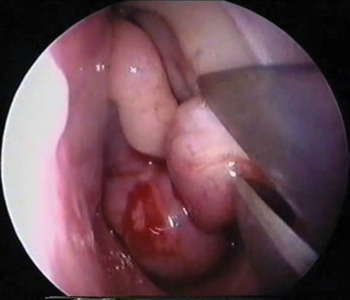
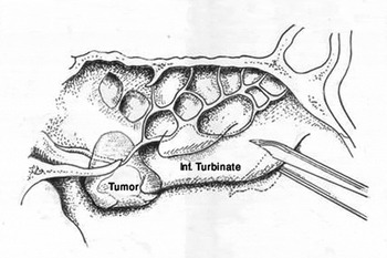
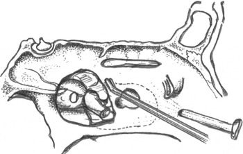
A partial inferior turbinectomy was performed to avoid trans-septal and canine fossa puncture (Figure 1). We then performed an uncinectomy, wide middle meatal antrostomy and sphenoethmoidectomy with removal of the lower half of the middle turbinate (Figure 2). The posterior and lateral wall of the maxillary sinus was then removed using a Hajek–Koeffler punch, in order to control the sphenopalatine artery (Figure 3). This artery was then followed proximally to achieve control of the maxillary artery with the help of bipolar electrocautery.
Stage 1: partial inferior turbinectomy.

Stage 2: partial middle turbinectomy plus middle meatal antrostomy and medial maxillectomy.

Stage 3: removal of the posterior and lateral wall of the maxillary sinus using a Hajek–Koeffler punch.

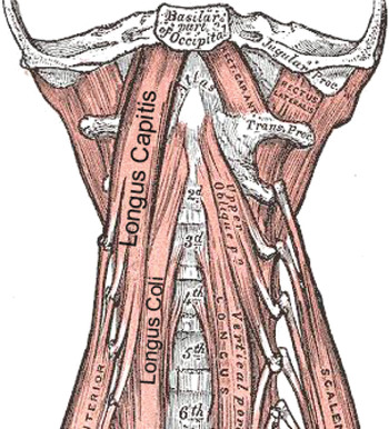
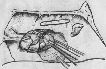
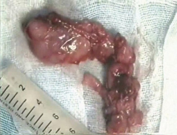
We then mobilised the endonasal tumour by separating it from the choana and posterior septum and freeing it from the sphenoid. The posterior region (i.e. the body of the sphenoid, pterygoid root and interpterygoid fossa) was exposed. The most difficult stage involved freeing the tumour from the posterior pharyngeal wall and the prevertebral muscles (i.e. the longus capitis and longus coli muscles) (Figure 4); bipolar cautery and surgical scissors were very useful tools for overcoming firm adhesions. Haemorrhage around the internal maxillary artery and internal carotid artery was another problem; this was controlled using bipolar cautery and Surgicel (oxidized cellulose) Johnson & Johnson Ethicon Surgicel 4″ × 8″ Absorbable Hemostats. When mobilising the pterygopalatine and infratemporal fossae tumour components, one surgeon grasped the tumour with Blakesley forceps and placed it under traction, allowing the other surgeon to dissect the tumour capsule (Figure 5). As the tumour was ‘rolled’ out of the infratemporal fossa, the medial and lateral head of the pterygoid muscles could be seen. Thorough examination of the infratemporal fossa, pterygoid canal and lateral wall of the sphenoid was mandatory in every case. The mobilised tumour was then delivered in toto through the nose (Figure 6).
Anatomical drawing showing the prevertebral muscles (longus capitis and longus coli) from which the tumour is dissected in stage 4.

Tumour removal during stage 5: one surgeon grasps the tumour while the other dissects it from the infratemporal fossa.

An angiofibroma tumour removed in toto.

In our 15 patients, the mean blood loss was 350 ml (range 400–650 ml). Only two young patients required blood transfusions.
Post-operative care
Patients were placed on systemic, broad spectrum antibiotics for at least two weeks, and also received saline nasal douches until the nose stopped crusting.
Post-operatively, nasal endoscopy was performed regularly every two weeks. Follow-up MRI scans were performed at two, six and 12 months. Any areas of enhancement were measured and watched closely at each follow-up scan, to ensure that they represented only post-operative inflammation.
Recurrence and revision surgery
Three of our 15 patients suffered a recurrence: one involving the lateral wall of the sphenoid, one involving the region around the pterygoid canal, and one involving the infratemporal fossa. In the latter two patients, revision surgery cleared the tumour. In the first patient, revision surgery was undertaken but further extension of the tumour was subsequently observed, from the lateral wall of the sphenoid to the cavernous sinus; this patient was thus referred for gamma knife surgery.
One patient showed excessive tearing due to nasolacrimal duct injury; this improved spontaneously within two months of surgery. No major post-operative complications were observed. Post-operative nasal crusting was extensive for up to three to six months, and regular endoscopic cleaning of the nasal cavities was essential.
Discussion
Surgical approaches to the infratemporal fossa involve complex, large scale procedures with significant potential morbidity.Reference Hartnick, Lacy and Myer12
The role of mid-facial degloving in modern rhinological practice has been described by Haward and Lund.Reference Howard and Lund13
Some benign tumours originating at the infratemporal fossa, and some benign and malignant sinonasal tract tumours extending to the infratemporal fossa, are amenable to resection by endoscopic transnasal or transantral techniques.Reference Kassam, Gardner, Synderman, Mintz and Carrau14 An endoscopic approach to the infratemporal fossa, for the treatment of invasive fungal sinusitis, has recently been published.Reference Goyal, Leung and Hwang15
An endoscopy-assisted antral window approach, for angiofibroma extending to the infratemporal fossa, has also been recently described.Reference Khalifa and Ragab16 Ong et al. have recently described a transantral, transinfratemporal fossa approach to the rostral middle fossa, which faces the temporal pole and proceeds in the direction of the endocranial, anterosuperior aspect of the greater wing of the sphenoid.Reference Ong, Gore, Donellan, Kertesz and Teo17 In their cadaveric study of skull base foraminae, Hartnick et al. used an external approach to enable endoscopic examination of the infratemporal fossa.Reference Hartnick, Myseros and Myer18 A recent endoscopic, transantral study of the infratemporal fossa examined the foramen rotundum, foramen ovale and foramen spinosum, and also identified the maxillary and mandibular nerves and the middle meningeal artery.Reference Herzallah, Germani and Casiano19
Both lateral and anterior external infratemporal fossa approaches may have complications, including facial nerve dysfunction, fascial deformities, inferior orbital nerve dysfunction, dental malocclusion and lacrimal dysfunction.Reference Janecka8
In 1996, Robinson et al. described a two-surgeon, transnasal approach for managing tumours extending into the infratemporal fossa.Reference Robinson, Patel and Wormald10 The development of this two-surgeon technique has overcome many of the difficulties and challenges of infratemporal fossa surgery. One advantage is better haemostasis, as the second surgeon can introduce high volume suction into the operative field while the first surgeon controls the bleeding vessel using bipolar cautery or clips. The two-surgeon technique has revolutionised our approach to infratemporal fossa tumours, and enabled larger and more extensive tumours to be resected.
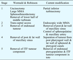
It is essential that surgery for tumours involving the infratemporal fossa is carefully planned. A step-wise process involving five stages has been described.Reference Wormald and Robinson9 Stage one involves uncinectomy, large middle meatal antrostomy, endoscopic medial maxillectomy, and sphenoethmoidectomy with removal of the lower half of the middle turbinate. Stage two involves the creation of trans-septal access for the second surgeon. Stage three comprises removal of the endonasal tumour. In stage four, the posterior and lateral walls of the maxillary sinus are removed to expose the pterygopalatine and infratemporal fossae. Finally, stage 5 comprises resection of the infratemporal fossa component of the tumour.
Our modification of the above process involves: (1) avoiding opening the septum (to avoid any affect on facial growth); (2) avoiding sublabial incision, and (3) meticulous tracing of the sphenopalatine artery proximally, in order to facilitate bipolar cautery of the maxillary artery and improve haemostasis; and (4) removing the tumour in toto where possible.
In the presented cases, control of the maxillary artery resulted in a marked decrease in bleeding, and could be considered a good alternative to embolisation; this decreases both the cost of the procedure and the risk of complications of angiography and embolisation.
In our experience, the most difficult part of surgical dissection involved the tumour adhesions to the posterior pharyngeal wall; these were managed with bipolar cautery and the use of cutting forceps and endoscopic scissors.
Table I shows the main differences between Wormald and Robinson's original two-surgeon approach and our modified approach.
Main differences: Wormald & Robinson vs Current modification
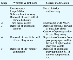
MMA = middle meatal antrostomy; post = posterior; lat = lateral; ITF = infratemporal fossa
The endoscopic approach to the infratemporal fossa is not a more conservative or less radical approach. In our opinion, the surgeon should know when to switch from an endoscopic to an external approach, and when to add another approach (e.g. sublabial or transantral) in addition to endoscopy.
When performing such surgery, specific attention should be given to the vidian canal, clivus and pterygoid plates, because small pseudopods of tumour may extend into these areas and can be overlooked. Surgeons should be fully aware of the risk of major complications, such as injury to the orbital apex, optic nerve, or carotid or cavernous sinuses, and should ensure they have a good understanding of the endoscopic anatomy of the pterygopalatine and infratemporal fossae.
• Angiofibroma extending into the pterygopalatine and infratemporal fossae can be well managed using an endoscopic, two-surgeon technique
• The presented modification of this technique avoids septal opening and sublabial incision, in order to prevent disruption of facial growth
• Bipolar cautery of the maxillary artery greatly improves haemostasis
• Careful tumour dissection from the pterygoid canal and lateral wall of the sphenoid, and careful inspection of pterygoid muscles post-dissection, may decrease the risk of residual tumour and recurrence
• Dissection of tumour adhesions from the posterior pharyngeal wall presented the greatest challenge
Meticulous examination of CT and MRI scans, with the help of a radiologist, is mandatory in order to fully evaluate the skull base, foraminae, carotid artery and lateral wall of the sphenoid. Special attention should also be paid to the pterygoid canals, orbital apex, and additional feeding arteries from the internal carotid artery.
Complications of the two-surgeon approach include infraorbital nerve dysfunction, nasolacrimal dysfunction and crusting due to medial maxillectomy. However, this approach avoids the risk of malocclusion and injury to lateral structures.Reference Wormald and Robinson9
Conclusion
The endoscopic, two-surgeon technique is an excellent approach for managing angiofibroma extending to the pterygopalatine and infratemporal fossae. Our modification of this technique avoids septal opening and sublabial incision and allows better control of the maxillary artery, improving haemostasis. Careful dissection of the tumour from the lateral wall of the sphenoid and the pterygoid canals, and careful inspection of the pterygoid muscles after tumour removal, may markedly decrease the risk of residual tumour and recurrence. In our experience, the most difficult part of tumour dissection involved the tumour adhesions to the posterior pharyngeal wall.