The real incidence and prevalence of intracardiac masses is difficult to obtain, as many are undetected in vivo. However, despite the lack of accurate epidemiological data from Africa, intracardiac masses are not rare in children and adolescents on this continent. They are usually non-malignant but cause significant morbidity and mortality. Discussion of unique aspects of aetiology, diagnosis, and management in underserved areas of Africa may be important to raise awareness in health professionals, allow timely diagnosis, and improve management and prognosis.
Description of cases
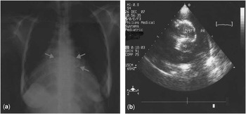
Case 1: A 20-year-old woman in New York Heart Association functional class III presented with recent onset of palpitations, chest pain, fever, congestive heart failure but no neurological signs. On examination, it was found that she had an irregular pulse (65 per minute), systolic murmur at the aortic and pulmonary areas, pulmonary congestion, and hepatomegaly. The laboratory tests revealed anaemia, moderate hypereosinophilia, and high erythrosedimentation rate. The renal and hepatic functions were normal. Examination of stool samples did not detect any parasites or eggs, and the serologic tests for cysticercosis and Human Immunodeficiency Virus were negative. The cutaneous tuberculin reaction was highly positive. First-degree atrioventricular block alternating with periods of type II second-degree block was present on the electrocardiogram. The chest radiography showed circular calcifications within the cardiac silhouette (Fig 1a), and the transthoracic echocardiography showed biventricular hypertrophy, preserved systolic function, and non-homogeneous masses of variable dimensions in the interventricular septum causing outflow tract obstruction (Fig 1b). No cysts were found on abdominal ultrasound or cerebral computer tomography. Endomyocardial biopsy and surgery were not considered because of the large size of the masses, risk of rupture, and location in the atrioventricular node area. Treatment for hydatid cyst and heart failure was started. In addition, treatment for tuberculosis was commenced in view of the highly reactive tuberculin skin test. This management was associated with remarkable clinical improvement and partial reduction of both the size of the cysts and the gradients across the outflow tracts. After 5 years, she is in class II under small doses of diuretics and vasodilators, despite persistence of conduction disturbances.
(a) Chest radiography of a 20-year-old woman, revealing increased size and abnormal shape of the heart, with round-shaped calcified structures visible (arrows). (b) On transthoracic echocardiography, thick-walled myocardial cysts suggestive of hydatic cysts are seen in the basal interventricular septum, and pressure gradient was measured across the outflow tracts.

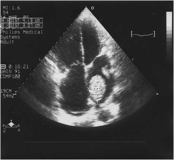
Case 2: A 14-year-old girl with previous admissions for heart failure was admitted in New York Heart Association class III with an acute neurological deficit. She was conscious, with flaccid left hemiplegia and mild cyanosis, but no fever. Her blood pressure was 100/50 millimetres of mercury, heart rate was 120 per minute; she had systolic and diastolic murmurs over the apex, systolic murmur over the third left intercostal space and a loud component of the second sound. There was hepatomegaly, splenomegaly, and pedal oedema. The electrocardiogram revealed sinus tachycardia and left atrial enlargement. Cardiomegaly and pulmonary congestion were seen on the chest radiography. Laboratory tests revealed mild anaemia, marked leukocytosis, and increased erythrosedimentation rate. The echocardiography confirmed severe mitral stenosis with left atrial thrombi (Fig 2), and mild aortic regurgitation with a large mobile vegetation attached to a cusp. The cerebral scan confirmed an extensive ischaemic stroke. Despite negative blood cultures, intravenous antibiotherapy was started in addition to diuretics and vasodilators, leading to remission of fever and heart failure after 1 week. She benefited from mechanical prostheses in mitral position, left atrial thrombectomy, removal of the aortic vegetation, and aortic valve plasty 4 weeks later. Although asymptomatic 5 years later, she continues to have neurological sequelae and has had marked fluctuations in her International Normalized Ratios for Prothrombin Time levels despite good compliance with anticoagulants.
Four-chamber view of severe mitral stenosis in a 14-year-old girl revealing thickened mitral leaflets, left atrial dilatation, and left atrial thrombi. AO = aorta; PA = pulmonary artery.

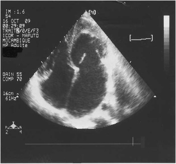
Case 3: A 15-year-old boy in New York Heart Association class III had a febrile episode with facial oedema 6 months earlier. The physical examination revealed tachycardia (134 per minute), low blood pressure (85/40 millimetres of mercury), facial oedema, third sound over the apical and tricuspid areas, moderate ascites but no pedal oedema. The electrocardiogram showed sinus rhythm, P mitrale, left ventricular hypertrophy, and diffuse repolarisation changes. The chest radiography showed that there was cardiomegaly with a bulge over the left heart border. He had severe hypereosinophilia (6.9×10⁹ per litre). On echocardiography, it was seen that the left ventricle had thickened hyperdense endocardium, was dilated and hyperkinetic in its basal portion, and obliterated at the apex (Fig 3); the mitral leaflets were thickened, moderate mitral regurgitation was present, and severe pulmonary hypertension was estimated from moderate tricuspid regurgitation. The diagnosis of left-sided endomyocardial fibrosis was made echocardiographically. Despite vigorous treatment, the boy died after 3 weeks from refractory heart failure.
Transthoracic images left ventricular apical obliteration underneath a thick endocardial fibrous plaque, from a 15-year-old boy with endomyocardial fibrosis. Notice ventricular cavity size reduction, thickening of valve leaflets, and left atrial dilatation.

Discussion
The cases reported show several aspects of intracardiac masses in young Africans. They highlight common clinical presentation and aetiology, as well as the challenges for the diagnosis and management of these clinical entities in resource-poor settings.
Intracardiac masses have diverse clinical presentation. The presenting symptoms and signs depend on the primary disease, as well as the size and position of the intracardiac masses, which may obstruct flow within the heart, embolise peripherally, or be associated with arrhythmias.Reference Shapiro 1 Embolisation results from fragmentation or dislodgement and migration of thrombus or vegetation, and causes systemic emboli (mainly cerebrovascular accidents) or pulmonary embolism. Valve obstruction mimics stenosis and may occur with large masses; in such cases, there may be chest pain, breathlessness, or syncope that may be related to posture. Intracavitary and intramyocardial masses may affect the cardiac rhythm, through direct infiltration of the conduction system or irritation of the myocardium, presenting as atrioventricular block, ventricular arrhythmia, and sudden death.Reference Shapiro 1
Like elsewhere,Reference Bruce 2 thrombi and vegetations are the most frequent intracardiac masses in Africa. Left atrial thrombi occur usually in the setting of rheumatic mitral stenosis, whereas right atrial and ventricular thrombi are associated with endomyocardial fibrosis in endemic areas. Large vegetations occur in uncorrected congenital defects and rheumatic heart valve disease, related to late diagnosis that is partially due to lack of awareness of parents and health personal. Concomitant left atrial thrombi and vegetations may occur in young patients with rheumatic heart valve disease and atrial fibrillation. Patients in whom the embolism may occur either from a vegetation or a thrombus, and which cannot be differentiated echocardiographically, may need both antibiotics and anticoagulation. There is, however, a dilemma when using anticoagulation in the presence of an ischaemic stroke, and treatment may have to be delayed.
The Human Immunodeficiency Virus epidemic in Africa has led to an increase in intracardiac masses; thrombi and bacterial vegetations are frequent, and marantic endocarditis occurs in 3–5% of these patients.Reference Rerkpattanapipat, Wongpraparat, Jacobs and Jotler 3 The incidence of Burkitt lymphoma, the most common childhood malignancy in Sub-Saharan Africa,Reference Rerkpattanapipat, Wongpraparat, Jacobs and Jotler 3 , Reference Ahmad and Molyneux 4 and that of primary cardiac lymphoma have also been increasing with the Human Immunodeficiency Virus infection and Acquired Immunodeficiency Virus Syndrome epidemics.Reference Rerkpattanapipat, Wongpraparat, Jacobs and Jotler 3 , Reference Orem, Mbidde, Lambert, Sanjose and Weiderpass 5 Cardiac involvement in Burkitt lymphoma usually causes heart failure; the chest radiography is usually abnormal, the echocardiography may show intracavitary and mural masses that may obstruct the valves, and mild to moderate pericardial effusion may be found.Reference Mocumbi, Paul, Maciel, Silva and Fereira 6
Finally, infections such as tuberculosis and cysticercosis should be considered as causes of cardiac masses. Cardiac tuberculosis is rarely described in the literature, but when reasonable doubt is present many clinicians who practice in areas where tuberculosis is endemic would perform a tuberculin skin test and if positive treat the patient for tuberculosis as was done in patient 2, despite the possibility of cysticercosis. Timely diagnosis of intracardiac masses is mandatory to improve outcomes. The suspicion of intracardiac masses should be raised by the presence of multiple cardiac murmurs in unusual areas of the chest, intermittent or variable degree atrioventricular block on the electrocardiogram, and abnormal heart shape on chest radiography. The level of suspicion is higher when neurologic or ischaemic events occur in patients with known heart disease or new cardiac murmur. The diagnosis is confirmed by transthoracic echocardiography, which allows identification and characterisation of the masses, definition of their location, attachment, shape, size, and mobility, as well as the presence and extent of haemodynamic disturbances.Reference Peters and Reinhardt 7 Where Magnetic Resonance Imaging is available, its use can increase reliability in characterising and differentiating cardiac masses.Reference Narin, Arman, Arslan, Simsek and Narin 8 The definite diagnosis is difficult in most African settings because of unavailability of cardiac catheterisation for biopsy, surgery for excision of the masses, and pathology services to evaluate the specimens, but diagnosis and appropriate management strategy is usually possible without the need for catheterisation. It can be achieved by integrating data such as epidemiological context, previous clinical history, careful physical examination, and echocardiographic data on location, size, and tissue characterisation of the mass. The management is also challenging because of late patient presentation, lack of awareness of the health personal, poor patient education, low socio-economic condition, and inaccessibility to coagulation testing.
Although rare in children, primary tumours must be considered in differential diagnosis of intracardiac masses, the most frequent being rhabdomyomas, fibromas, and myxomas.Reference Al-Mateen, Hood, Trippel, Insalaco, Otto and Vitikainen 9 Atrial myxoma must be excluded in children with acute neurological deficits, especially if there are embolic lesions suggestive of infective endocarditis and/or retinal artery occlusion.Reference Al-Mateen, Hood, Trippel, Insalaco, Otto and Vitikainen 9
Our reports and this review of literature show that although definite data on incidence and prevalence of intracardiac masses in the young African are lacking, they occur in clinical practice. Clinical features, radiographic changes, and electrocardiogram abnormalities may be the key for initial suspicion for the general practitioner, but transthoracic echocardiography is important to confirm the diagnosis and help with management.