Introduction
Organoids, multi-cellular aggregates which recapitulate multiple aspects of a single organ, present promising models for in vitro organ development, disease modeling, and drug screening. During organoid formation, cells differentiate and self-organize according to the biochemical and mechanical cues encountered. Every organ in the human body has a very specific set of these cues that result in precise control over cell fate. To create organoids, which resemble the organ of specific interest, be it liver or heart, scientists must learn what these cues are and how to provide them in vitro in order to recapitulate the natural microenvironment. Matrigel is a naturally derived material that has been shown to promote organoid growth and self-assembly; however, its xenogeneic source and batch-to-batch variation, limit its use for in vivo applications and systematic studies of tumor and organ development.[Reference Nowak, Freudenberg, Tsurkan, Werner and Levental1] Moreover, since Matrigel contains a variety of extracellular matrix (ECM) proteins, cytokines, and growth factors, it is difficult to determine which components are essential for a specific developmental event. Bioengineered hydrogels allow researchers to mimic specific in vivo conditions in a controlled manner, where biochemical and mechanical properties can be varied systematically and independently from each other. Recently, hydrogels have been used to investigate the role of the ECM on the formation of self-organized multi-cellular aggregates.[Reference Ranga, Girgin, Meinhardt, Eberle, Caiazzo, Tanaka and Lutolf2] By tuning matrix stiffness, degradability, and type and amount of biologically active ligand, the optimal conditions for proliferation, differentiation, and self-organization can be identified. Interestingly, the optimal hydrogel compositions vary significantly for different cell types, reflecting the advantage of specifically bioengineered matrices over Matrigel.
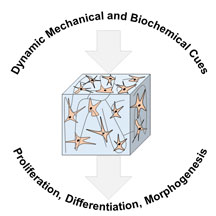
Currently, researchers have a good grasp on the tools available to develop hydrogels with static mechanical and biochemical properties. However, in the natural tissue environment, mechanical and biochemical signals are presented in a spatiotemporal manner. Elasticity of tissues and organs change during organ development, disease progression, and recovery after injury.[Reference Moeendarbary, Weber, Sheridan, Koser, Soleman, Haenzi, Bradbury, Fawcett and Franze3] During embryonic development cells perceive spatiotemporally resolved mechanical forces, which affect their differentiation and morphogenesis giving rise to the formation of individual organs. These mechanical forces are communicated to cells/tissues on various scales, starting with force generation on individual cells through force transmission to neighboring cells and eventual force integration within tissue to promote collective events and activate large-scale changes. Detailed explanations about the role of mechanical forces on organ development and their mechanisms of action can be found in recent reviews.[Reference Mammoto, Mammoto and Ingber4, Reference Heisenberg and Bellaïche5] Even within a fully developed, uninjured organ, mechanical properties are not uniform. Using an advanced atomic force microscopy mapping technique, Bouchonville et al. revealed that brain tissue rigidity changes as steeply as 12 kPa/μm.[Reference Bouchonville, Meyer, Gaude, Gay, Ratel, Nicolas, Reinhart-King, Margulies, Dembo, Boettiger, Hammer, Weaver, Shi and Robinson6] While the mean elasticity of the human-derived pituitary gland tissue was 9.5 kPa, areas with moduli as high as 25.9 kPa and as low as 3.5 kPa were detected. Organs and tissues also receive and interpret a range of biochemical signals that vary in space and time; presentation and removal of biologically relevant molecules affect cell differentiation and morphology. To study cell response to spatial and temporal changes in the cellular microenvironment, dynamic hydrogels with universal chemistries that can be adapted for multiple cell types must be developed. In this perspective, we showcase hydrogels with tailored mechanical and biochemical properties, with an emphasis on hydrogels that enable spatiotemporal control over the aforementioned properties, and their effects on cell behavior.
Influence of mechanical properties in bioengineered hydrogels
Tissue elasticity varies from organ to organ, increasing from ~1 kPa for soft tissues like the brain to ~500 kPa for cartilage and ~20 GPa for hard tissues such as cortical bone.[Reference Budday, Sommer, Birkl, Langkammer, Haybaeck, Kohnert, Bauer, Paulsen, Steinmann, Kuhl and Holzapfel7–Reference Bilston10] Hydrogels are particularly useful to mimic soft tissues. They enable in vitro culture of multiple cell types by mimicking in vivo matrix elasticity of various tissues and organs. Hydrogel stiffness can be tuned independently of biochemical properties, which enable the analysis of cell responses to specific mechanical changes. It is widely accepted that stem cell differentiation, maturation, and morphogenesis are influenced by matrix elasticity. Using collagen-modified polyacrylamide hydrogels, Engler et al. revealed that lineage preference of naive mesenchymal stem cells (MSCs) could be modulated by the stiffness of the culture matrix.[Reference Engler, Sen, Sweeney and Discher11] When cultured on soft, medium, and stiff hydrogels, MSCs exhibited increased expression of neurogenic, myogenic, and osteogenic lineage markers, respectively. It was later identified that stem cells sense the mechanical properties of their environment by adhering to and pulling on the ECM components of the scaffold.[Reference Trappmann, Gautrot, Connelly, Strange, Li, Oyen, Cohen Stuart, Boehm, Li, Vogel, Spatz, Watt and Huck12] In synthetic hydrogels comprised of the components which cannot support cell adhesion on their own, ECM proteins or cell adhesive peptide sequences are covalently integrated into the matrix. Consequentially, forces exerted by cells on the proteins/peptides are extended toward the entire scaffold and thus cells are able to detect the bulk scaffold elasticity.[Reference Wen, Vincent, Fuhrmann, Choi, Hribar, Taylor-Weiner, Chen and Engler13] During the past decade, the importance of the substrate stiffness for successful directed stem cell differentiation has been demonstrated for different kinds of stem cells. For example, Leipzig and Shoichet demonstrated that differentiation of NSPC (neural stem/progenitor cells) into three central nervous system lineages: neurons, oligodendrocytes, and astrocytes depended on the stiffness of the hydrogels used for their culture.[Reference Leipzig and Shoichet14]
Substrate stiffness not only affects stem cell differentiation, but also affects cell maturation. Cells typically differentiate and mature more effectively when cultured on substrates that resemble the mechanical properties of the natural tissue.[Reference You, Park, Shin, Patel, Raghunathan, Kim, Murphy, Tae and Revzin15] Yu and co-workers showed that the hydrogel that best resembled the mechanical properties of the adult liver led to the formation of the most adult-like hepatocytes from human pluripotent stem cell-derived hepatocytes (hpst-Hep).[Reference Mittal, Tasnim, Yue, Qu, Phan, Choudhury, Tan and Yu16] Hydrogels with elastic moduli of 20, 45, and 140 kPa were prepared, with the 20 kPa hydrogel having the most similar stiffness to that of the liver.[Reference Nava, Mazza, Furrer, Villiger and Reinhart17, Reference Huwart, Peeters, Sinkus, Annet, Salameh, ter Beek, Horsmans and Van Beers18] Albumin production, a measurement that correlates well with hepatocyte maturity, was the highest in cells cultured on the softest hydrogel and declined with increasing scaffold stiffness. The expression of key enzymes involved in drug metabolism, CYP1A2, and CYP3A4, also correlates to hepatocyte maturity and is higher in the adult liver compared with that of the fetus. When the expression of these enzymes was investigated in hpst-Hep cells, it was found that expression levels were the highest for cells cultured on the softest scaffold.
During organoid formation multi-cellular self-organization is as important as cell differentiation and maturation. Therefore, it is essential to create scaffolds that enable cell migration. It has been shown that the elasticity of the scaffold affects the progression of vascular morphogenesis. Gerecht et al. studied the influence of substrate stiffness on the tubulogenesis of endothelial progenitor cells (EPCs) cultured on poly(ethylene glycol)-diacrylate (PEGDA) crosslinked hyaluronic acid (HA)-gelatin hydrogels.[Reference Hanjaya-Putra, Yee, Ceci, Truitt, Yee and Gerecht19] The number, length and thickness of the formed tubes increased with decreasing scaffold stiffness. EPCs cultured on softer substrates readily assembled into chains and formed the longest tubes with the largest open lumen spaces. However, tube formation on all scaffolds was only possible in the presence of high vascular endothelial growth factor (VEGF) concentrations. VEGF activated the production of matrix metalloproteinases (MMPs), which enabled cell-mediated scaffold remodeling necessary for cell migration. The use of hydrogels with MMP-cleavable crosslinkers will be further explored in the following sections.
In the studies described, cells were cultured on top of the hydrogels. Burdick et al. recently identified that stem cell response to the mechanical properties of the scaffold depends on culture dimensionality.[Reference Caliari, Vega, Kwon, Soulas and Burdick20] Cell spreading increased when MSCs were cultured on top of stiffer HA-based hydrogels [two-dimensional (2D)], but the opposite trend was observed for the cells encapsulated within the hydrogels [three-dimensional (3D)]. Cells cultured within stiff, highly crosslinked hydrogels did not spread and displayed predominantly rounded morphology. In another study, Burdick and coworkers showed, however, that cell spreading within covalently crosslinked hydrogels can be induced by the incorporation of proteolytically cleavable crosslinks.[Reference Khetan, Guvendiren, Legant, Cohen, Chen and Burdick21] Furthermore, Mooney and coworkers showed, using murine MSCs, that during 3D encapsulation lineage fate did not correlate with cell morphology as it did in previous studies on 2D surfaces.[Reference Huebsch, Arany, Mao, Shvartsman, Ali, Bencherif, Rivera-feliciano and Mooney22] Together, these studies emphasize that influence of scaffold properties on cell behavior must be considered with respect to the culture dimensionality.
Temporal control of mechanical properties
Organoid formation from stem cells involves multiple phases: cell proliferation, differentiation, migration, and self-assembly, all of which occur over different time scales. For optimal organoid formation within a single scaffold, its mechanical properties must be tunable in order to match (or adapt to) each of the biologic phases. Lutolf and co-workers showed that changes in matrix stiffness were required for organoid formation from intestinal stem cell spheroids.[Reference Gjorevski, Sachs, Manfrin, Giger, Bragina, Ordóñez-Morán, Clevers and Lutolf23] Initially when cells were grown in soft hydrogels (shear modulus of 0.2 kPa), their proliferation was impeded and optimal cell expansion was achieved by using stiffer hydrogels (shear modulus of approximately 1.3 kPa). However, these stiffer hydrogels did not support cell differentiation or organoid formation. To achieve spheroid growth and morphogenesis, hydrogels had to be softened following cell expansion from 1.3 kPa to approximately 0.2 kPa. This was achieved by using mechanically dynamic hydrogels with hydrolytically degradable components. The degree of hydrolysis, and therefore the final modulus, was controlled by the ratio between the two hydrogel precursors: mechanically static vinylsulfone-functionalized poly(ethylene glycol) (PEG) and hydrolytically degradable acrylate-functionalized PEG. In the aqueous media, the ester functionality of the latter is hydrolyzed, which results in hydrogel softening.
There are various methods to achieve temporal control over mechanical properties of hydrogels. In the following sections, we present studies in which temporal mechanical changes in hydrogels are achieved by chemical, light, magnetic, or thermal stimuli.
Chemically induced gradual mechanical change
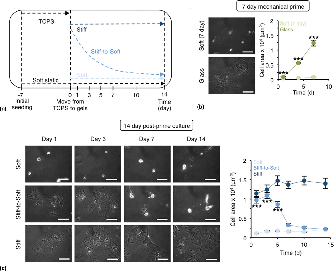
Incorporation of certain functional groups within precursor materials or crosslinkers provides hydrogels with dynamic mechanical properties. The most commonly used strategy to induce temporal changes of mechanical properties is hydrolysis. During hydrolysis, water labile functionalities (e.g. esters) undergo substitution reactions with water molecules, leading to bond dissociation and softening of the hydrogel due to a decrease in crosslink density. The rate and degree of hydrogel dissociation can be adjusted by controlling the concentration of hydrolytically labile bonds. Burdick and co-workers studied hepatic stellate cell behavior during fibrosis regression using water-labile hydrogels.[Reference Caliari, Perepelyuk, Soulas, Lee, Wells and Burdick24] To mimic the tissue softening during fibrosis regression, an HA hydrogel system with a hydrolytically degradable PETMA [pentaerythritol tetrakis(mercaptoacetate)] as crosslinker was synthesized; the elastic modulus gradually decreased over 14 days from ~17 to 3 kPa [Fig. 1(a)]. To understand the role of elastic modulus on hepatic stellate cell spreading, cells were cultured on both static (stiff and soft) and dynamic (gradually softening) hydrogels. Hepatic stellate cells differentiated toward the myofibroblast phenotype, with the characteristic spread morphology, when cultured on hard tissue culture polystyrene (TCPS) for 7 days [Fig. 1(b)]. The pre-differentiated cells transferred onto the stiff, static hydrogel retained their elongated morphology, whereas cells cultured on soft, static hydrogel became more rounded. Cells transferred onto dynamically softening hydrogels gradually altered their morphology from elongated to rounded concomitant with the changing mechanical environment [Fig. 1(c)].
(a) Schematic representation of the experimental set-up to study fibrosis regression using gradually softening hydrogels. (b) During 7-day mechanical prime on glass/TCPS, hepatic stellate cells differentiate toward the myofibroblast phenotype with characteristic spreading. (c) Morphological and phenotypical variations in hepatic stellate cells cultured on soft, gradually softening (stiff-to-soft), and stiff hydrogels. Scale bars: 100 µm. Reproduced from Ref. Reference Caliari, Perepelyuk, Soulas, Lee, Wells and Burdick24 with permission from The Royal Society of Chemistry.

In addition to the carboxylic ester functionality, hydrolysis susceptible hydrazine bonds can be used to create gradually softening hydrogels. Recently, Maynard and co-workers combined non-reversible oxime and reversible hydrazine chemistries to form hydrogels with tunable degradability.[Reference Boehnke, Cam, Bat, Segura and Maynard25] Interestingly, the authors noted that their hydrogels degraded more rapidly in the cell culture media than in the buffer solution and that the degradation time was decreased even further in the presence of cells. This observation underscores the importance of testing materials in physiologically relevant conditions as mechanical testing is usually done in buffer solutions, which ignores the influence of enzymes, proteins, and hormones typically found in the culture media. In principle, due to similar chemistry, gradually degrading materials developed for controlled cargo delivery can be adapted for the use as gradually softening scaffolds in cell culture.[Reference Kharkar, Kloxin and Kiick26]
In other cases, such as simulating the transition from mesoderm to adult myocardium, gradual hydrogel stiffening is sought. Young and Engler showed that the chicken heart undergoes a ninefold increase in the elastic modulus, from ~0.9 kPa at 36 h post-fertilization to ~8.2 kPa at 408 h, as a result of the mesoderm to adult myocardium transition.[Reference Young and Engler27] They were able to synthesize a hydrogel that had a similar mechanical change through the time-dependent Michael-type addition reaction between thiol-modified HA and acrylate-functionalized PEG crosslinker. Alternatively, hydrogel stiffening can be achieved by light-triggered secondary crosslinking; however, it leads to more abrupt changes in the mechanical properties.
Light-induced externally controlled mechanical change
Photo-activated reactions enable externally controlled mechanical changes, which are useful for studying the mechanical memory of cells. Depending on the chemical structure of the hydrogel, light irradiation can induce softening or stiffening of the scaffold. Anseth and co-workers decreased the Young's modulus of the hydrogel from 10 to 2 kPa upon ultraviolet (UV) irradiation by using hydrogel precursors with photolabile o-nitrobenzylether groups.[Reference Yang, Tibbitt, Basta and Anseth28] The authors used this system to investigate the mechanical memory of human mesenchymal stem cells (hMSCs) based on expression of the transcriptional activator Yes-associated protein (YAP). When hMSCs are cultured on stiff substrates, YAP is activated in the nucleus. In hMSC cultured on soft substrates, YAP is deactivated and relocates to the cytoplasm. By inducing hydrogel softening at different time points of hMSCs culture, authors showed that YAP remained in the nucleus when hMSCs were cultured on soft hydrogels after being cultured on stiff substrates for an extended period of time; however, when cells were cultured on the stiff substrate for a shortened period of time below a certain threshold, cells were able to adapt to the new softer environment and YAP was deactivated. Alternatively, Guvendiren and Burdick performed a delayed hydrogel stiffening using a sequential crosslinking technique, where initial gelation was obtained by an addition reaction and the delayed secondary crosslinking was induced by light-triggered radical polymerization.[Reference Guvendiren and Burdick29] Hydrogel stiffening from ~3 to 30 kPa was performed during hMSC culture and was used to study their differentiation. Substrate stiffening at earlier stages of hMSCs culture led to predominantly osteogenic cell differentiation, while stiffening at later time points led to the development of nearly equally mixed osteogenic/adipogenic cell populations. When the mechanical change was introduced at the later time point, some cells already exhibited an adipogenic phenotype and the subsequent change in hydrogel stiffness did not influence their phenotype. Cells, which were undifferentiated prior to hydrogel stiffening, adapted to the new mechanical environment and differentiated toward the osteogenic lineage.
Irradiation can also be used to release compounds that promote mechanical changes of hydrogels. For example, Suggs and co-workers developed liposomes, which released calcium-chelating agents upon irradiation with near-infrared light, reducing the initial crosslink density and softening alginate-based hydrogels, which are typically crosslinked with divalent ions such as calcium.[Reference Stowers, Allen and Suggs30] By incorporating calcium ions instead of the chelating agent, authors used the same method to dynamically stiffen the gels. With this approach, hydrogel storage modulus was varied between 10 and 5000 Pa. The dynamic tuning of hydrogel stiffness depended on the initial calcium concentration, liposome concentration, and irradiation time.
Magnetic field-induced reversible mechanical change
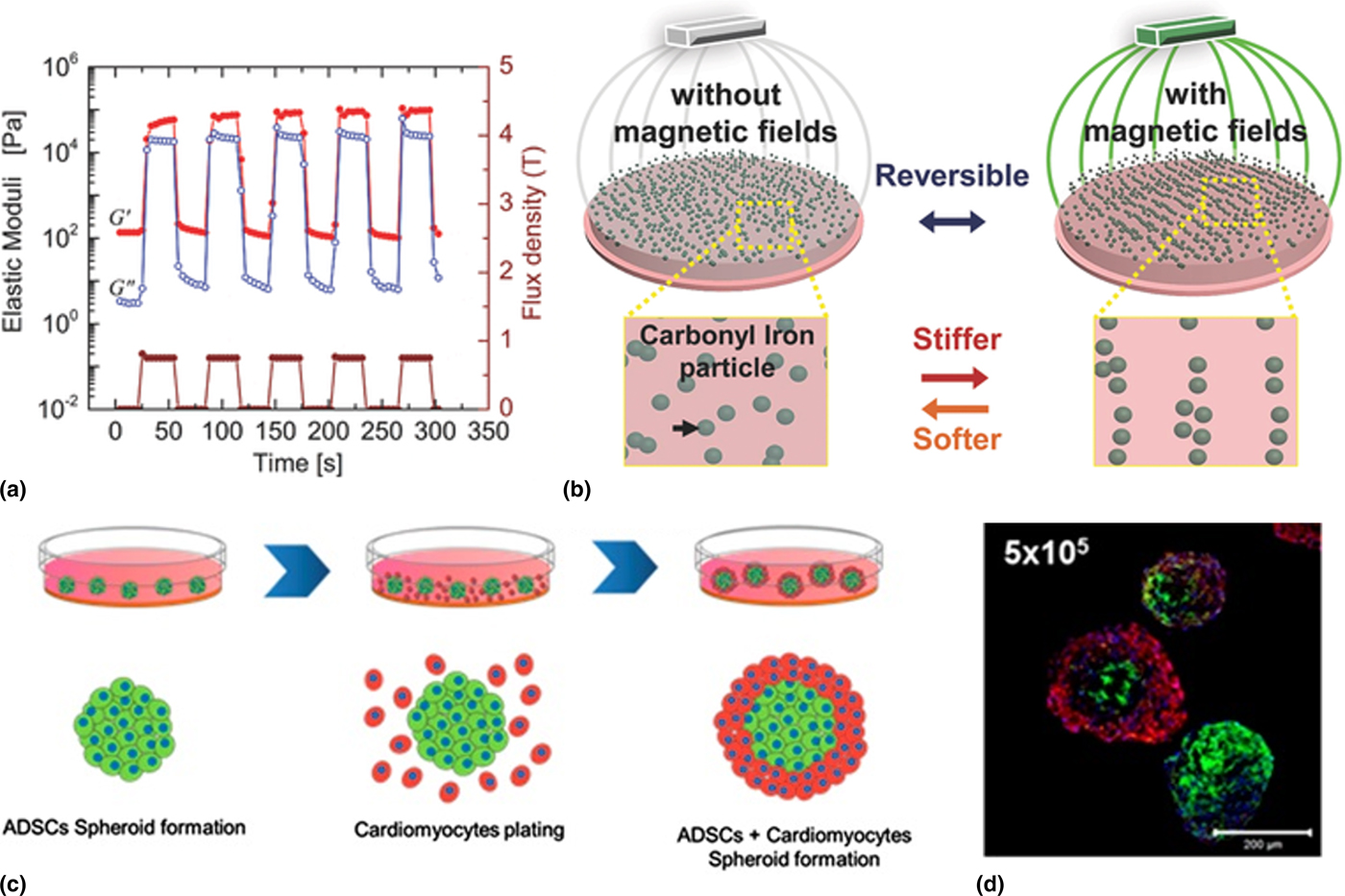
The incorporation of magnetic particles within hydrogels enables the use of magnetic fields to control the scaffold elastic modulus. Magnetic field-induced elasticity changes are reversible, enabling repeated variations of hydrogel modulus. Kilian and co-workers designed a polyacrylamide-based hydrogel with embedded magnetic carbonyl iron particles.[Reference Abdeen, Lee, Bharadwaj, Ewoldt and Kilian31] Application of a magnetic field caused the alignment of the embedded particles, which led to hydrogel stiffening from 0.1 to ~90 kPa [Fig. 2(a)].[Reference Ubaidillah, Sutrisno, Purwanto and Mazlan32, Reference Mitsumata, Ohori, Honda and Kawai33] Once the magnetic field was removed, the particles returned to a random distribution and the initial hydrogel stiffness was regained [Fig. 2(b)]. The authors showed that the hydrogel could be reversibly stiffened at least five times without significant changes to its highest or lowest modulus. Unlike reversible elastic changes induced by magnetic fields, light-mediated secondary crosslinking results in the formation of permanent covalent linkages and cannot be reversed.
(a) Pulsed magnetic field from 0 to 0.75 Tesla changes hydrogel stiffness from 0.1 to ~90 kPa. (b) Schematic representations of the function principle of magnetoactive hydrogels. Reproduced from Ref. Reference Abdeen, Lee, Bharadwaj, Ewoldt and Kilian31 by permission of John Wiley & Sons, Inc. Copyright by John Wiley Sons, Inc. (c) Schematic representation of a cell co-culture experiment using thermoactive hydrogels. (d) Confocal fluorescence microscopy images of ADSC/cardiomyocytes forming concentric cell layers, blue (cell nuclei), green (ADSCs), red (cardiomyocytes). Adapted from Ref. Reference Cho, Li, Shim, Cho, Nurunnabi, Park, Lee, Moon, Kim, Kang and Huh37 using Creative Commons CC-BY license.

In addition to its reversible properties, the single magnetoactive hydrogel covered a broad elastic modulus range from 0.1 to 90 kPa, depending on the magnetic field applied. This system was used to study the influence of temporal changes in substrate stiffness on the differentiation of MSCs. It is worth mentioning that prior to cell seeding, hydrogels had to be further modified with fibronectin to facilitate cell adhesion. In addition to polyacrylamide-based systems, PDMS [poly(dimethylsiloxane)] was used to develop magnetoactive hydrogels.[Reference Mayer, Rabindranath, Boerner, Hoerner, Bentz, Salgado, Han, Boese, Probst, Shamonin, Monkman and Schlunck34] Notably, these hydrogels required extensive post-fabrication treatment to produce cell adhesive surfaces suitable for cell culture. Thus, magnetoactive materials provide an interesting platform for reversible mechanical changes, but their compatibility with biologic systems must be further improved in order to use this technology for organoid culture. While the use of the magnetoactive hydrogels is straightforward for top seed experiments, possible cell–particle interactions should be taken into account when cells are encapsulated.
Temperature-controlled reversible mechanical change
Thermoresponsive hydrogels that undergo temperature-induced gel–liquid transition provide an elegant method for long-term 3D cell culture and subsequent cell extraction. Lei and Schaffer used a thermoresponsive hydrogel composed of commercially available poly(N-isopropylacrylamide) (PNIPAAm)–PEG for human pluripotent stem cell (hPSC) expansion and differentiation.[Reference Lei and Schaffer35] The polymer solution is a liquid at 4 °C but solidifies into a hydrogel at 37 °C. Thus, cells can be seeded at low temperature, grown suspended in a solid gel at 37 °C, and subsequently harvested or passaged by cooling the gel. hPSCs cultured in this scaffold formed dense multi-cellular spheroids and expanded by tenfold at 4 days after seeding, while in conventional static-suspension culture only a threefold expansion was achieved. The hPSCs cultured in this scaffold could be continuously propagated for 280 days. Notably, the high expansion rate, pluripotency marker expression, and spheroid size distribution remained consistent over these numerous passages, indicating the suitability of this system for long-term expansion. Later the PNIPAAm–PEG system was successfully adapted to the scalable production of glioblastoma tumor-initiating cells.[Reference Li, Lin, Wang, Qiu, Kidambi, Deleyrolle, Reynolds and Lei36] The ability to easily harvest intact multi-cellular spheroids from thermoresponsive hydrogels by just lowering the temperature was used by Huh and co-workers to study cell co-culture.[Reference Cho, Li, Shim, Cho, Nurunnabi, Park, Lee, Moon, Kim, Kang and Huh37] After adipose-derived stromal cell (ADSC) spheroid formation, the hydrogel was cooled down and neonatal cardiomyocytes were added to the liquefied substrate [Fig. 2(c)]. Subsequently, the temperature was increased to 37 °C resulting in both cells being encapsulated within the hydrogel. By adjusting the ratio between the two cell types, spheroids with concentric ADSCs/cardiomyocytes layers could be formed [Fig. 2(d)]. Co-culture can recapitulate the cell–cell interactions in natural tissue, and thermoresponsive hydrogels may offer a feasible method to systematically integrate additional cell types for co-culture experiments.
Spatial control of mechanical properties
Cells respond to the mechanical properties of their environment on the micron scale and hence sense the mechanical heterogeneity of organs.[Reference Wolfenson, Meacci, Liu, Stachowiak, Iskratsch, Ghassemi, Roca-Cusachs, O'Shaughnessy, Hone and Sheetz38, Reference Meacci, Wolfenson, Liu, Stachowiak, Iskratsch, Mathur, Ghassemi, Gauthier, Tabdanov, Lohner, Gondarenko, Chander, Roca-Cusachs, O'Shaughnessy, Hone and Sheetz39] To further bridge the gap between complex in vivo microenvironments and engineered hydrogels for organoid culture, researchers are beginning to design scaffolds that mimic the mechanical heterogeneity of tissues using exogenous and endogenous methods.
Light-induced mechanical changes
Over the past decade a variety of techniques were developed to create scaffolds with user-defined mechanical gradients as detailed in recent reviews.[Reference Whang and Kim40, Reference Tingting, Wanqian and Li41] Here we highlight an example where mechanical gradients are used to study stem cell behavior. Tse and Engler studied MSC behavior along a physiologically relevant gradient of 1 kPa/mm (from 1 to 14 kPa) in an attempt to mimic the migration of MSCs from bone marrow to injured tissue.[Reference Tse and Engler42] A gradient in elastic modulus was achieved by using a gradient photomask, which controlled the amount of irradiation applied to crosslink acrylamide-based hydrogel precursor materials. MSCs seeded on top of the hydrogels migrated toward the stiffer regions of the scaffold with few cells remaining on regions where the stiffness fell below 6 kPa. Although after 21 days of culture the majority of cells were located in regions stiffer than 10 kPa, their phenotype differed from that of cells continuously cultured on the homogeneously stiff (11 kPa) hydrogel. While MSCs cultured on the homogeneously stiff hydrogels differentiated toward a myogenic phenotype, those cultured on the gradually stiffening substrate expressed both myogenic and neural phenotypes. This suggests that cells initially seeded on the softer regions possessed memory of the softer mechanical environment where they typically differentiated to neural cells. Differentiation toward a mixed phenotype was also possible by temporally changing the bulk mechanical properties of the hydrogel[Reference Guvendiren and Burdick29]; however, this technique did not promote cell migration.
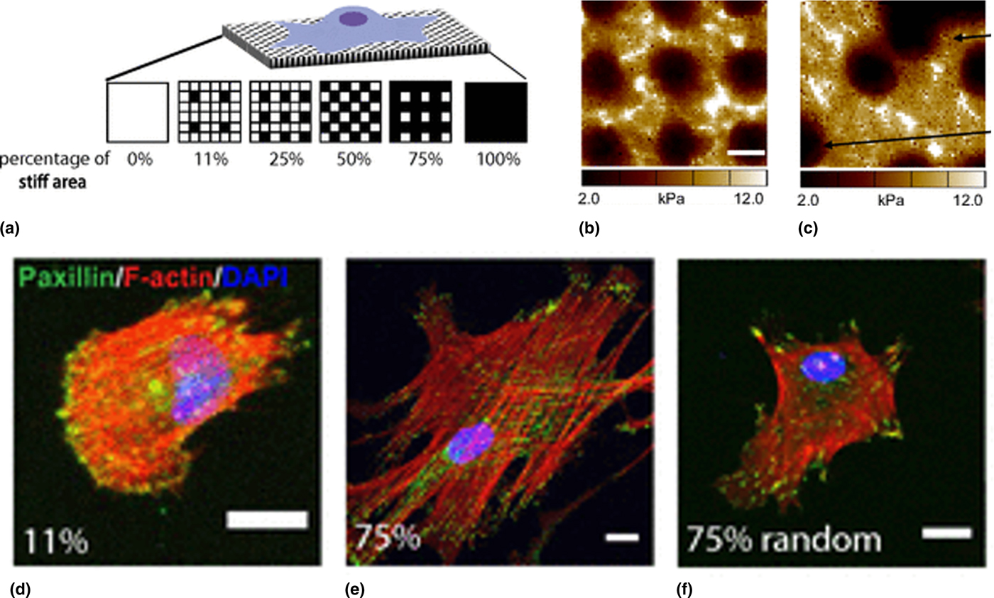
Stiffness gradients spanning multiple millimeters provide a great platform to study cell migration in response to gradual mechanical changes. In addition to cell sensitivity to mechanical changes on the macroscopic scale, recent studies proposed that cells are able to sense their mechanical environment with micron-scale precision.[Reference Wolfenson, Meacci, Liu, Stachowiak, Iskratsch, Ghassemi, Roca-Cusachs, O'Shaughnessy, Hone and Sheetz38, Reference Meacci, Wolfenson, Liu, Stachowiak, Iskratsch, Mathur, Ghassemi, Gauthier, Tabdanov, Lohner, Gondarenko, Chander, Roca-Cusachs, O'Shaughnessy, Hone and Sheetz39] Cells sense micron-scale changes in elasticity and even respond to the spatially oriented mechanical cues. Yang et al. designed hydrogels with either regularly alternating [Figs. 3(a) and 3(b)] or randomly placed [Fig. 3(c)] stiff and soft 2 × 2 µm2 squares to determine whether hMSCs respond to sub-cellular differences in scaffold elasticity or if they simply sense average substrate stiffness.[Reference Yang, DelRio, Ma, Killaars, Basta, Kyburz and Anseth43] Increasing the number of stiff squares on the regularly patterned hydrogels increased cell spreading, led to more elongated cells, and promoted YAP activation similar to the hMSCs cultured on the homogeneously stiff substrate [Figs. 3(d) and 3(e)]. For hMSCs cultured on the random patterns, the increase in the number of stiff regions did not affect the cells. They exhibited decreased spreading and deactivated YAP in the cytoplasm, resembling cells cultured on the homogenously soft substrate [Fig. 3(f)]. Furthermore, while hMSCs cultured on regularly pattered gels differentiated toward the osteogenic lineage, hMSCs cultured on the randomly pattered hydrogel remained largely undifferentiated and continued to exhibit the MSC marker, CD105.
(a) Schematic representations of the patterning of hydrogels on the subcellular scale; (b) regularly alternating stiffness pattern, scale bar 2 µm; (c) randomly alternating stiffness pattern; (d) rounded cell on the hydrogel with 11% of regularly patterned stiff areas; (e) cell with a spread morphology on the hydrogel with 75% of regularly patterned stiff areas; (f) cell with decreased spreading on the hydrogel with 75% of randomly patterned stiff areas. Scale bars 20 µm. Reproduced from Ref. Reference Yang, DelRio, Ma, Killaars, Basta, Kyburz and Anseth43.

Enzymatically induced mechanical changes
By developing hydrogels susceptible to cell-mediated changes, scientists can study how cells influence and remodel their own microenvironment, thus gaining deeper insights into native cell behavior. Incorporation of MMP cleavable crosslinkers into hydrogels enables cell-mediated hydrogel degradation, migration, and self-assembly.[Reference Lutolf, Lauer-Fields, Schmoekel, Metters, Weber, Fields and Hubbell44] Proteolytically driven cell invasion into HA-based hydrogels was shown by Fisher et al.[Reference Fisher, Anandakumaran, Owen and Shoichet45] In this study, invasive MDA-MB-231 breast cancer cells migrated twice as far into the gels crosslinked with an MMP degradable peptide sequence (GPQG-IWGQ) compared with the gels with a non-degradable (GAGGAG) crosslinker. This demonstrated that these cells actively remodel and degrade the hydrogel scaffold by MMP secretion. Such dynamic cellular remodeling by MMPs was quantitatively characterized by Schultz et al.[Reference Schultz, Kyburz and Anseth46] The migration of hMSCs within PEG hydrogels containing MMP-degradable GPQG-IWGQ peptide sequences was analyzed by a multiple particle tracking microrheology technique. In a short time frame after cell seeding, a degradation gradient was measured in the pericellular region, with areas of increased degradation observed farther away from the cell. The identified gradient indicated that cell-secreted enzymes diffused away from the cell faster than they could cleave the peptides. For the longer timescale study, after initial cell attachment and spreading, the pericellular regions changed from an elastic gel into a viscoelastic fluid in which cells could rapidly migrate, reaching a speed of up to 140 µm/h. Using a fluorescently labeled peptide, authors showed that, with time, hMSCs irreversibly remodeled their environment leaving permanent migration paths eroded into the substrate.
Physically induced mechanical changes
To study endogenous matrix remodeling, scaffolds should be susceptible to cell-induced changes. Chemically crosslinked hydrogels that have MMP-degradable crosslinkers enable enzymatically induced bond cleavage; however, if the rest of the scaffold is non-degradable, there is an inherent limitation. Physically crosslinked hydrogels are in dynamic equilibrium with their soluble components and thus are easily remodeled; however, they often suffer from poor stability. An alternative is provided by hydrogels with mobile crosslinkers that are both stable and susceptible to mechanical remodeling. Tong and Yang used modified hydrophilic α-cyclodextrins containing polyrotaxane (SCPR-VS) to develop the first sliding hydrogel with encapsulated cells.[Reference Tong and Yang47] Chemically crosslinked bonds, located on cyclic α-cyclodextrins, can rotate and slide along the PEG backbone, thus providing the hydrogel with mechanical freedom. The incorporation of α-cyclodextrins did not change the bulk mechanical properties or stability (over 30 days) from the covalent equivalent, where PEG chains were directly bonded to each other. This mobility enabled encapsulated hMSCs cells to extend protrusions through the rearrangement of crosslinking molecules and adhesive ligands. Cells cultured within the hydrogel with permanent crosslinks did not form protrusions, indicating that the flexible network was essential to induce cell spreading. Although sliding hydrogels do not promote cell morphogenesis to the same extent as MMP-degradable or hydrolytically susceptible hydrogels, combining these approaches might lead to the development of materials that provide greater opportunity for cellular remodeling.
Naturally derived ECM components and native tissues, unlike chemically crosslinked synthetic hydrogels, are viscoelastic and exhibit partial stress relaxation—that is, matrix deformation upon applied strain.[Reference Liu and Bilston48–Reference McDonald, Dooley, McDonald, Schuijers, Ward and Grills50] Materials with fast and slow rates of stress relaxation require short and long strain durations, respectively. In viscoelastic scaffolds, cells can mechanically remodel their local environment by applying traction forces.[Reference Chaudhuri, Gu, Darnell, Klumpers, Bencherif, Weaver, Huebsch and Mooney51] Furthermore, recent studies showed that the viscoelastic properties of scaffolds influence cell behavior independent of bulk scaffold stiffness. By varying the molecular weight of polysaccharides, Mooney et al. developed alginate-based hydrogels with variable rates of stress relaxation.[Reference Chaudhuri, Gu, Klumpers, Darnell, Bencherif, Weaver, Huebsch, Lee, Lippens, Duda and Mooney52] Relaxation rate-dependent differences in spreading and proliferation of 3T3 fibroblasts encapsulated within the hydrogels were observed; cells encapsulated within hydrogels with long relaxation rates remained rounded and showed suppressed proliferation compared to cells cultured in hydrogels with short relaxation rates, which spread and proliferated. Notably, the initial elastic modulus of all hydrogels was 9 kPa, indicating that differences in cell shape and proliferation were induced by matrix susceptibly to mechanical remodeling.
Modulation of biochemical properties in bioengineered hydrogels
It has been demonstrated that the incorporation of biochemical features can control cell fate in 3D culture when combined with optimized mechanics. These include: adhesive peptides, growth factors, and co-culture of multiple cell types. PEG is commonly used as a scaffold because it is non-adsorptive to proteins and non-adhesive to cells, allowing researchers to evaluate how different components of the microenvironment affect cell fate.[Reference Tao, Wang, Lin, Shen and Li53] HA, heparin, and PNIPAAm are also used in engineered hydrogels due to their role in biologic signaling, affinity-mediated growth factor release, and thermoresponsive properties, respectively.[Reference Burdick and Prestwich54–Reference Nguyen, Payré, Fitremann, Lauth-De Viguerie and Marty57] Cell-adhesive peptide ligands and matrix metalloprotease-degradable crosslinking molecules are ubiquitous features across 3D scaffold design. Notwithstanding the advances in 3D cell culture, it remains somewhat of an art. Slight variation in peptide ligand sequence, rate of degradation, growth factor presentation, and co-culture conditions can all impact cell fate.
Heparin-based hydrogels for growth factor release
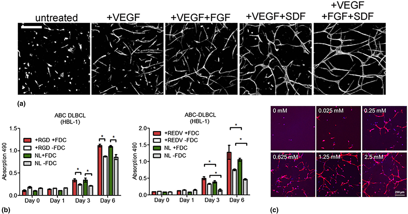
Covalently functionalizing proteins to hydrogel scaffolds can result in a loss of protein bioactivity. Heparin is a sulfated glycosaminoglycan (GAG) that binds proteins reversibly via electrostatic interactions, and researchers have harnessed this natural affinity for growth factor release within hydrogels. For example, Chwalek et al. developed a heterocellular angiogenesis model by designing a hydrogel system composed of star-PEG and heparin to support endothelial cell (EC) morphogenesis into capillary structures.[Reference Chwalek, Tsurkan, Freudenberg and Werner58] The incorporation of VEGF, basic fibroblast growth factor (bFGF), and stromal-derived growth factor (SDF-1α) was investigated. The simultaneous introduction of all three growth factors (5 µg/mL each of VEGF, bFGF, and SDF-1α) resulted in capillary networks with significantly increased branch length, density, and stability (at 10 days versus 4 days) when compared with VEGF alone [Fig. 4(a)]. The effects of co-culture were also examined. Using 10% MSCs, ECs formed stable capillary networks for over 4 weeks, whereas with more than 10% MSCs, the formation of EC tube structures was inhibited. This long-term culture period of ECs and MSCs (at 10%) resulted in lumens within the EC tubes, with diameters matching those of human capillaries in vivo. Co-culture with different cell types caused dramatic differences in capillary density, suggesting that functional differences in vivo are determined by proximity and recruitment of specific mural cells.
(a) The influence of VEGF, bFGF, and SDF incorporation into hydrogels with heparin and star-PEG on branch length, density, and stability of endothelial cells. Adapted from Ref. Reference Chwalek, Tsurkan, Freudenberg and Werner58 with permission from Nature Publishing Group. (b) Effect of RGD and REDV ligand presentation on B cell proliferation. Adapted from Ref. Reference Tian, Ahn, Schneider, Yang, Roman-Gonzalez, Melnick, Cerchietti and Singh64 with permission from Elsevier. (c) Influence of immobilized RGD ligand concentration on the proliferation of smooth muscle cells in PEG-based hydrogels. Adapted from Ref. Reference Lin, Marchant, Zhu and Kottke-Marchant63 with permission from Elsevier.

While heparin-based hydrogels are advantageous for preservation of growth factor bioactivity and in vivo compatibility, inconsistent release profiles and batch-to-batch variability complicate their use for 3D cell culture applications. Hettiaratchi et al. demonstrated that the release rate of bone morphogenetic protein-2 (BMP-2) from heparin-based microparticles increased in the presence of bovine serum due to competitive protein binding.[Reference Hettiaratchi, Chou, Servies, Smeekens, Cheng, Esancy, Wu, McDevitt, Guldberg and Krishnan59] In another study by Hettiaratchi et al., it was shown that <25% of bound BMP-2 was released from heparin microparticles over 28 days.[Reference Hettiaratchi, Miller, Temenoff, Guldberg and McDevitt60] In this case, BMP-2 bioactivity was conserved. However, the xenogenic source of heparin, porcine intestinal mucosa, has inherent batch-to-batch variability, and provides an incentive to engineer synthetic heparin-like materials.[Reference Chalovich and Eisenberg61, Reference Christman, Vázquez-Dorbatt, Schopf, Kolodziej, Li, Broyer, Chen and Maynard62] Furthermore, varying concentrations of heparin may have detrimental effects on cell survival. Kottke-Marchant and co-workers demonstrated that increasing heparin concentration in PEGMA/PEGDA hydrogels decreased the spreading and proliferation of smooth muscle cells (SMC) in vitro.[Reference Lin, Marchant, Zhu and Kottke-Marchant63]
Integrin–peptide interactions affect cell fate
While hydrogels are often functionalized with peptide ligands, integrin expression is not always characterized. Evaluating integrin expression has the potential to guide hydrogel design and explain organoid phenotype. For example, Levental and co-workers discussed the role of integrin binding in culturing mammary epithelial cells (MECs).[Reference Nowak, Freudenberg, Tsurkan, Werner and Levental1] Polarized acini formed in soft formulations of star-PEG and heparin hydrogels, but only when an MMP-degradable crosslinker was used. In MMP-degradable gels, laminin-332 (LN-332) was secreted by cells and distributed around the basal surface after 4 days of culture. When a blocking antibody was used against α6β4, an integrin involved in LN-332 binding, cell growth was arrested. The authors hypothesized two explanations for MEC morphogenesis that are not mutually exclusive: (1) secreted LN-332 binds to heparin and activates integrins on MECs; and (2) heparin promotes adhesion and survival by binding cell integrins.
Studying integrin expression levels can guide peptide selection. The role of integrin-matrix interactions on cell aggregation and proliferation was investigated in the context of non-Hodgkin's lymphoma by Singh and co-workers[Reference Tian, Ahn, Schneider, Yang, Roman-Gonzalez, Melnick, Cerchietti and Singh64] A co-culture system with lymphoma and follicular dendritic cells (FDCs) was designed using maleimide-functionalized star-PEG, MMP2/9-degradable peptide crosslinkers (GCRDVPMS↓MRGGDRCG), and adhesive peptides containing fibronectin-derived RGD or REDV sequences which bind to integrins αvβ3 and α4β1, respectively. It was found that RGD peptides increased the aggregation of B-cell lymphoma cell line, HBL-1. Although RGD peptides increased the clustering of HBL-1 cells, REDV peptides increased proliferation [Fig. 4(b)]. This was further demonstrated through cell cycle analysis, which revealed a 28% reduction in the DNA synthesis phase when RGD peptides were used instead of REDV peptides.
Studies investigating the effects of different peptide ligands may benefit from considering integrin-peptide binding affinities. Gould and Anseth investigated the effects of peptide ligand sequence and MMP activity on valvular interstitial cell (VIC) fate in an eight-arm-PEG hydrogel.[Reference Gould and Anseth65] VICs cultured with either RGDS or P15 (collagen-1-derived) peptides exhibited significantly higher MMP activity when compared with those cultured with VGVAPG (elastin-derived). The percentage of cells that stained positive for α-SMA (α-smooth muscle actin) was the highest when VGVAPG was used, suggesting that the myofibroblast phenotype is associated with low MMP activity and elongation. However, the authors of this study note that binding strengths between peptides and integrins are not evaluated. Significant differences in binding affinities may weaken conclusions made about an MMP activity-dependent phenotype, and should be considered for future studies of this nature.
Biochemical cue concentration depicts cell phenotype
Although adhesive peptides and MMP-degradable crosslinkers are typically incorporated into 3D cell culture, achieving the appropriate concentrations for desirable phenotypes is non-trivial. Enemchukwu et al. investigated the effects of altering biochemical cue concentration on epithelial morphogenesis in a star-PEG hydrogel with MMP-degradable crosslinkers and RGD peptide ligands.[Reference Enemchukwu, Cruz-Acuna, Bongiorno, Johnson, Garcia, Sulchek and Garcia66] Polarity and lumen formation of MDCK (Madin–Darby canine kidney) epithelial cells depended on RGD concentration, whereas proliferation and self-assembly into multi-cellular structures were not affected. High concentrations of RGD (>250 µM) gave rise to cysts that contained lumen and internal apical polarity, and at 2000 µM RGD, hydrogels exhibited a phenotype that closely matched that observed with collagen, a common matrix material for epithelial cell culture. A study by Lin et al. demonstrated that there is an optimal concentration of immobilized RGD ligand that increases the proliferation of SMC in vitro.[Reference Lin, Marchant, Zhu and Kottke-Marchant63] Concentrations between 0.625 and 1.25 mM maximized proliferation in PEG, while >1.25 mM resulted in decreased proliferation [Fig. 4(c)]. Furthermore, MMP-degradable crosslinker concentration had implications on cell proliferation and spreading. Limited cell spreading was observed when slowly MMP-degradable crosslinkers were used when compared with rapid MMP-degradable crosslinkers; yet, increasing the concentration of rapid MMP-degradable crosslinkers decreased cell proliferation and spreading. Thus, a balance of rate and concentration of MMP-degradable crosslinkers was required.
The types and concentrations of growth factors, peptides, crosslinkers, and co-cultured cells are all factors that can be optimized in bioengineered hydrogels to achieve a desirable phenotype; however, control over bulk biochemical cues is insufficient for engineering more sophisticated tissue-specific organoids. Organs have multiple cell types and intricate architectures. Spatiotemporal control over biochemical cues in hydrogel systems will be necessary to study dynamic processes that occur during organogenesis, disease, and injury.
Temporal control of biochemical properties
Temporal control over biochemical cues is required for dynamic control of cell adhesion and differentiation. Photocaging is a technique where bioligands are capped with photocleavable moieties, rendering them inactive. Focused illumination activates bioligands and promotes cell interactions. Photocaging and photocleavable molecules have been engineered in hydrogels to control the attachment and removal of bioactive ligands as well as to temporally control cell phenotype. In vivo activation of biochemical ligands also impacts cell adhesion, inflammation, fibrous encapsulation, and vascularization.[Reference Lee, García, Paez, Singh, Phelps, Weis, Shafiq, Shekaran, Del Campo and García67]
Temporal control of bioactive ligands to promote differentiation
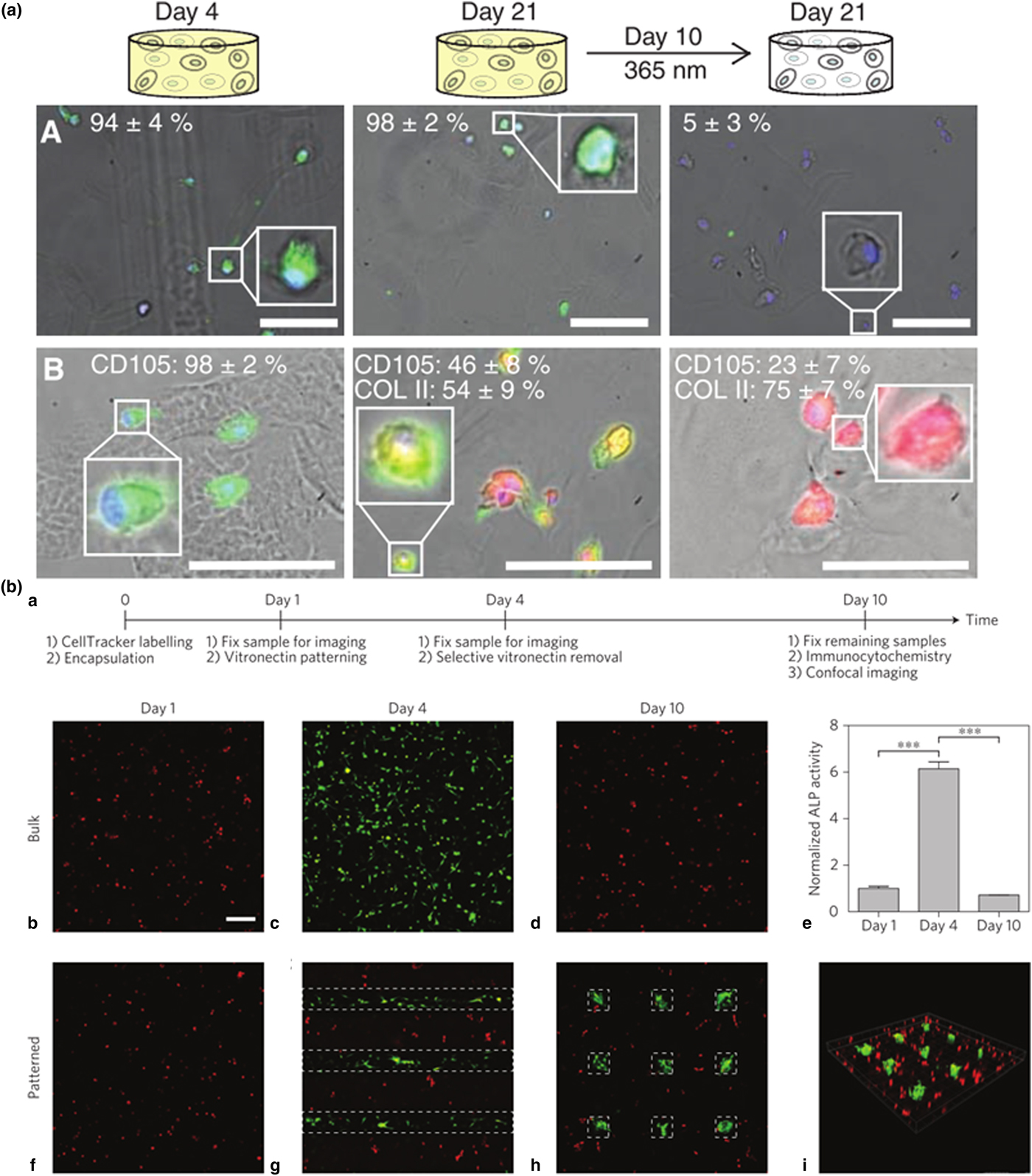
Controlled presentation and removal of biochemical cues impact cell differentiation. For example, dynamically tuning the presence and release of adhesive ligands is required for modeling in vivo differentiation of hMSCs, as demonstrated by Anseth and co-workers.[Reference Kloxin, Kasko, Salinas and Anseth68] A nitrobenzyl ether-derived photolabile moiety bound to an acrylate monomer (photodegradable acrylate, PDA) was bound to an RGDS peptide. This compound was copolymerized with PEG-diacrylate (PEGDA) to form a hydrogel with photolabile RGDS. The hMSC viability and chondrogenic differentiation was influenced by RGDS presentation. When hMSCs differentiate into chondrocytes in vivo, fibronectin is downregulated between 7 and 10 days after which cells upregulate GAG and type II collagen (COLII), indicators of chondrogenic differentiation. This pattern of fibronectin downregulation was mimicked through timely controlled RGDS removal. PEG-only hydrogels (without RGDS functionalization) resulted in significantly lower hMSC viability compared with hydrogels with RGDS. When RGDS was photocleaved on day 10 in culture, hMSC viability was not affected; however, GAG production increased fourfold with statistical significance compared to hydrogels with constant RGDS or no RGDS. A decrease in CD105 expression (hMSC marker) and increase in COLII production was observed, indicating chondrocyte differentiation [Fig. 5(a)].
(a) hMSC expression of integrin αvβ3 (green) when cultured in RGDS (yellow gel) or cleaved RGDS (white gel) after 4 or 21 days (panel A). Cleaving RGDS on day 10 showed a decrease in CD105 (green) and an increase in COLII (red) production by day 21, indicating chondrocyte differentiation (panel B). Adapted from Ref. Reference Kloxin, Kasko, Salinas and Anseth68 with permission from Science Publishing Group. (b) hMSCs show markers of osteocyte differentiation after vitronectin is patterned into a PEG-based hydrogel. Osteocalcin (OC) (green) and ALP activity increase in areas where VTN is photopatterned. Adapted from Ref. Reference DeForest and Tirrell69 with permission from Nature Publishing Group.

Orthogonal photocaging enables light-mediated attachment and subsequent removal of biologically active molecules to promote differentiation. DeForest and Tirrell designed a PEG hydrogel system with spatiotemporal control over full-length protein attachment and subsequent removal.[Reference DeForest and Tirrell69] The chemistries for bioconjugation and detachment are orthogonal and cytocompatible, and the reactions can be performed in the presence of live cells. To yield the initial scaffold, azide-functionalized peptide crosslinkers and azide-functionalized 2-(2-nitrophenyl)propoxycarbonyl (NPPOC)-photocaged alkoxyamine moieties underwent strain-promoted azide-alkyne cycloaddition (SPAAC) with cyclooctyne functionalized star-PEG. Upon exposure to UV light, uncaged alkoxyamines reacted with aldehyde-functionalized proteins resulting in the protein-modified scaffold. The incorporation of a second photocleavable moiety, o-nitrobenzyl ester, between the aldehyde functionality and the protein, enabled protein detachment with UV exposure. Vitronectin (VTN) was patterned to investigate hMSC differentiation into osteocytes; osteocalcin (OC) immunostaining and alkaline phosphatase (ALP) activity were evaluated as indicators of osteogenic differentiation. VTN was introduced on day 1 and cells showed an increase in OC staining and ALP activity by day 4. Upon VTN removal on day 4, OC staining and ALP activity decreased and returned to original levels by day 10 [Fig. 5(b)].
Spatial control of biochemical properties
The complex architecture of organ systems gives researchers incentive to develop techniques for spatial tuning of biochemical cues in 3D culture systems. Spatial manipulation of biologically relevant molecules enables studies of cell adhesion, aggregation, spreading, and invasion within user-defined and intricate geometries.[Reference Goubko, Majumdar, Basak and Cao70–Reference Mosiewicz, Kolb, van der Vlies, Martino, Lienemann, Hubbell, Ehrbar and Lutolf74] Concentration gradients of bioactive molecules enable studies of cell migration, development, and growth.[Reference Owen, Fisher, Tam, Nimmo and Shoichet75, Reference Gao, Konno and Ishihara76] Ultimately, simplified and adaptable systems will be required for widespread implementation whether for research or clinical application.
Light-induced patterning of biochemical cues
Photocaging can be used for biochemical ligand presentation; upon irradiation, active proteins are exposed to cells to elicit a biochemical response. However, proteins can be damaged upon irradiation, which impacts their bioactivity and stability. A unique technique was developed by Lutolf and co-workers to immobilize proteins on hydrogels with minimal effect on protein stability and bioactivity whereby uncaged hydrogel-bound peptides were bound to proteins through enzyme-mediated conjugation.[Reference Mosiewicz, Kolb, van der Vlies, Martino, Lienemann, Hubbell, Ehrbar and Lutolf74] Specifically, transglutaminase factor XII (FXIIIa) catalyzed the reaction between an amine-donor FXIIIa substrate AcFKG (K-peptide) bound to the PEG hydrogel and an amine-acceptor peptide NQEQVSPL (Q-peptide) bound to the desired protein. K-peptide was caged with nitroveratryloxycarbonyl (Nvoc) to enable spatially controlled enzyme-mediated photopatterning [Fig. 6(a)]. VEGF, Protein A (for tethering to Fc-tagged proteins), RGD peptide, recombinant fibronectin fragment FN9–10, and platelet-derived growth factor B (PDGF-BB) were all immobilized to the PEG hydrogel using this method. Multicellular clusters of MSCs were incorporated into MMP-cleavable PEG hydrogels, and a defined area of the hydrogel was patterned with RGD, FN9–10, or PDGF-BB. Cell invasion into the RGD, FN9–10 and PDGF-BB patterned regions was significantly greater compared with unpatterned regions, demonstrating bioactivity after immobilization and photo-illumination.
(a) Enzyme-mediated conjugation scheme of biochemical cues. FXIIIa catalyzes the reaction between K- and Q-peptides following photo-activation of the K-peptide. Adapted from Ref. Reference Mosiewicz, Kolb, van der Vlies, Martino, Lienemann, Hubbell, Ehrbar and Lutolf74 with permission from Nature Publishing Group. (b) Results of simultaneously patterning Barstar–SHH–488 (green) and biotin–CNTF–633 (red) into an agarose-based hydrogel using two-photo patterning. Adapted from Ref. Reference Wylie, Ahsan, Aizawa, Maxwell, Morshead and Shoichet78 with permission from Nature Publishing Group. (c) Glioblastoma and human astrocyte clusters magnetically combined into a single spheroid in a hydrogel containing gold nanoparticles, iron oxide nanoparticles, and M13-derived phage particles. Adapted from Ref. Reference Souza, Molina, Raphael, Ozawa, Stark, Levin, Bronk, Ananta, Mandelin, Georgescu, Bankson, Gelovani, Killian, Arap and Pasqualini81 with permission from Nature Publishing Group.

Two-photon irradiation is advantageous for fine-tuning the spatial control and concentration of biologically relevant molecules in 3D culture systems. Complex patterns incorporating one or more biologically relevant molecules can be achieved with two-photon irradiation.[Reference Wosnick and Shoichet77, Reference Wylie, Ahsan, Aizawa, Maxwell, Morshead and Shoichet78] Recent studies demonstrate the advantages of two-photon irradiation for fine-tuning the spatial control of biologically relevant molecules in 3D culture systems. Wosnick and Shoichet functionalized agarose with 6-bromo-7-hydroxy coumarin (Bhc)-protected thiol moieties to spatially control bioconjugation.[Reference Wosnick and Shoichet77] Aizawa et al. demonstrated that, with a two-photon microscope, exposed thiols in an agarose hydrogel could be reacted with maleimide-modified biomolecules such as vascular-endothelial growth factor-A (VEGF-A).[Reference Aizawa, Wylie and Shoichet79]
Orthogonal immobilization chemistries are advantageous for precise spatial control of multiple bioligands. Wylie et al. took advantage of two high-binding, orthogonal reactions—streptavidin–biotin and barnase–barstar—to immobilize two proteins simultaneously in spatially defined volumes with distinct protein concentrations.[Reference Wylie, Ahsan, Aizawa, Maxwell, Morshead and Shoichet78] Agarose, a transparent hydrogel, was modified with Bhc-protected thiol moieties that were modified after exposure to multi-photon irradiation with maleimide–streptavidin and maleimide–barnase, which have high affinities to biotin and barstar, respectively. Ciliary neurotrophic factor (CNTF) expressed recombinantly with biotin, and sonic hedgehog (SHH) with barstar, were simultaneously immobilized to agarose [Fig. 6(b)]. These recombinant proteins remained bioactive and when immobilized as a gradient guided retinal stem cell progenitors into the gels. While two-photon irradiation has the advantages of improved resolution and penetration depth, molecules with low uncaging efficiencies limit subsequent bioconjugation. For example, Bhc protecting groups can undergo photoisomerization reactions following irradiation. Mahmoodi et al. developed 6-bromo-7-hydroxy-3-methylcoumarin-4-ylmethyl (mBhc) to cage thiol moieties.[Reference Mahmoodi, Fisher, Tam, Goff, Anderson, Wissinger, Blank, Shoichet and Distefano80] HA-based hydrogels modified with mBhc-protected thiols had a fourfold greater uncaging efficiency than Bhc-protected thiols, which impacts the success of biochemical photopatterning.
Magnetic fields for spatial control over cell distribution
While spatially defined aggregates with multiple cell types are not common in current tissue engineering and regenerative medicine approaches, strategic co-culture of multiple cell types will be required for complex organogenesis models and intricate organoid culture. Magnetically controlled systems enable user-defined control of cell distribution. For example, with magnetic levitation, a magnet is used to guide cell distribution of a system comprised of magnetic particles intercalated with cells. Souza et al. designed a hydrogel containing gold nanoparticles, iron oxide nanoparticles, and M13-derived phage particles in order to form human glioblastoma multi-cellular aggregates with defined geometries and cell–cell contacts.[Reference Souza, Molina, Raphael, Ozawa, Stark, Levin, Bronk, Ananta, Mandelin, Georgescu, Bankson, Gelovani, Killian, Arap and Pasqualini81] The shape of the magnet determined the geometry of the cell cluster. Moreover, separate glioblastoma and human astrocyte clusters were magnetically combined into a single spheroid [Fig. 6(c)]. In another study by Bratt-Leal et al., magnetic nanoparticles were incorporated into the extracellular space of embryoid bodies (EBs).[Reference Bratt-Leal, Kepple, Carpenedo, Cooke and McDevitt82] Cell response to magnetic fields was dose dependent, and EBs labeled with different fluorescent tags could form aggregates with user-defined patterns.
Conclusions
We have highlighted the importance of spatiotemporal control over mechanical and biochemical properties in engineered hydrogels. While controlling the bulk properties of the hydrogel has implications on cell viability, aggregation, proliferation, and differentiation, spatiotemporal control enables guided cell growth and helps to recapitulate intricate cell networks that are required for studying biologically relevant processes. We expect future efforts to be focused on translating this knowledge to organoid development, for the applications of disease modeling and drug screening.
Research efforts that focus on developing hydrogel systems with stable and orthogonal chemistries for controlling mechanical and biochemical cues separately are required to understand the key factors of the cellular microenvironment that guide cell fate and organ formation. “Plug and play” chemistries that enable modular design with multiple combinations of biologically relevant components will enable biologists to answer fundamental questions pertaining to organ development and disease modeling. For hydrogel platforms to have widespread use in the research community, they must be versatile, easy to work with, and affordable. In the coming years, we expect a push toward the development of high-throughput screening platforms for drug development and personalized medicine, where patient-derived iPSCs could be used for individual drug screening. There are already platforms for screening with 3D culture conditions, such as microfluidic devices and assays developed in a conventional 96-well plate format.[Reference Mabry, Schroeder, Payne and Anseth83, Reference Chan, Zhang, Ho, Chiu, Jung and Leong84]. We expect hydrogel systems to not only advance the understanding of fundamental biologic processes, but advance the development of novel therapeutics and in vivo regenerative medicine technologies.
Acknowledgments
We are grateful for funding from the Canadian Institutes of Health Research (Foundation grant to M.S.S.), the Natural Sciences and Engineering Research Council (Discovery grant to M.S.S.), the QEII-GSST graduate student scholarship (to L.C.B.) and the Canada First Research Excellence Fund, Medicine by Design (to M.S.S.).