Introduction
The thalassaemias are a group of inherited disorders of haemoglobin synthesis. Patients with thalassaemia major suffer from refractory anaemia requiring regular blood transfusions, predisposing them to haemosiderosis. Desferrioxamine mesylate is an iron chelating agent used to counteract this. A side effect is auditory neurotoxicity, resulting in high frequency sensorineural hearing loss, which is reversible on early withdrawal.
Case report
The patient was a six-year-old boy who had been recently commenced on low-dose desferrioxamine therapy for transfusion-dependent beta-thalassaemia major, as his ferritin levels were starting to rise. An audiogram was performed at the time, which revealed a right-sided hearing loss, particularly in the low and high frequencies, with an air–bone gap on unmasked bone conduction. The patient was referred to our department for further assessment and advice on whether to stop the desferrioxamine.
Clinical examination revealed a normal left ear. The right tympanic membrane was slightly retracted but intact. A vague impression of a whitish opacity was noted in the posterior aspect of the drum, leading to a suspicion of congenital cholesteatoma.
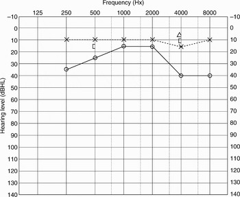
Repeat audiometry confirmed a conductive loss on the right, on the basis of masked bone conduction (Figure 1). Tympanometry was normal.
Pure tone audiogram O = right air conduction; X = left air conduction; [=right masked bone conduction; Δ = unmasked bone conduction.

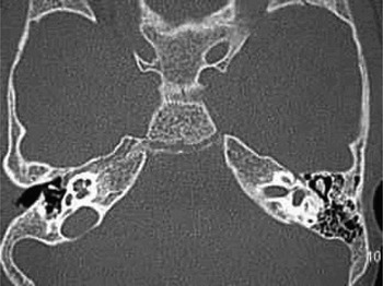
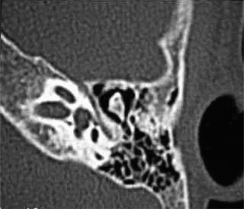
Computed tomography of the patient's temporal bones was arranged. The axial scans showed a widespread abnormality of bone, with widening of the right marrow cavity, consistent with thalassaemia. The middle-ear cleft was well ventilated and there was no evidence of congenital cholesteatoma. Both the incus and malleus were noted to have widened marrow cavities, leading to the conclusion that the patient's conductive hearing loss was due to marrow proliferation in the ossicular chain (Figures 2 and 3).
Axial computed tomography scan of temporal bones, demonstrating marrow expansion of right ossicular chain.

Magnified view of right temporal bone axial computed tomography scan, demonstrating marrow expansion of ossicular chain.

Discussion
The thalassaemias are a group of inherited disorders of haemoglobin synthesis. Their clinical presentation varies widely, ranging from asymptomatic forms to severe or even fatal entities. In these disorders, the production of either alpha or beta chains of haemoglobin may be reduced, resulting in alpha and beta-thalassaemia, respectively.
Beta-thalassaemias may be categorised by phenotypic severity into major, intermediate or minor forms. They can also be classified according to genotype. In beta0 thalassaemia, no beta chains are produced. In beta+ thalassaemia, beta chain production is impaired. Patients with thalassaemia major are homozygous for either beta0 or beta+ alleles.
In beta-thalassaemia major, defective beta chain production results in a relative excess of alpha chains. These are unstable and precipitate within red cell precursors, causing their destruction within the bone marrow. Anaemia ensues, with consequent marrow proliferation stimulated by erythropoietin production. Extramedullary haemopoeisis occurs, resulting in hepatosplenomegaly.
Patients with thalassaemia major present within the first year of life with failure to thrive, intermittent infection, refractory anaemia and hepatosplenomegaly. In addition, massive expansion of bone marrow results in bony deformities. Children may have mongoloid facies, frontal bossing of the skull and dental malocclusion.Reference Murphy, Kumar and Clark1
Due to the need for repeated blood transfusions, iron overload commonly occurs. Pathological quantities of metabolically active iron are released intracellularly in the form of haemosiderin and free iron, catalysing the formation of free radicals, which damage membrane phospholipids and cause cell death, leading to organ failure.Reference Giardina and Grady2 This is treated with subcutaneous desferrioxamine mesylate, an iron chelating agent.
One of the side effects of desferrioxamine mesylate is auditory neurotoxicity. High frequency sensorineural hearing loss may result as a consequence of such therapy.
The potential toxicity of desferrioxamine mesylate is well recognised in the literature. In 1979, De Virgiliis et al. reported a high incidence of hearing loss in thalassaemia major patients.Reference De Virgiliis, Argiolu, Sanna, Cornacchia, Cossu and Cao3 They identified iron overload as a possible mechanism for this high frequency hearing loss. They also commented that other potential possibilities were chronic hypoxia or expansion of the temporal bone as a result of marrow hyperplasia, in turn causing a narrowing of the internal auditory canal.
Following on from this report, in 1986 Olivieri et al. published a study of 89 patients receiving daily desferrioxamine, and reported a link between high dose desferrioxamine and neurotoxicity.Reference Olivieri, Buncic, Chew, Gallant, Harrison and Keenan4 They noted that clinical improvement ensued on withdrawal of the drug. The affected group were younger, had lower serum ferritin values, and were self-administering higher doses of desferrioxamine per kilogram of body weight.
In 1988, Cases et al. reported five cases of visual and auditory neurotoxicity in patients with end-stage renal failure receiving desferrioxamine; these patients improved clinically on withdrawal of the drug.Reference Cases, Kelly, Sabater, Campistol, Torras and Montoliu5
Several other studies have further strengthened the relationship between desferrioxamine and neurotoxicity.Reference Gallant, Boyden, Gallant, Carley and Freedman6–Reference Karimi, Asadi-Pooya, Khademi, Asadi-Pooya and Yarmohammadi9 In 2002, Karimi et al. evaluated the incidence of sensorineural hearing loss in beta-thalassaemia major patients undergoing regular chelation therapy with desferrioxamine. They concluded that the extent of ototoxicity caused by the drug was determined not only by the total amount of drug given, but also by the maximal plasma concentration.Reference Karimi, Asadi-Pooya, Khademi, Asadi-Pooya and Yarmohammadi9
• This paper reports a case of conductive hearing loss as a consequence of marrow expansion involving the ossicular chain, in a child with beta-thalassaemia treated with desferrioxamine therapy; this phenomenon has not been previously reported in the literature
• The use of desferrioxamine mesylate is associated with auditory neurotoxicity, resulting in a sensorineural hearing loss. This effect is reversible with early withdrawal of desferrioxamine
• Hearing loss can also result from marrow proliferation involving the ossicular chain as a consequence of the disease process. This hearing loss is conductive in nature
• The authors emphasise that it is important to distinguish between the two causes of hearing impairment, so as to avoid unnecessary withdrawal of desferrioxamine in the treatment and prevention of haemosiderosis in transfusion-dependent thalassaemia patients
Despite the amount of attention received by desferrioxamine, there are reports in the literature of thalassaemic patients with conductive hearing loss. Onerci et al. studied the audiological and impedancemetric findings of 34 thalassaemic patients, 27 of whom had thalassaemia major and seven thalassaemia intermedia.Reference Onerci, Aslan, Gϋmrϋk, Aksoy, Belgin and Özçelik10 The thalassaemia major group were administered desferrioxamine subcutaneously and received blood transfusions, albeit on an irregular basis. Oncerci et al. found that the majority of ears studied in the thalassaemia major group had a conductive hearing loss or a mixed pattern hearing loss. Futhermore, no patients in this group were found to have a pure sensorineural deficit. An air–bone gap was noted in the majority of patients in both groups, associated with normally shaped, stiff amplitude and normal pressure tympanograms with a high degree of static compliance. This study was interesting as it suggested that the pathological process of the disease process itself could account for stiffness in the middle-ear sound transmission system.
Prior to this, De Virgiliis et al. had in fact reported that 12 patients from their original cohort of 75 children suffering from beta-thalassaemia major had exhibited a mild, bilateral conductive hearing impairment, which they attributed to bony hypertrophy of the turbinates and adenotonsillar hypertrophy, leading to eustachian tube dysfunction.Reference De Virgiliis, Argiolu, Sanna, Cornacchia, Cossu and Cao3 It is interesting to note that, as this was in the early days of computed tomography (CT) scanning, no CT studies of the temporal bone were performed on this group of patients.
In addition, Sheikha et al. reported a case of three Saudi Arabian siblings suffering from thalassaemia intermedia with features of severe marrow expansion, one of whom presented with a left-sided conductive hearing loss and CT findings suggestive of a cholesteatoma.Reference Sheikha, Kameswaran, Okafor and al-Saigh11 On surgical exploration, the mastoid and middle-ear cavities were found to be filled with vascular tissue. The ossicular chain was surrounded by this material but intact. Histological analysis later confirmed haemopoietic tissue consistent with marrow expansion. Interestingly, ossicular chain hypertrophy due to marrow expansion was not noted.
The present case demonstrates that marrow expansion in thalassaemia major cases can involve the ossicular chain itself and result in a pure conductive hearing loss as a consequence of the disease process. Because so much attention has been placed on desferrioxamine as the culprit in cases of hearing loss in transfusion-dependent patients, other differential diagnoses may be easily overlooked. As a result, there may be situations in which desferrioxamine is withdrawn prematurely and inappropriately in the hope of reversing such hearing loss. The authors feel it is important to be aware of, and to exclude, marrow proliferation involving the ossicles as a cause of reduced hearing in such patients. This would include pure tone audiometry with masked bone conduction studies as standard testing.