Various infections may develop during hematopoietic stem cell transplantation (HSCT) in patients with hematological disease; infection control is essential for these patients. Thus, HSCT is commonly performed in biologically clean rooms in Japan to prevent lung disease such as invasive aspergillosis as well as to avoid contact and fecal–oral infection. After HSCT, patients are highly immunocompromised, and pulmonary nontuberculous mycobacterium (NTM) infection has been reported.Reference Weinstock, Feinstein, Sepkowitz and Jakubowski1
Hospital water, water-related devices, moist environments, and aqueous solutions can serve as reservoirs of waterborne pathogens. The hospital environment may allow for contamination by waterborne pathogens such as Legionella, NTM, and other gram-negative bacteria.Reference Decker and Palmore2 The complex structure of hospital water systems often leads to stagnation, corrosion, and biofilm formation.Reference Kanamori, Weber and Rutala3 At the end of the water supply, many handwashing stations are used for hand hygiene for both patients and healthcare staff; therefore, other than the water itself, aerators and/or sinks may be contaminated, leading to the development of outbreaks.Reference Weinbren4
We report a pseudo-outbreak of Mycobacterium chimaera due to biofilms in aerators on the faucets of handwashing machines (HWMs) in our HSCT center.
Methods
Setting
Our HSCT center is composed of 28 beds; each room has a handwashing machine. The air cleanness is regulated at ISO class 7 (ISO 14644-1) levels on the whole floor. HSCT or chemotherapy, which can cause persistent neutropenia, is performed on the floor. A few pediatric patients may be present on the ward. The HWM in each room is a Mitsubishi Chemical MRE-SFE-1K (Tokyo, Japan). This machine filters water from the central water supply through a 0.1-µm filter and then irradiates the water with ultraviolet light at 100–280 nm inside the faucet to prevent backward contamination. Therefore, the machine is designed to supply sterile water. At the facet of the machine, water is delivered through a metallic aerator to ensure a straight and evenly pressured shower-like stream of water. Patients turn the HWM on or off by holding their hands above the infrared sensor.
Case definition and patient data
Case patients were defined as patients with no respiratory symptoms related to NTM with sputum samples positive for acid-fast bacillus as detected by culture or polymerase chain reaction (PCR) at the HSCT center.
Detection of Mycobacterium from environmental and clinical samples
Mycobacterial cultures from clinical specimens were prepared by inoculating a 200-μL aliquot of decontaminated samples onto 2% Ogawa medium (Kyokuto Pharmaceutical Industrial, Tokyo, Japan) or into an MP Mycobacterium bottle at 37°C in BacT/ALERT3D (BioMérieux, Marcy-l’Étoile, France) for 8 or 6 weeks, respectively. Environmental water samples from each HWM (50 mL/sample), swab samples from each HWM, and water samples from a tea server (50 mL/sample) were incubated in 2% Ogawa medium or MGIT (Becton Dickinson Biosciences, Franklin Lakes, NJ, USA). To identify the M. avium complex, all specimens and environmental samples were evaluated using Cobas TaqMan MAI PCR tests with a Cobas TaqMan 48 Analyzer (Roche Diagnostics, Basel, Switzerland). Culture isolates were classified by internal transcribed spacer sequencing to differentiate M. chimaera as previously described.Reference Wallace, Iakhiaeva and Williams5
Variable numbers of tandem repeats (VNTR) analysis
Primer sets for 16 M. intracellulare VNTR loci were used in VNTR analysis as described previously.Reference Ichikawa, van Ingen and Koh6 Template DNA, dNTP mixture, primer sets, and 2× GC buffer containing ExTaq (Takara Bio, Shiga, Japan) were subjected to PCR assay. The amplified DNA products were subjected to electrophoresis to determine their sizes. Template DNA from M. intracellulare ATCC 13950, which is most closely related to M. chimaera, was used as a control in each experiment. Phylogenetic analysis was estimated by DendroUPGMA from the distance matrix data of the allele profiles of VNTR.
Results
Pseudo-outbreak description and surveillance
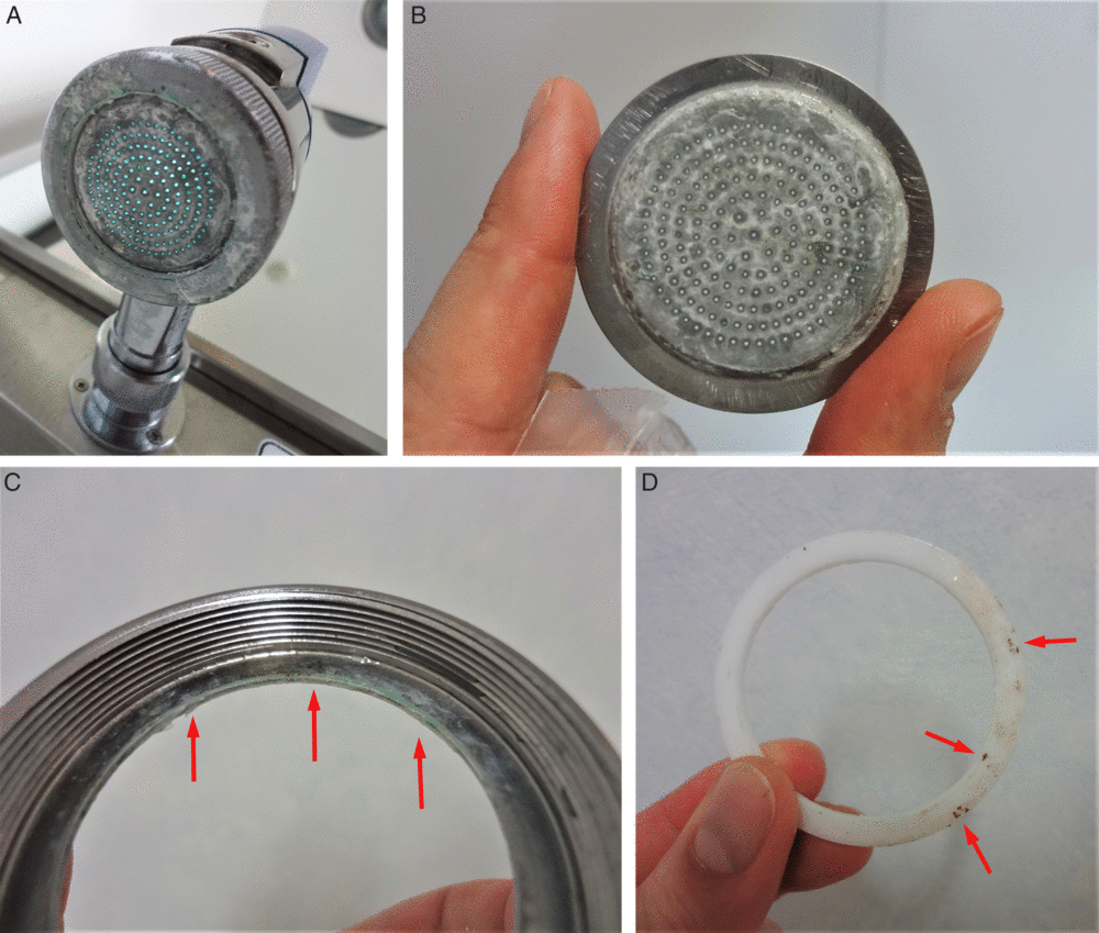
The number of patients in whom Mycobacterium was detected in the sputum increased from 2 during the last year to 11 in the last 6 months. None showed respiratory symptoms, and their sputum was routinely sampled at the onset of fever. Because we first suspected waterborne transmission, we examined the tea dispenser shared by all patients in the ward. The bacterium was not isolated from 4 water samples tested from the central supply and water tank of the dispenser. Next, 28 surface biofilm sites on the aerators and flat washers in HWMs were collected using sterile swabs (Fig. 1A–D). Mycobacteria was detected in 19 of 28 samples (67.9%) from HWMs. A total 4 of 8 clinical mycobacteria (50%) and 13 of 19 environmental mycobacteria (68.4%) were identified as representative M. intracellulare using a commercial kit and as M. chimaera based on internal transcribed spacer sequences. Cultures for other bacteria and fungi were negative.
(A, B) Aerators of hand washing machine contaminated by biofilm. (C, D) Parts of faucets, packing for aerators were polluted by biofilm (red arrows).

Genetic relationships between clinical and environmental isolates
The VNTR typing revealed that 4 isolates of Mycobacteria in 4 of 8 related patients and 13 samples from 1 flat washer and 12 aerators of HWMs were genetically identical M. chimaera. All isolates were clustered, showing the same VNTR profile but appeared to be a distinct branch of M. intracellulare ATCC 13950 of the resulting phylogenetic tree (Supplemental Fig. 1 online).
Intervention outcome and follow-up
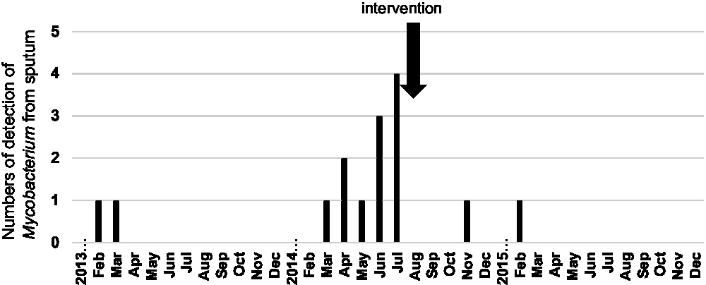
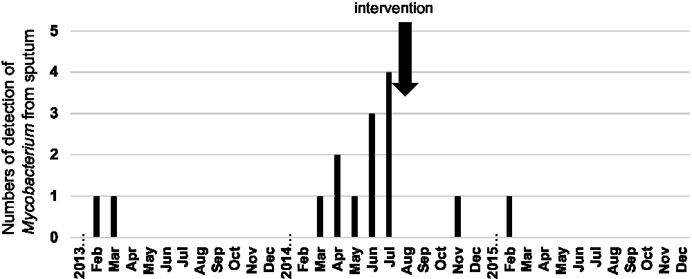
We determined that the aerator of the HWM in each room with biofilm was the cause of the Mycobacterium pseudo-outbreak in the patient sputum. We communicated with facilities maintenance personnel including officers and mechanics, and we improved the procedure for managing the units to incorporate routine work to replace aerators and their related parts every 6 months. After these actions were taken, mycobacterial detection in the sputum decreased very rapidly (Fig. 2), and a similar pseudo-outbreak has not occurred for >5 years.
Monthly prevalence of Mycobacteriumcultures in the sputum from January 2013 through December 2015. After intervention against the pseudo-outbreak, the number of Mycobacterium was decreased.
Discussion
We experienced a pseudo-outbreak that developed because of biofilm formation on the aerators of HWMs. Relapse of the pseudo-outbreak has not been observed since regular replacement of the aerator was implemented.
Biofilm formation on aerators by NTM appears to be common, as recently reported.Reference Feazel, Baumgartner, Peterson, Frank, Harris and Pace7 NTM can survive under high shear stress, low nutrition, and low mineral contents, even with chloride disinfectant.Reference Carson, Petersen, Favero and Aguero8 Although post-HSCT patients reportedly have high rates of NTM colonization in the sputum,Reference Weinstock, Feinstein, Sepkowitz and Jakubowski1 we predicted that our pseudo-outbreak developed from contaminated water use in patient daily living activities for the following reasons: (1) factory-recommended regular replacement of aerators had not been conducted for 5 years, (2) patients frequently use HWMs for mouth washing or drinking, (3) NTM detection rapidly decreased after regular replacement of the aerators, and (4) VNTR analysis showed that most NTM isolates were genetically identical to M. chimaera.
Mycobacterium chimaera related to M. intracellulare recovered from respiratory samples was first reported in patients with confirmed lung infection.Reference Tortoli, Rindi and Garcia9 Cardiac surgery-related outbreaks of M. chimaera infection in heater-cooler units have also been reported.Reference Sommerstein, Ruegg, Kohler, Bloemberg, Kuster and Sax10 Healthcare-related M. chimaera outbreaks are likely caused by water in hospital facilities because water reservoirs provide favorable temperature and environmental conditions, which are considered potential sources of infection.
Hospital facilities and equipment are often managed by engineering and facilities maintenance personnel who are not always familiar with the association between the hospital environment and human infections. Additionally, hospital staff do not always evaluate whether all machines in the hospital are maintained appropriately. Miscommunication among hospital staff, engineering, and facility maintenance personnel may have also caused our pseudo-outbreak.
In conclusion, the waterborne pseudo-outbreak of M. chimaera in our HSCT center likely resulted from biofilm present on the faucet outlets of HWMs. In the HSCT unit, regular maintenance of faucet parts must be performed to prevent biofilm formation by NTM.
Financial support
This work was supported in part by MEXT/JSPS KAKENHI (grant no. JP16K09120).
Conflicts of interest
All authors report no conflicts of interest relevant to this article.
Supplementary material
To view supplementary material for this article, please visit https://doi.org/10.1017/ice.2019.268.