INTRODUCTION
Reduction of the host's reproductive capacity is a common outcome of parasitic infections (Poulin, Reference Poulin2007). This phenotypic change may reflect non-adaptive side-effects of infection, defensive host adaptations to mitigate the impact of parasitism, or adaptive manipulations of the parasite to promote its own propagation (Ewald, Reference Ewald1980; Heins and Baker, Reference Heins and Baker2003; Hurd, Reference Hurd2001, Reference Hurd2009; Lafferty and Kuris, Reference Lafferty and Kuris2009). In vertebrates, examples of reduced fecundity are best documented in fishes parasitized by larval cestodes (plerocercoids), namely in three-spined sticklebacks (Gasterosteus aculeatus) and in roach (Rutilus rutilus) infected by Schistocephalus solidus and Ligula intestinalis, respectively (Arme, Reference Arme1997; Heins and Baker, Reference Heins and Baker2008; Heins et al. Reference Heins, Baker, Toups and Birden2010; Hoole et al. Reference Hoole, Carter and Dufour2010). Both parasites are diphyllobothridean cestodes characterized by complex life cycles, involving a free-swimming coracidium, a procercoid in a copepod (first intermediate host), a plerocercoid in fish (second intermediate host), and an adult worm in piscivorous birds (final host) (Dubinina, Reference Dubinina1980). Most of the parasite's growth takes place as a plerocercoid in the body cavity of a fish, and plerocercoids can attain a considerable size causing an energetic drain on the host (Dubinina, Reference Dubinina1980; Barber et al. Reference Barber, Wright, Arnott and Wootton2008). Despite striking similarities between S. solidus and L. intestinalis with regard to life cycle and host involvement, their effects on host reproduction differ. Both are generally considered as parasitic castrators which selectively target host reproductive energy and severely suppress host reproduction (Lafferty and Kuris, Reference Lafferty and Kuris2009). Impacts of S. solidus infection on stickleback reproduction involve delayed gametogenesis (Tierney et al. Reference Tierney, Huntingford and Crompton1996; Heins and Brown-Peterson, Reference Heins and Brown-Peterson2010), reduced egg size (Heins and Baker, Reference Heins and Baker2003), or decreased expression of secondary sexual characteristics and reduced courtship behaviour (MacNab et al. Reference MacNab, Katsiadaki and Barber2009). There is increasing evidence that the effects of S. solidus infection on host reproduction are variable and correlated with the parasite load (Tierney et al. Reference Tierney, Huntingford and Crompton1996; Bagamian et al. Reference Bagamian, Heins and Baker2004; Heins and Baker, Reference Heins and Baker2003, Reference Heins and Baker2008; Heins et al. Reference Heins, Baker, Toups and Birden2010; Heins and Brown-Peterson, Reference Heins and Brown-Peterson2010). These data are consistent with the hypothesis that simple nutrient depletion is the cause of the parasite's impact on host reproduction (Ewald, Reference Ewald1980; Hurd, Reference Hurd2001, Reference Hurd2009). In fact, Schultz et al. (Reference Schultz, Topper and Heins2006) demonstrated no change in the reproductive effort (the proportion of energy devoted to reproduction) in response to S. solidus-infection in female sticklebacks and consequently concluded that reproductive curtailment is a side-effect of reduced host energy reserves caused by the demands of the parasite. In roach on the other hand, infection by L. intestinalis always seems to prevent host reproduction by inhibiting gonad development at an early stage of gametogenesis (Arme, Reference Arme1997; Hoole et al. Reference Hoole, Carter and Dufour2010), and it has been shown that parasitization disrupts the endocrine system controlling reproductive function (Kloas et al. Reference Kloas, Urbatzka, Opitz, Wuertz, Behrends, Hermelink, Hofmann, Jagnytsch, Kroupova, Lorenz, Neumann, Pietsch, Trubiroha, Van Ballegooy, Wiedemann and Lutz2009; Geraudie et al. Reference Geraudie, Boulange-Lecomte, Gerbron, Hinfray, Brion and Minier2010; Trubiroha et al. Reference Trubiroha, Kroupova, Wuertz, Frank, Sures and Kloas2010). In particular, both genders of L. intestinalis-infected roach are characterized by a low expression of gonadotropins in the pituitary (Carter et al. Reference Carter, Pierce, Dufour, Arme and Hoole2005; Trubiroha et al. Reference Trubiroha, Wuertz, Frank, Sures and Kloas2009). The gonadotropins, follicle-stimulating hormone (FSH) and luteinizing hormone (LH), are heterodimeric glycoprotein hormones that consist of a common glycoprotein-hormone α-subunit (αGSU) non-covalently linked to a specific β-subunit (FSHβ or LHβ) (Levavi-Shivan et al. Reference Levavi-Sivan, Bogerd, Mananos, Gomez and Lareyre2010). Both are the key-hormones for reproduction in all vertebrates, regulating gonad development and the synthesis of sex steroids. FSH is considered to play a major stimulatory role during ovarian follicle growth and testicular spermatogenesis while LH is mainly involved in final gamete maturation (Lubzens et al. Reference Lubzens, Young, Bobe and Cerda2010; Schulz et al. Reference Schulz, de Franca, Lareyre, LeGac, Chiarini-Garcia, Nobrega and Miura2010). Congruently, recent studies under laboratory conditions suggest the involvement of FSH rather than LH in mediating effects of parasitization by L. intestinalis early during gonad development in roach (Trubiroha et al. Reference Trubiroha, Wuertz, Frank, Sures and Kloas2009). Further impacts of infection on reproductive physiology in roach comprise, for example, low plasma concentrations of the sex steroids 17β-oestradiol (E2), testosterone (T), and 11-ketotestosterone (11-KT) as well as decreased expression and plasma levels of vitellogenin (VTG), the hepatic precursor of yolk proteins (Geraudie et al. Reference Geraudie, Boulange-Lecomte, Gerbron, Hinfray, Brion and Minier2010; Trubiroha et al. Reference Trubiroha, Kroupova, Wuertz, Frank, Sures and Kloas2010). The described endocrine disruption appears to become manifested in a density-independent manner (Arme, Reference Arme1997; Hoole et al. Reference Hoole, Carter and Dufour2010) which indicates adaptive host manipulation by L. intestinalis. Irrespective of these studies on host endocrinology, nothing is known about the role played by environmental factors in the curtailment of roach reproduction following L. intestinalis-infection. Scientists are becoming increasingly aware of the profound effects nutrition can have on the outcome of parasitic infections (Smith, Reference Smith2007). In this context, laboratory studies allow controlling for variables such as food supply and can provide insights into the mechanisms and the evolutionary significance that underlie host phenotypes observed in the field.
The aim of the present study was to investigate whether long-term maintenance of roach under laboratory conditions with unlimited food supply can modulate or abolish the effects of parasitization by L. intestinalis on the reproductive physiology of its host. We investigated morphological features and reproductive parameters of ad libitum-fed infected and uninfected roach that were held in the laboratory for 2 years. The reproductive parameters studied comprise quantitative histological data on gonad development, plasma levels of the sex steroids E2, T, and 11-KT, as well as the expression of gonadotropin subunits (αGSU, FSHβ, LHβ) in the pituitary. Furthermore, the expression of hepatic VTG and of sex steroid receptors was investigated. The results are compared to field data from roach parasitized by L. intestinalis.
Materials and Methods
Animals and sampling
In November/December 2006, roach were collected by electrofishing from Lake Grosser Mueggelsee (52° 260′ N, 13° 390′ E) (Berlin, Germany). Visual inspection revealed that several fish were parasitized by plerocercoids of L. intestinalis. Immediately after catching, mass and length of a subsample of 1+ and 2+ fish were recorded. The following morphological parameters (females and males pooled; mean±s.d) were used as a basis for comparison of growth after 2 years of maintenance: uninfected n=12; total length=10·5±1·0 cm; somatic mass=8·78±3·0 g; infected n=11; total length=11·2±1·2 cm; somatic mass=10·9±4·0 g; parasitization index=7·5±4·1% (range 1·0–12·7%). Gonad histology showed that all fish were immature, except for 2 uninfected males that had entered spermatogenesis. Approximately 80 roach collected during this survey, including infected individuals, were transferred to the laboratory for investigation of the effects of L. intestinalis in fish receiving unrestricted food supply, in order to distinguish between direct effects of the tapeworm on gonad maturation and indirect effects caused by limited possibilities of food uptake. Roach were maintained in an aerated 1000 L tank under natural photoperiod and at a constant flow of tap water at 15±2°C temperature. Fish were fed daily ad libitum with commercial trout pellets (DAN-Ex 1750, Dana Feed) and living Chaoberus spp. larvae. Roach are classified as single-spawners, and gametogenesis is initiated in summer and proceeds throughout winter (Rinchard and Kestemont, Reference Rinchard and Kestemont1996) with spawning occurring in late April/early May at Lake Mueggelsee (personal observation). Investigations of roach held in the laboratory under the same conditions as described above showed that gametogenesis progressed in parallel to the situation in the field (personal observation). Spontaneous spawning in captivity, however, has not been observed.
After 2 years of maintenance, 59 remaining roach were sacrificed in November 2008. Blood was collected by puncture of the caudal vein using heparinized syringes and was centrifuged to obtain plasma. Liver tissue and pituitaries were removed for gene expression analyses. Tissue and plasma samples were stored at −80°C until further processing. Upon sampling, fish total length, fish total mass as well as fish somatic mass (eviscerated bodies excluding parasite mass) were measured to the nearest 1 mm and 0·1 g, respectively. Roach gonads and parasites when present were weighed to the nearest 0·01 g using precision scales. The number of parasites per fish was also recorded. Fish condition factor was calculated as CF=fish somatic mass×100/(fish total length)3, and the gonadosomatic index as GSI=fish gonad mass/fish somatic mass×100. Parasitization index was calculated as PI=parasite mass/fish somatic mass× 100. For histological analysis, samples of roach gonads were preserved in Bouin's fixative (Sigma) for 12 h, dehydrated in a graded series of ethanol and subsequently embedded in paraffin. Samples were sectioned at a thickness of 5 μm and stained with haematoxylin-eosin (H&E) or Goldner's modification of the Masson trichrome stain. All experimental procedures were conducted in compliance with the institutional guidelines for the care and use of animals.
Histological analysis of gonads
Gonads were analysed under an Axiovert 200 microscope (Zeiss) equipped with a Show View II digital camera (Olympus). Image analysis was performed using AnalySIS software (Soft Imaging Systems). Staging of roach gonads was conducted as described previously (Rinchard and Kestemont, Reference Rinchard and Kestemont1996; Nolan et al. Reference Nolan, Jobling, Brighty, Sumpter and Tyler2001). In females, the following stages of ovarian follicles were distinguished: primary growth (vacuole-free cytoplasm), early cortical alveolus (cortical alveoli occupy 1–3 rings in the cytoplasm), late cortical alveolus (cortical alveoli occupy more than 3 rings in the cytoplasm), vitellogenic (appearance of yolk globuli) and atretic. Ovaries were analysed based on the relative frequencies of follicles in the respective stage. Relative frequencies were calculated by dividing the number of follicles in a certain stage by the total number of follicles counted (a minimum of 100 follicles was counted per female). Early and late cortical alveolus, vitellogenic, as well as atretic follicles were summarized to be recruited into secondary growth and the percentage of follicles in secondary growth was calculated by dividing the number of secondary growth follicles by the total number of follicles. Furthermore, the ratio of atretic follicles to secondary growth follicles was calculated. For analysis of testes, pictures of 2 randomly selected fields of vision (200x magnification; area: 23,600 μm2) were taken and the area occupied by cysts containing spermatogonia A or spermatogonia B was measured (no other germ cell stages were present). The area occupied by spermatogonia B is expressed as a percentage of the total area investigated.
Hormone assays
Blood plasma was extracted twice with diethyl ether (Roth) as described previously (Trubiroha et al. Reference Trubiroha, Kroupova, Wuertz, Frank, Sures and Kloas2010) and concentrations of E2, T, and 11-KT were determined by specific enzyme-linked immunoassays (Cayman Chemicals) according to the manufacturer's instructions. Absorption at 412 nm was measured in a 96-well plate reader (Infinite M200, Tecan).
RNA extraction and reverse transcription
RNA extraction and reverse transcription was conducted as described previously (Trubiroha et al. Reference Trubiroha, Wuertz, Frank, Sures and Kloas2009, Reference Trubiroha, Kroupova, Wuertz, Frank, Sures and Kloas2010) with minor modifications. Briefly, total RNA from individual pituitary glands was extracted using the RNeasy Mini-Kit (Qiagen) including on-column treatment with DNase I (Qiagen). RNA from liver samples was extracted using Trizol (Invitrogen) followed by treatment with DNase I (AmpGrade; Invitrogen). The concentration of total RNA samples was measured by UV absorption using a NanoDrop ND-1000 spectrophotometer (NanoDrop Products, Thermo Fisher Scientific), and high integrity of the RNA was verified with an Agilent 2100 Bioanalyzer. Total RNA of pituitary (0·3 μg) and liver samples (1 μg) was reversely transcribed as reported previously (Trubiroha et al. Reference Trubiroha, Wuertz, Frank, Sures and Kloas2009; Reference Trubiroha, Kroupova, Wuertz, Frank, Sures and Kloas2010) using Affinity Script (Agilent) and AMV reverse transcriptase (Finnzymes), respectively.
Gene expression analysis by real-time PCR
Real-time PCR assays were carried out in an Mx3005P qPCR Cycler (Stratagene) using gene-specific primers as described previously (Trubiroha et al. Reference Trubiroha, Wuertz, Frank, Sures and Kloas2009, Reference Trubiroha, Kroupova, Wuertz, Frank, Sures and Kloas2010). Relative target transcript abundance was calculated by the comparative CT method (Pfaffl, Reference Pfaffl2001) and was normalized to the expression of ribosomal protein L8 (rpL8). Expression of target genes is presented as mean±s.d. relative to the expression value of uninfected females.
Statistical analysis
Normally distributed parameters (log transformed) of uninfected and infected roach of the same sex were analysed by Student's t-test, whereas not normally distributed data were evaluated using Mann-Whitney U-test. The level of statistical significance was set at P<0·05. Statistical analyses were performed using the software package SPSS 14.0.
RESULTS
Morphological and parasitological parameters
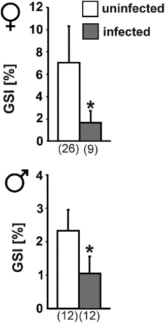
Summarized data on fish and parasites are given in Table 1. Length and somatic mass of infected female and male roach was significantly higher compared to their uninfected conspecifics (Table 1). No difference in CF was detected between uninfected and infected roach (Table 1) and both groups deposited perivisceral fat. PI of infected roach and number of parasites per fish was not significantly different between male and female roach. GSI was significantly lower in infected roach of both genders (Fig. 1).
Gonadosomatic index (GSI) of female and male roach, uninfected and infected by Ligula intestinalis. The number of individual samples is given in parentheses. Data are presented as mean±s.d. Asterisks indicate significant differences between uninfected and infected roach of the same gender (P<0·05).

Morphological and parasitological features of the sampled roach

a CF, condition factor; b PI, parasitization index.
Gonad histology
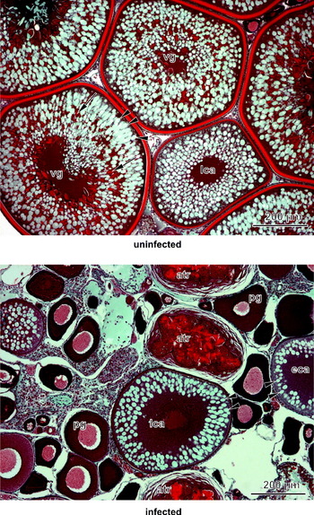
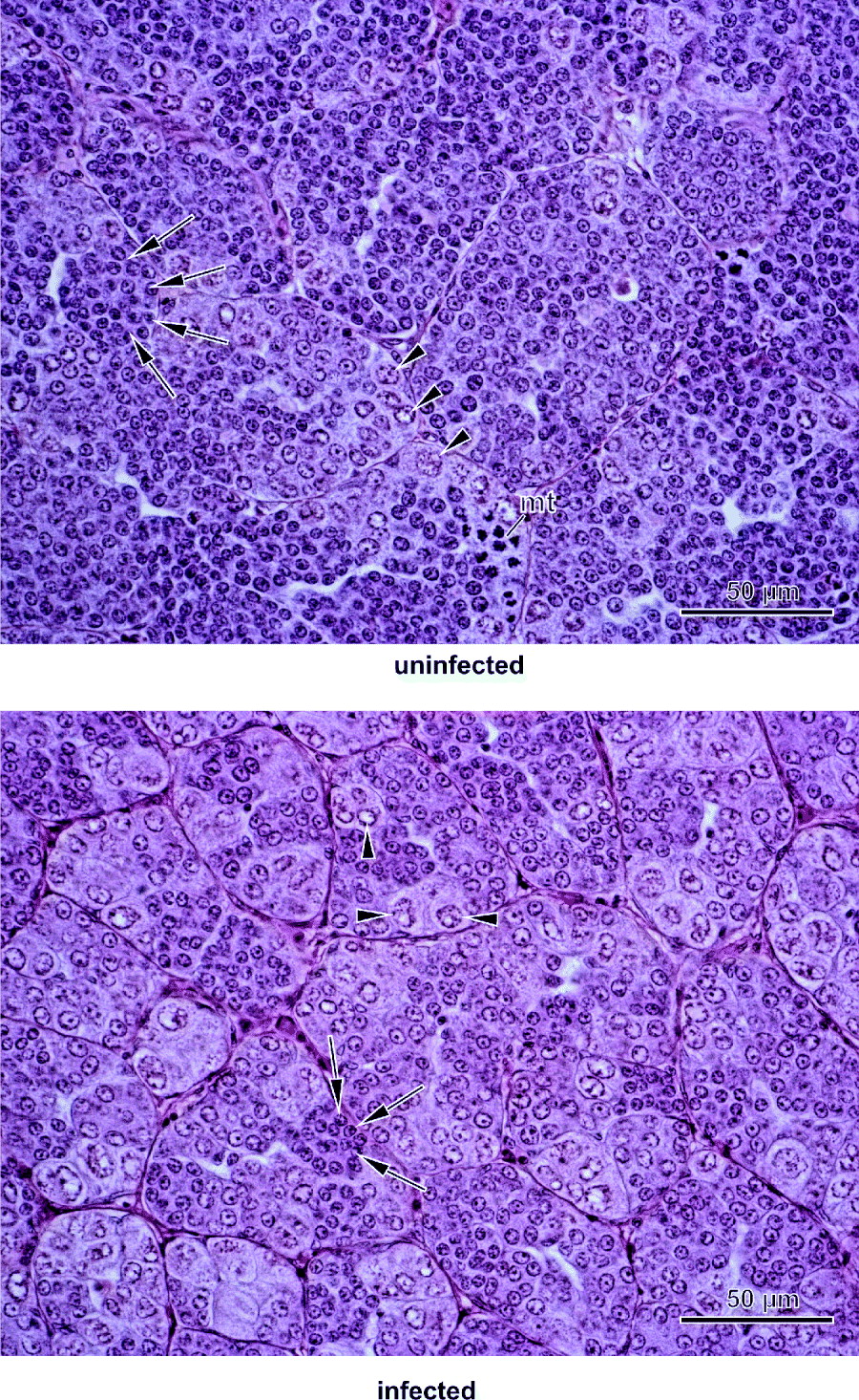
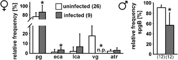
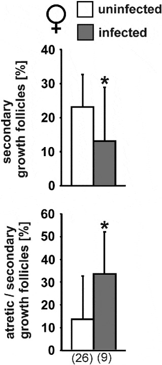
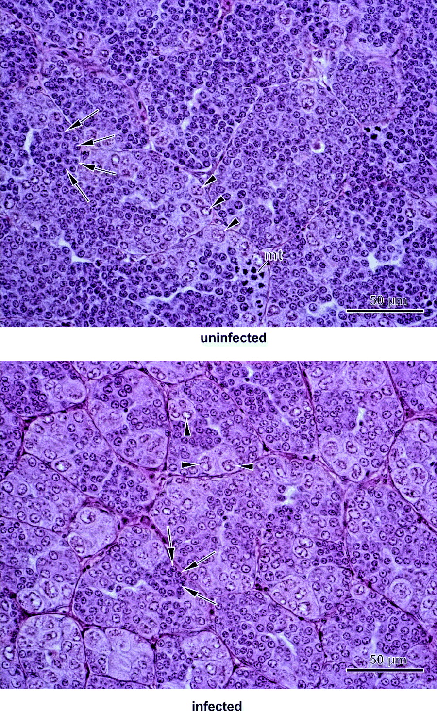
Ovaries of uninfected female roach contained follicles in the primary growth stage and developing follicles in secondary growth. Vitellogenic follicles were the most advanced stages found in the ovaries of uninfected females (Fig. 2). Infected females were also able to recruit follicles into secondary growth with the most advanced follicles observed being in the late cortical alveolus stage. Ovaries of infected females contained a significantly higher percentage of follicles in the primary growth and in the early cortical alveolus stage compared to uninfected conspecifics, whereas no vitellogenic follicles were present (Figs 2 and 4). The relative frequency of follicles which were recruited into secondary growth was significantly higher in uninfected females (uninfected 23·2%, infected 13·2%) (Fig. 5). Both uninfected and infected females contained atretic follicles in their ovaries but the percentage of atretic follicles in relation to follicles in secondary growth was significantly higher in infected females (uninfected 13·6%, infected 33·7%) (Fig. 5). In males, testes of uninfected individuals comprised mainly spermatogonia B (90·8% of testes area) (Figs 3 and 4). In testes of infected males, spermatogonia B were also present but at a significantly lower percentage (56·4% of testes area) than in uninfected conspecifics (Figs 3 and 4).
Histological sections of ovaries of female roach uninfected and infected by Ligula intestinalis. Sections were cut at 5 μm thickness and stained with Goldners modification of Masson's trichrome stain to facilitate distinguishing between the carbohydrate-containing cortical alveoli (blue/green, arrowheads) from the yolk globules (red, arrows). atr, atretic follicle; eca, early cortical alveolus follicle; lca, late cortical alveolus follicle; pg, primary growth follicle; vg, vitellogenic follicle.

Histological sections of testes of male roach uninfected and infected by Ligula intestinalis. Sections were cut at 5 μm thickness and stained by haematoxylin and eosin. mt, mitotic spermatogonia; arrows, spermatogonia B; arrow heads, spermatogonia A.
Relative frequency of germ cell types in female and male roach uninfected and infected by Ligula intestinalis. The number of individual samples is given in parentheses. Data are presented as mean±s.d. Asterisks indicate significant differences between uninfected and infected roach regarding the relative frequency of a certain cell type (P<0·05). Note that vitellogenic follicles were not present (n.p.) in infected females. atr, atretic follicles; eca, early cortical alveolus follicles; lca, late cortical alveolus follicles; pg, primary growth follicles; spgB, spermatogonia B; vg, vitellogenic follicles.

Effects of parasitization by Ligula intestinalis on follicle recruitment into secondary growth and atresia of secondary follicles in female roach. Data are presented as mean±s.d. The number of individual samples is given in parentheses. Asterisks indicate significant differences between uninfected and infected individuals (P<0·05).

Expression of gonadotropin subunits in the pituitary of roach
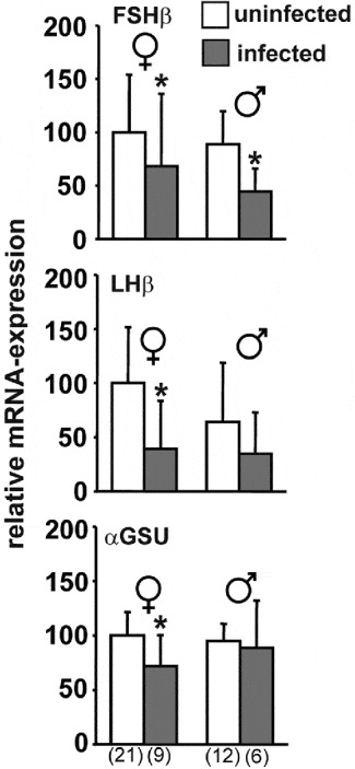
Analysis by real-time RT-PCR revealed significantly lower levels of FSHβ mRNA in L. intestinalis-infected roach of both genders (Fig. 6). In infected females and males, mRNA expression of FSHβ reached only 68·1% and 51·9% of levels in uninfected conspecifics, respectively. Parasitization also had a significant negative impact on the levels of LHβ and αGSU mRNA in females (60·8% and 72·0%, respectively, of levels in uninfected females), whereas in males no significant difference was detected in the expression of these two genes. Nevertheless, LHβ mRNA expression tended to be lower (54·0%; P<0·06) in infected male roach (Fig. 6).
Pituitary mRNA expression of follicle-stimulating hormone β-subunit (FSHβ), luteinizing hormone β-subunit (LHβ), and glycoprotein-hormone α-subunit (αGSU) in uninfected and Ligula intestinalis-infected female and male roach. Data are presented as mean±s.d. The number of individual samples is given in parentheses. Asterisks indicate significant differences between uninfected and infected roach of the same gender (P<0·05).

Plasma sex steroid levels
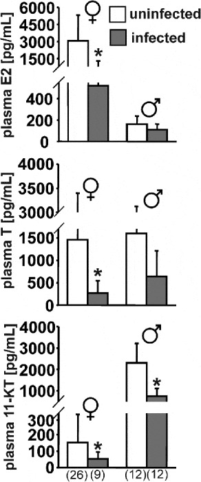
Infected females had significantly lower plasma concentrations of E2, T, and 11-KT, reaching only 16·9%, 18·4%, and 35·2% of the levels in uninfected conspecifics, respectively (Fig. 7). In males, only 11-KT was significantly reduced in infected fish (32·9% compared to uninfected males) but there was a trend toward lower levels of E2 (67·8%; P<0·06) and T (43·1%; P<0·06).
Plasma levels of 17β-oestradiol (E2), testosterone (T), and 11-ketotestosterone (11-KT) in uninfected and Ligula intestinalis-infected female and male roach. Data are presented as mean±s.d. The number of individual samples is given in parentheses. Asterisks indicate significant differences between uninfected and infected roach of the same gender (P<0·05).

Expression of VTG and receptors for sex steroids in roach liver
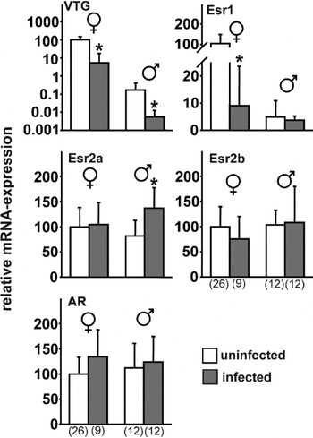
The expression of VTG mRNA in the liver was significantly lower upon parasitization in both genders and reached 5·3% and 2·8% of levels in uninfected females and males, respectively (Fig. 8). Expression of sex steroid receptors was affected in a gender-specific manner by L. intestinalis infection. Compared to uninfected conspecifics, oestrogen receptor 1 (Esr1, synonymous to ERα) mRNA was significantly reduced in infected females (reaching 9·0% of levels in uninfected females) but not in males. Oestrogen receptor 2a (Esr2a, synonymous to ERβ2) mRNA expression was significantly higher (167%) in infected males but not in infected females compared to uninfected conspecifics. The liver expression of oestrogen receptor 2b (Esr2b, synonymous to ERβ1) and androgen receptor (AR) mRNA was not significantly different between uninfected and infected roach of both genders.
Liver mRNA expression of oestrogen receptors (Esr1, Esr2a, and Esr2b), androgen receptor (AR), and vitellogenin (VTG) in uninfected and Ligula intestinalis-infected female and male roach. Data are presented as mean±s.d. The number of individual samples is given in parentheses. Asterisks indicate significant differences between uninfected and infected roach of the same gender (P<0·05).

DISCUSSION
After long-term maintenance under laboratory conditions with unlimited food supply, infected roach of both genders had lower GSI than their uninfected conspecifics. In females, this was a consequence of inhibited gonad development, reduced follicle recruitment into secondary growth and increased follicular atresia, whereas in males, the lower GSI values reflected inhibited spermatogonial proliferation. Although the negative effects of L. intestinalis-infection on gonadal growth and development in roach were significant, the present histological data are in contrast to observations under field conditions. Usually oogenesis is arrested in the primary growth stage in infected females (Arme, Reference Arme1968; Arme and Owen, Reference Arme and Owen1968; Trubiroha et al. Reference Trubiroha, Wuertz, Frank, Sures and Kloas2009; Geraudie et al. Reference Geraudie, Boulange-Lecomte, Gerbron, Hinfray, Brion and Minier2010), and in infected males, spermatogonia B are more sparsely observed (occupying only about 20% of the testes area – unpublished data). Under field conditions, the gonads of roach parasitized by L. intestinalis resemble those of hypophysectomized fish. Based on molecular biological investigations, depressed gonadal growth and development in infected fish is thought to be a result of inhibited gonadotropin expression in the pituitary (Carter et al. Reference Carter, Pierce, Dufour, Arme and Hoole2005; Trubiroha et al. Reference Trubiroha, Wuertz, Frank, Sures and Kloas2009). Similarly in the present study, the expression of pituitary gonadotropins was negatively affected by L. intestinalis. Nevertheless, the differences between uninfected and infected individuals in the present study were not as pronounced as under field conditions when mRNA expression of FSHβ and LHβ in infected roach did not exceed 10% and 30%, respectively, of the levels in uninfected conspecifics (Trubiroha et al. Reference Trubiroha, Wuertz, Frank, Sures and Kloas2009). In teleosts, gonadotropins regulate important aspects of gametogenesis via the induction of gonadal E2 and 11-KT, in particular, vitellogenesis in females and spermatogenesis in males (Lubzens et al. Reference Lubzens, Young, Bobe and Cerda2010; Schulz et al. Reference Schulz, de Franca, Lareyre, LeGac, Chiarini-Garcia, Nobrega and Miura2010). Consistent with the low expression of gonadotropins, significantly reduced plasma concentrations of sex steroids, namely E2 in females and 11-KT in males, were detected in infected roach in the present study. Similar observations have been made in bream (Abramis brama) (Hecker and Karbe, Reference Hecker and Karbe2005) and roach (Geraudie et al. Reference Geraudie, Boulange-Lecomte, Gerbron, Hinfray, Brion and Minier2010; Trubiroha et al. Reference Trubiroha, Kroupova, Wuertz, Frank, Sures and Kloas2010) under field conditions. Again, the negative effect of parasitization on sex steroids was less severe in the present study compared to previous field observations, where plasma concentrations of E2 in females and 11-KT in males reached only 7–8% of levels in uninfected conspecifics (Trubiroha et al. Reference Trubiroha, Kroupova, Wuertz, Frank, Sures and Kloas2010).
In general, the expression of gonadotropin subunits in the pituitary and concentrations of sex steroids in plasma correlated well with the histological data, which showed a more progressed stage of gametogenesis in infected roach kept under long-term laboratory conditions compared to field observations. Interestingly, the recruitment as well as the maintenance of secondary growth follicles was impaired in females infected by L. intestinalis in the present study. This might be a consequence of the lower expression of FSHβ and LHβ in these individuals, since FSH plays a key role in follicle recruitment, and gonadotropins in general constitute important anti-apoptotic/anti-atretic factors in the ovary (Wood and Van Der Kraak, Reference Wood and Van der Kraak2002; Lubzens et al. Reference Lubzens, Young, Bobe and Cerda2010). It should be noted here that mature oocytes and sperm have been reported recently in a small number of roach infected by L. intestinalis during the spawning season, even under field conditions (Geraudie et al. Reference Geraudie, Boulange-Lecomte, Gerbron, Hinfray, Brion and Minier2010). The reason for this exceptional observation remains unknown but these data together with the present results show that the phenotype of L. intestinalis-infected roach with regard to reproductive physiology can vary under certain circumstances.
Investigations on gene expression in the liver demonstrated significantly lower mRNA levels of VTG in parasitized roach of both genders compared to uninfected conspecifics. In infected females, this is most probably a result of their lower E2 plasma concentrations, since in oviparous vertebrates hepatic VTG synthesis is induced by oestrogens (Wahli, Reference Wahli1988). Our findings are in agreement with field data for female bream (Hecker and Karbe, Reference Hecker and Karbe2005) and roach (Geraudie et al. Reference Geraudie, Boulange-Lecomte, Gerbron, Hinfray, Brion and Minier2010; Trubiroha et al. Reference Trubiroha, Kroupova, Wuertz, Frank, Sures and Kloas2010). Similar to previous findings in the field (Trubiroha et al. Reference Trubiroha, Kroupova, Wuertz, Frank, Sures and Kloas2010), we measured considerably lower levels of liver VTG mRNA in infected male roach in the present study. Interestingly, plasma E2 concentrations were not significantly lower in infected compared to uninfected males after long-term maintenance under laboratory conditions. This indicates an effect of L. intestinalis on liver VTG mRNA expression in males which is probably not related to oestrogen levels. Lower VTG mRNA in infected males could, nevertheless, also be a consequence of their reduced plasma concentrations of androgens, since VTG induction by exposure to low concentrations of methyltestosterone has been demonstrated in male zebrafish (Danio rerio) (Andersen et al. Reference Andersen, Goto-Kazeto, Trant, Nash, Korsgaard and Bjerregaard2006). Furthermore, VTG has been shown to be involved in immune function in fish (Li et al. Reference Li, Zhang and Liu2008) and it would be interesting to address whether reduced VTG expression in infected roach is potentially associated with immune interactions between L. intestinalis and its host.
Oestrogen-induced synthesis of hepatic VTG in oviparous vertebrates is mediated via nuclear oestrogen receptors. Recent studies in rainbow trout (Oncorhynchus mykiss) (Leanos-Castaneda and Van Der Kraak, Reference Leanos-Castaneda and Van De Kraak2007) and goldfish (Carassius auratus auratus) (Nelson and Habibi, Reference Nelson and Habibi2010) showed that the Esr2 subtypes are crucial for vitellogenesis in fishes, with Esr1 being auto-induced via Esr2 to sensitize the liver to further stimulation of VTG synthesis by oestrogens. Auto-induction of Esr1 mRNA has been consistently documented in teleosts, including roach, whereas the regulation of Esr2 subtypes by oestrogens is more complex and may depend on species, sex, and reproductive stage (Menuet et al. Reference Menuet, Le Page, Torres, Kern, Kah and Pakdel2004; Filby and Tyler, Reference Filby and Tyler2005; Katsu et al. Reference Katsu, Lange, Urushitani, Ichikawa, Paull, Cahill, Jobling, Tyler and Iguchi2007; Lange et al. Reference Lange, Katsu, Ichikawa, Paull, Chidgey, Coe, Iguchi and Tyler2008). Thus, in accordance with a previous field investigation (Trubiroha et al. Reference Trubiroha, Kroupova, Wuertz, Frank, Sures and Kloas2010), the lower mRNA expression of hepatic Esr1 in infected female roach in the present study is likely to be a result of their lower plasma E2 compared to uninfected conspecifics. In contrast to Esr1 mRNA, the expression of Esr2a, Esr2b, and AR differed in some cases from the situation described in the field where hepatic Esr2a mRNA in both genders (and Esr2b mRNA in males) was higher in infected individuals and AR mRNA was down-regulated in response to parasitism in females (Trubiroha et al. Reference Trubiroha, Kroupova, Wuertz, Frank, Sures and Kloas2010). Nevertheless, we detected a higher expression of Esr2a in infected males also in the present study. Whether this is a result of changes in plasma sex steroids upon infection or a response to other parasite factors remains yet unknown.
Differences in the outcome of parasitism on host reproduction between field and laboratory studies have been reported in several host-parasite systems, including the three-spined stickleback infected by plerocercoids of S. solidus (Candolin and Voigt, Reference Candolin and Voigt2001; Barber and Svensson, Reference Barber and Svensson2003). Under favourable laboratory conditions, no effect of S. solidus on reproduction in male sticklebacks was observed (Candolin and Voigt, Reference Candolin and Voigt2001). In experimentally infected females, Barber and Svensson (Reference Barber and Svensson2003) even reported an increase in GSI compared to uninfected conspecifics. On the other hand, we demonstrate here for the first time that long-term maintenance under laboratory conditions with unlimited food access can attenuate but not abolish the negative impact of parasitization by L. intestinalis on reproductive physiology in roach. In the present study, CF did not differ significantly between infected and uninfected individuals and perivisceral fat was abundant in both groups, showing that long-term energy reserves were deposited. As described for the three-spined stickleback infected by S. solidus, reproductive parameters of the host should be negatively related to parasite burden if inhibition of gametogenesis is a simple side effect of nutrient deprivation (Hurd, Reference Hurd2001; Hall et al. Reference Hall, Becker and Caceres2007; Heins and Baker, Reference Heins and Baker2008). In roach parasitized by L. intestinalis however, even small plerocercoids that are unlikely to pose significant energetic demands cause an arrest of host gametogenesis (Arme, Reference Arme1968; Arme and Owen, Reference Arme and Owen1968). Consistent with previous investigations (Arme, Reference Arme1968; Arme and Owen, Reference Arme and Owen1968; Trubiroha et al. Reference Trubiroha, Wuertz, Frank, Sures and Kloas2009), no negative correlation was detected between PI and any of the reproductive parameters in roach after long-term maintenance under laboratory conditions (data not shown). Therefore, it is concluded that the observed impact of L. intestinalis on roach gametogenesis is not due to energetic drains upon parasitization but reflects a specific strategy of the parasite, which is manipulating the allocation pattern of host energy away from reproduction (Hurd, Reference Hurd2001, Reference Hurd2009; Lafferty and Kuris, Reference Lafferty and Kuris2009). Such a strategy seems advantageous for the parasite because the high energy costs required for reproduction of roach might then be available for the growing plerocercoid. This would concomitantly minimize the harm exerted on host somatic tissues due to general nutritional drains, thereby avoiding a decrease in host viability (Hurd, Reference Hurd2001; Ebert et al. Reference Ebert, Carius, Little and Decaestecker2004; Lafferty and Kuris, Reference Lafferty and Kuris2009). In addition, preventing host reproduction may provide the temporal storage of energy reallocated from reproduction already during early infection before the parasite can fully exploit these resources. If the energy liberated from reproduction is higher than the demands of the parasite plus the energetic costs for the host in mounting an immune defence, this in turn can lead to enhanced growth (gigantism) of the infected host under certain circumstances (Ebert et al. Reference Ebert, Carius, Little and Decaestecker2004; Hall et al. Reference Hall, Becker and Caceres2007). In the present study, data relevant in this context were not collected and therefore no conclusions about the influence of L. intestinalis on the growth of the investigated roach can be drawn.
Gametogenesis in fish is triggered by body energy stores, and the pituitary gonadotropins as well as gonadal steroidogenesis are influenced by metabolic hormones (e.g. Campbell et al. Reference Campbell, Dickey, Beckman, Young, Pierce, Fukada and Swanson2006). Given the large biomass L. intestinalis can attain relative to the host, it seems possible that the reproductive dysfunction of infected roach under field conditions is in part mediated by energetic drains, acting in concert with the specific endocrine-disrupting effects of the parasite. Thus, unlimited food supply was possibly a predominant factor for the more advanced gonadal development of infected roach in the laboratory as compared to the field. Notwithstanding the experimental feeding regime, changes in additional confounding variables under laboratory conditions are potentially involved. For example, roach are known to release steroid hormones into the water (Lower et al. Reference Lower, Scott and Moore2004) and the action of pheromones in fishes is not restricted only to the final reproductive events such as synchronization of spawning (Van Weerd et al. Reference Van Weerd, Sukkel, Bongers, Van der Does, Steynis and Richter1991; Huertas et al. Reference Huertas, Scott, Hubbard, Canario and Cerda2006). Fishes infected with L. intestinalis do not exhibit normal shoaling patterns (Orr, Reference Orr1966; Loot et al. Reference Loot, Brosse, Lek and Guegan2001) and thus, pheromonal stimuli that would normally be received within the group-shoaling situation are probably absent from parasitized individuals. Under the present conditions, i.e. simultaneous maintenance of infected and uninfected fish in one tank, infected roach could also inevitably receive pheromonal stimuli from their uninfected conspecifics, which in turn might stimulate gametogenesis in parasitized individuals to some degree. However, nothing is known about the responsiveness of infected roach towards pheromonal/hormonal stimuli and further studies involving exposure experiments would help to get a better picture of the state of the endocrine system in roach parasitized by L. intestinalis.
In summary, we characterized for the first time the influence of L. intestinalis on reproductive physiology of roach kept for a long time under laboratory conditions with unlimited food supply. Effects of parasitism involved inhibited gametogenesis in both genders of roach, accompanied by reduced expression of pituitary gonadotropin subunits and lowered plasma concentrations of sex steroids. Furthermore, a strong negative impact of parasitization in both genders on hepatic VTG mRNA was observed. In general, the effect of L. intestinalis on reproductive physiology appeared attenuated under laboratory conditions compared to the phenotype found in the wild. Still, there was a clear deleterious impact of parasitization on host gametogenesis despite the fact that infected individuals were apparently in a good condition as indicated by CF and the deposition of perivisceral fat. Our observation is in contrast to studies on the related stickleback-S. solidus host-parasite system, where the negative impact of parasitism on host reproduction has been documented to be a side-effect of nutritional drains and can become abolished under favourable conditions. It is concluded here that arrested gametogenesis in roach upon infection by L. intestinalis in the wild is mediated only in part by environmental factors or nutritional drains of parasitism. The present results support the hypothesis that the cestode L. intestinalis selectively inhibits roach reproduction via endocrine disruption.
ACKNOWLEDGEMENT
This study was supported by CEFIC via Grant EMSG 42. H. K. received a Post-doctoral Fellowship of the Alexander von Humboldt Foundation. Technical support by Matthias Kunow and Eric Eckmann is gratefully acknowledged. We would also like to thank anonymous reviewers for valuable comments on a former version of this manuscript.