Introduction
The presence of respiratory disease in pigs is commonly associated with significant production losses and is considered one of the main obstacles for the swine industry. If a thorough diagnostic investigation is conducted, results indicate that clinical respiratory disease in pigs is often polymicrobial, resulting from a combined infection with one or more viruses and bacteria. Respiratory pathogens can be further divided into primary pathogens, capable of inducing severe lesions in respiratory tissues as a result of their own virulence, and secondary or opportunistic infectious pathogens that typically need help from other co-infecting pathogens or cofactors to induce substantial lesions in the respiratory system. Moreover, based on their relevance to the swine industry in a particular region or population, respiratory pathogens in pigs can perhaps also be divided into pathogens of major and minor importance or relevance (Table 1). Compared to other domestic animals, pigs are unique because they are typically housed in large groups in relatively small spaces, thereby providing ideal conditions to maintain pathogens for extended time periods. Pathogens involved in respiratory disease in pigs vary significantly among farms, production sites, regions and countries, making generalizations about porcine respiratory disease complex (PRDC) treatment and control difficult (Gutiérrez-Martín et al., Reference Gutiérrez-Martín, Rodríguez-Delgado, Alvarez-Nistal, De La Puente-Redondo, García-Rioja, Martín-Vicente and Rodríguez Ferri2000; Harms et al., Reference Harms, Halbur and Sorden2002). In some regions of the world, establishment of an accurate diagnosis is often lacking due to limited availability of the most appropriate diagnostic tools which compromises implementation of the most appropriate treatment and control regimens.
Commonly recognized pathogens of major or minor relevance to the pig industry based on their ability to induce respiratory disease and lesions by themselves (major relevance) or under circumstances such as a concurrent infection with other pathogens (minor relevance)

The interactions that occur on the cellular and molecular levels during concurrent infection of pigs with two or more respiratory pathogens are multifaceted and complex. The standard paradigm of an initial primary viral infection followed by opportunistic bacterial infection certainly occurs, but the interactions can be much more complex involving multiple viruses and bacteria and different sequences of infection. The modes of action of pathogens can, when combined, be additive or synergistic. Because of the complicated nature of mixed infections, studying their interactions can be a laborious task. Under experimental conditions, it is often difficult to reliably reproduce clinical respiratory disease involving a single pathogen; it is even more difficult to design experimental studies that examine the interactions among two or more pathogens.
PRDC
The term ‘porcine respiratory disease complex (PRDC)’ is now regularly used to describe mixed respiratory infections of multiple etiologies (Halbur, Reference Halbur1998). The clinical picture of PRDC is often characterized by decreased feed intake, fever, cough and dyspnea ultimately resulting in decreased feed efficiency and decreased growth rate. Morbidity rates associated with PRDC may range from 30 to 70% and mortality rates between 4 and 6% or even higher in affected farms (Halbur, Reference Halbur1998). Macroscopic lesions highly depend on the pathogen involved and can vary from no visible lesions to nearly 100% multifocal tan mottling or consolidation of the lung. Frequently, lesions are located in the cranioventral parts of the lung, where consolidation, discoloration and failure of the lung tissue to collapse may be observed (Harms et al., Reference Harms, Halbur and Sorden2002; Martínez et al., Reference Martínez, Peris, Gómez and Corpa2009; Fraile et al., Reference Fraile, Alegre, López-Jiménez, Nofrarías and Segalés2010). Similarly, microscopic lesions may vary according to the pathogens involved, but bronchopneumonia, sometimes in combination with interstitial pneumonia is often reported (Harms et al., Reference Harms, Halbur and Sorden2002; Kim et al., Reference Kim, Chung and Chae2003).
Factors involved in PRDC
The severity of PRDC manifestation in pigs depends upon the interactions and synergy of infectious factors (both viral and bacterial pathogens), environmental factors, type of production system and management, and pig-specific factors (genetics, age and immunological status).
Infectious factors
Many respiratory pathogens are ubiquitous; thus, it is not uncommon for multiple pathogens to circulate in a herd at any given time. New pathogens commonly enter a herd through aerosol, fomites, vectors and in many cases through the introduction of infected animals. The pathogens frequently associated with PRDC and abbreviations used in this review are summarized in Table 1.
Viral pathogens
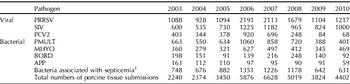
Porcine reproductive and respiratory syndrome virus (PRRSV) replicates in macrophages and dendritic cells located in the tonsil, upper respiratory tract and lungs, resulting in viremia as early as 12 h post infection which can persist for several weeks or months (Wills et al., Reference Wills, Doster, Galeota, Sur and Osorio2003). It has been shown that PRRSV induces cell lysis (Thanawongnuwech et al., Reference Thanawongnuwech, Thacker and Halbur1997), apoptosis (Labarque et al., Reference Labarque, Van Gucht, Nauwynck, Van Reeth and Pensaert2003), changes in function of infected pulmonary alveolar and intravascular macrophages (Thanawongnuwech et al., Reference Thanawongnuwech, Thacker and Halbur1997, Reference Thanawongnuwech, Thacker and Halbur1998b), damage to the mucociliary apparatus (Thanawongnuwech et al., Reference Thanawongnuwech, Halbur, Ackermann, Thacker and Royer1998a) and changes in T-cell subpopulations (Shimizu et al., Reference Shimizu, Yamada, Kawashima, Ohashi, Shimizu and Ogawa1996), ultimately resulting in increased susceptibility to opportunistic bacterial pneumonia and septicemia. Based on data from case submissions (Table 2) to the Iowa State University Veterinary Diagnostic Laboratory (ISU-VDL), PRRSV is the number 1 pathogen detected in swine respiratory disease in the USA. However, the overall trend for PRRSV-induced pneumonia based on percentage of total number of porcine tissue cases received for the last 8 years indicates that PRRSV-associated respiratory disease is slowly declining (Fig. 1). This may be due to recent PRRSV elimination efforts by individual pork producers and systems, and regional PRRS control and eradication projects. Alternatively, this may be due to overall higher numbers of growing pigs that are being vaccinated against PRRSV in the USA, which is mainly due to increased efforts to vaccinate pigs from negative farms before or shortly after they have been sent to finishing units located in pig dense areas (Loula, Reference Loula2011). As with all prevalence estimates based on submissions to veterinary diagnostic labs the data presented in this review should be evaluated with caution due to potential influence of factors unrelated to pig health. For instance, it is well known that the increased media attention and public awareness during the 2009 flu pandemic negatively impacted submission rates of pigs and pig tissues to veterinary diagnostic labs which may be incorrectly interpreted as an improvement in the overall health of pigs in the Midwest.
Trends in pathogens associated with respiratory disease in case submissions to the Veterinary Diagnostic Laboratory at Iowa State University. Data presented as percentage of total number of porcine tissue cases received.

Trends in pathogen detection over time in swine respiratory case submissions to the Veterinary Diagnostic Laboratory at Iowa State University

1 APYO, HPS, SALM, SSUIS and ASUIS.
Similar to PRRSV, swine influenza virus (SIV) also has a tropism for the respiratory system where it attaches to and replicates in epithelial cells of the upper respiratory tract (Brown et al., Reference Brown, Alexander, Chakraverty, Harris and Manvell1994). Infection then spreads to bronchi and bronchioles resulting in loss of cilia, extrusion of mucus, exudation of neutrophils and macrophages, and necrosis and metaplasia of airway epithelium (Brown et al., Reference Brown, Alexander, Chakraverty, Harris and Manvell1994). It has been demonstrated that levels of certain cytokines, tumor necrosis factor α (TNFα), interferon α (IFNα), and interleukin-1 (IL-1), are all elevated 18–24 h after inoculation with SIV and correlate with neutrophil infiltration as well as onset and degree of clinical signs (Karasin et al., Reference Karasin, Olsen, Brown, Carman, Stalker and Josephson2000; Charley et al., Reference Charley, Riffault and Van Reeth2006). SIV-induced damage to the respiratory tract reduces the ability to clear the infection and also decreases macrophage function (Dea et al., Reference Dea, Bilodeau, Sauvageau, Montpetit and Martineau1992; Rekik et al., Reference Rekik, Arora and Dea1994; Thacker, Reference Thacker2001). SIV is the second most commonly identified viral respiratory pathogen based on data from case submissions to the ISU-VDL, and the overall 8-year-trend indicates that the incidence of SIV-induced pneumonia cases remains relatively steady (Fig. 1).
Pseudorabies virus (PRV) replicates in alveolar macrophages and monocytes, and causes necrotic rhinitis and tracheitis with disrupted ciliary activity in the respiratory mucosa. PRV-induced damage to the respiratory tract mucosa may contribute to decreased clearance and increased adherence of other pathogens (Narita et al., Reference Narita, Kawashima, Matsuura, Uchimura and Miura1994). Macrophage functions such as phagocytosis, killing of phagocytized bacteria, antibody-dependent and -independent cytotoxicity, IFNα production, phagosome–lysosome fusion and oxygen production after stimulation with opsonized zymosan were reduced in PRV-infected macrophages (Fuentes and Pijoan, Reference Fuentes and Pijoan1986; Iglesias et al., Reference Iglesias, Pijoan and Molitor1989, Reference Iglesias, Pijoan and Molitor1992). Moreover, direct destruction of monocytes and macrophages, which are involved in both innate and active immunity, may also contribute to the degree of immunosuppression (Fuentes and Pijoan, Reference Fuentes and Pijoan1986; Iglesias et al., Reference Iglesias, Pijoan and Molitor1989, Reference Iglesias, Pijoan and Molitor1992). In the USA, PRV has been eradicated from the domestic pig population; however, since it is still circulating in the wild pig population in certain regions, PRV-associated respiratory disease has to remain on the respiratory disease complex differential diagnosis list.
Porcine circovirus type 2 (PCV2) is now widely accepted to be an important pathogen in PRDC. PCV2-infection has been associated with mild interstitial pneumonia and may induce attenuation and ulceration of the airway epithelium and cause prominent peribronchiolar and peribronchial fibroplasia (Kim et al., Reference Kim, Chung and Chae2003). PCV2 antigen has been demonstrated in the cytoplasm of pulmonary macrophages, the bronchiolar epithelium and endothelial cells, which is suggestive that PCV2 may be contributing to the severity and duration of PRDC (Harms et al., Reference Harms, Halbur and Sorden2002). PCV2-associated respiratory disease in the USA reached its peak during 2006 and 2007 (Table 2). In support of the important role of PCV2 in PRDC, implementation of PCV2 vaccination in PRDC affected herds substantially lowers the incidence of respiratory disease and pulmonary co-infections (Fachinger et al., Reference Fachinger, Bischoff, Jedidia, Saalmuller and Elbers2008). The efficacy of PCV2 vaccination is also clearly seen in the overall trend for PCV2-induced pneumonia at the ISU-VDL, which indicates a dramatic decline of PCV2-associated pneumonia from 2008 to 2010 (Fig. 1). To better mimic what is going on under field conditions and to have an improved model to test the efficacy of different treatment strategies, poly-viral experimental inoculations have gained importance in recent years in PCV2 research. Researchers have experimentally inoculated pigs concurrently with PCV2 and PRRSV (Opriessnig et al., Reference Opriessnig, Madson, Prickett, Kuhar, Lunney, Elsener and Halbur2008, Sinha et al., Reference Sinha, Shen, Schalk, Beach, Huang, Halbur, Meng and Opriessnig2010), PCV2, PRRSV and SIV (Opriessnig et al., Reference Opriessnig, Patterson, Madson, Pal and Halbur2009a), or PCV2, PRRSV and porcine parvovirus (PPV) (Shen et al., Reference Shen, Beach, Huang, Halbur, Meng and Opriessnig2010; Opriessnig et al., Reference Opriessnig, Shen, Pal, Ramamoorthy, Huang, Lager, Beach, Halbur and Meng2011b) to better mimic the field situation and evaluate vaccine efficacy.
Porcine respiratory coronavirus (PRCV) replicates primarily in the epithelium of the lower respiratory tract, in type 2 pneumocytes, and to a lesser extent in bronchi and bronchiolar epithelium (Atanasova et al., Reference Atanasova, Van Gucht, Barbé, Lefebvre, Chiers and Van Reeth2008). PRCV induces disruption of the mucociliary apparatus and decreases pulmonary alveolar macrophage function, which reduces the ability of macrophages to clear bacteria (Charley et al., Reference Charley, Riffault and Van Reeth2006).
Porcine cytomegalovirus (PCMV) replicates in and destroys cells of the nasal mucosa and tubuloalveolar glands where it is associated with large intranuclear inclusion bodies (Narita et al., Reference Narita, Kawamura, Shirai and Haritani1987). PCMV-induced damage to the upper respiratory tract epithelium and mucociliary apparatus likely makes pigs more susceptible to bacterial pathogens (Edington et al., Reference Edington, Watt and Plowright1977; Narita et al., Reference Narita, Kawamura, Shirai and Haritani1987).
Paramyxoviruses (PMV) such as porcine rubulavirus and Nipha virus play a role in respiratory disease in certain geographic regions where they are associated with economically relevant disease outbreaks in affected pig populations (Hooper et al., Reference Hooper, Zaki, Daniels and Middleton2001; Rodríguez-Ropón et al., Reference Rodríguez-Ropón, Hernández-Jauregui, Sánchez-Torres, Favila-Castillo, Estrada-Parra, Moreno-López and Kennedy2003). A PMV different from porcine rubulavirus and Nipha virus was associated with interstitial pneumonia in pigs in the USA (Janke et al., Reference Janke, Paul, Landgraf, Halbur and Huinker2001) indicating that this may be another contributor to PRDC. However, since it is identified only on rare occasions, it is likely of minor relevance.
Torque teno sus virus (TTSuV) was only recently associated with respiratory disease in pigs. When gnotobiotic pigs were infected with TTSuV genogroup 1 (TTSuV-1), the pigs developed mild interstitial pneumonia (Krakowka and Ellis, Reference Krakowka and Ellis2008) implying a potential role for this virus in PRDC. In support of this, a high prevalence of TTSuV infection (54–82%) was detected in Japanese pigs which were 60 days and older, and suffered from PRDC or porcine circovirus-associated disease (Taira et al., Reference Taira, Ogawa, Nagao, Tuchiya, Nunoya and Ueda2009). When serum samples from 253 pigs from 83 farms were tested for TTSuV-1 and TTSuV-2 DNA, there was a correlation between the diagnosis of PRDC and the detection of TTSuV (Lang et al., Reference Lang, Söllner, Barz, Ladinig, Langhoff, Weissenböck, Kekarainen, Segalés and Ritzmann2011). Moreover, under experimental conditions using gnotobiotic pigs, combined PCV2 and TTSuV-1 infection resulted in systemic clinical disease (Ellis et al., Reference Ellis, Allan and Krakowka2008) and combined PRRSV and TTSuV-1 infection resulted in clinical disease resembling porcine dermatitis and nephropathy syndrome (Krakowka et al., Reference Krakowka, Hartunian, Hamberg, Shoup, Rings, Zhang, Allan and Ellis2008). The relevance of these findings is unknown as TTSuV is widely distributed in swine populations. There is only limited data available from field and experimental studies and much more needs to be learned about this virus and its importance in PRDC.
Bacterial pathogens
Actinobacillus pleuropneumoniae (APP) resides in tonsils, necrotic lung tissue and the nasal cavity (Willson et al., Reference Willson, Falk and Klashinsky1987). There is a marked difference in virulence between serotypes and strains within serotypes. Virulence factors include three exotoxins (ApxI, ApxII and ApxIII), which play a role in inducing the characteristic necrosis and hemorrhage (Kamp et al., Reference Kamp, Stockhofe-Zurwieden, van Leengoed and Smits1997). The capsule of APP is also capable of inhibiting phagocytosis (Sakano et al., Reference Sakano, Shibata, Samegai, Taneda, Okada, Irisawa and Sato1993; Kamp et al., Reference Kamp, Stockhofe-Zurwieden, van Leengoed and Smits1997). Based on diagnostic case submissions to the ISU-VDL, APP-associated respiratory disease is of minor importance compared to other bacterial infections (Table 2) and remains consistently at a low level based on overall percentages of cases diagnosed as APP (Fig. 1).
Mycoplasma hyopneumoniae (MHYO) attaches to the cilia and surface of epithelium by adhesion proteins, resulting in clumping, damage and loss of cilia, epithelial cell death and reduced function of the mucociliary apparatus (DeBey and Ross, Reference DeBey and Ross1994; Young et al., Reference Young, Thacker, Erickson and Ross2000). The loss of cilia is thought to be important in the increased incidence of secondary bacterial infections associated with MHYO. Infection with MHYO is commonly associated with immunostimulation and immunosuppression (Kishima and Ross, Reference Kishima and Ross1985; Kishima et al., Reference Kishima, Ross and Kuniyasu1985; Maes et al., Reference Maes, Verdonck, Deluyker and de Kruif1996), non-specific mitogenic effects on swine lymphocytes (Messier and Ross, Reference Messier and Ross1991), or cytokines secreted by activated macrophages (Messier and Ross, Reference Messier and Ross1991; Asai et al., Reference Asai, Okada, Ono, Irisawa, Mori, Yokomizo and Sato1993; Thacker et al., Reference Thacker, Thacker, Kuhn, Hawkins and Waters2000a; Thanawongnuwech et al., Reference Thanawongnuwech, Young, Thacker and Thacker2001). Among bacterial causes of respiratory disease in pigs in the USA, MHYO is the second most common following Pasteurella multocida (PMULT) (Table 2) and despite widespread vaccination, mycoplasmal pneumonia remains a major concern for pig producers. Recent studies indicate that Mycoplasma hyorhinis (MHYOR) may also be important in PRDC (Lin et al., Reference Lin, Chen, Yeh and Weng2006; Hansen et al., Reference Hansen, Pors, Jensen, Bille-Hansen, Bisgaard, Flachs and Nielsen2010), but more research in this area is needed.
Bordetella bronchiseptica (BORD) is known to cause attenuation of the respiratory epithelium and loss of cilia (Sakano et al., Reference Sakano, Sakurai, Furutani and Shimizu1984). The dermonecrotic toxin of BORD causes turbinate atrophy and pneumonic lesions characterized by necrosis, hemorrhage, neutrophil accumulation and fibrosis (Sakano et al., Reference Sakano, Okada, Taneda, Ono and Sato1992; Horiguchi, Reference Horiguchi2009). Thus, BORD infection can result in delayed clearance mechanisms, increased accumulation of mucus and exposure of submucosal areas. BORD is also cytotoxic for alveolar macrophages in vitro (Brockmeier et al., Reference Brockmeier, Palmer, Bolin and Rimler2001; Brockmeier and Register, Reference Brockmeier and Register2007). In the USA, BORD is most commonly seen in younger pigs and has a relatively low prevalence in diagnostic submissions to the ISU-VDL (Table 2 and Fig. 1).
Pasteurella multocida (PMULT) is generally considered to be an opportunistic invader that is rapidly cleared from the lungs of normal pigs. PMULT has a polysaccharide capsule that aids in resisting phagocytosis and the Tox-A protein, which is an essential virulence factor for progressive atrophic rhinitis (Lax and Grigoriadis, Reference Lax and Grigoriadis2001; Horiguchi, Reference Horiguchi2009; Wilson and Ho, Reference Wilson and Ho2010). The detection of Tox-A is frequently used for confirmation of pathogenicity of PMULT isolates (Lichtensteiger et al., Reference Lichtensteiger, Steenbergen, Lee, Polson and Vimr1996; Takada-Iwao et al., Reference Takada-Iwao, Uto, Mukai, Okada, Futo and Shibata2007). PMULT remains the most common respiratory bacterium identified in cases of PRDC at the ISU-VDL (Table 2 and Fig. 1).
Haemophilus parasuis (HPS),Streptococcus suis, Actinobacillus suis, Arcanobacterium pyogenes andSalmonella enterica serovars Choleraesuis and Typhimurium may induce lung damage either directly (rare) or in association with septicemia. HPS is commonly isolated from nasal secretions and tracheal swabs of healthy pigs (Bertschinger and Nicod, Reference Bertschinger and Nicod1970). The septicemic form of HPS infection is better known as Glässer's disease. Streptococcus suis (SSUIS) invades tonsils and reaches the lymph nodes via lymphatics (Cloutier et al., Reference Cloutier, D'Allaire, Martinez, Surprenant, Lacouture and Gottschalk2003). Infected monocytes may distribute the organism throughout the body including the brain (Zheng et al., Reference Zheng, Zhao, Zhang, Kang, Chen and Jin2009). The capsule of SSUIS inhibits phagocytosis and antigens associated with virulence include muramidase-release protein (MRP), capsid and extracellular factor (EF) (Smith et al., Reference Smith, Damman, van d, Wagenaar, Wisselink, Stockhofe-Zurwieden and Smits1999; Tanabe et al., Reference Tanabe, Bonifait, Fittipaldi, Grignon, Gottschalk and Grenier2010). Similar to APP, Actinobacillus suis (ASUIS) can be carried in tonsils and nostrils of healthy pigs (Ross et al., Reference Ross, Hall, Orning and Dale1972) and infection likely occurs via the upper respiratory tract. Arcanobacterium pyogenes (APYO) is another common pathogen on the membranes of the upper respiratory tract and is frequently associated with suppurative inflammation and abscess formation. Finally, Salmonella enterica (SALM), most commonly serovars Choleraesuis and Typhimurium, can also be associated with respiratory disease in pigs.
Parasites
Disease conditions such as parasitism can also negatively impact on the pig's ability to respond to respiratory pathogens (Tjørnehøj et al., Reference Tjørnehøj, Eriksen, Aalbaek and Nansen1992). Especially for pigs raised outdoors, lungworms (Metastrongylus spp.) are commonly encountered with respiratory disease. Adult lungworms, ranging from 14 to 55 mm and localized within the terminal bronchioles, elicit bronchitis, bronchoalveolitis, alveolar emphysema and atelecatsis (Stewart and Hoyt, Reference Stewart, Hoyt, Straw, Zimmermann, D'Allaire and Taylor2006). Ascarid larvae also migrate through lungs and when exposure is high in naïve pigs ascarids can cause severe respiratory disease and lesions. Ascaris suum has also been shown to negatively impact MHYO vaccination in pigs (Steenhard et al., Reference Steenhard, Jungersen, Kokotovic, Beshah, Dawson, Urban, Roepstorff and Thamsborg2009).
Non-infectious factors
Various non-infectious factors are significant contributors to respiratory disease by increasing transmission and spread of the pathogens, by creating unfavorable conditions that result in increased stress on the animal, or by directly damaging the respiratory tract (Sorensen et al., Reference Sorensen, Edwards, Noordhuizen and Gunnarsson2006; Martínez et al., Reference Martínez, Peris, Gómez and Corpa2009). The non-infectious factors contributing to the disease process can by divided broadly into three groups including environment, management (system, herd size and nutrition) and pig factors (age, genetics, breed and immune status), which have been summarized previously (White, Reference White2011). Commonly, severe environmental stressors such as, but not limited to, extreme temperature changes, elevated humidity, aerial pollutants and excessive ammonia are known to predispose pigs to development of PRDC (Done, Reference Done1991). Feed quality is also important as dust in feed can interfere with the pig's respiratory tract defense. In addition, molds or mycotoxins in feed can either directly damage lungs or compromise immunity. Nutritional deficiencies can also affect the immune system.
The age and breed of the pig at the time of infection can also be of importance. PRDC is predominantly seen in grow–finish pigs, typically around 12–24 weeks of age, and may be associated with waning of passively acquired antibodies (Thanawongnuwech et al., Reference Thanawongnuwech, Thacker and Halbur1998b; Cho et al., Reference Cho, Dee, Deen, Trincado, Fano, Jiang, Faaberg, Murtaugh, Guedes, Collins and Joo2006; Pomorska-Mól et al., Reference Pomorska-Mól, Markowska-Daniel, Rachubik and Pejsak2011). To the authors’ knowledge, to date no specific resistance factors to respiratory pathogens have been identified in pigs. However, evidence of increased susceptibility of certain breeds, or lines within breeds, to disease induced by certain viruses (PRRSV and PCV2) has been reported (Halbur et al., Reference Halbur, Rothschild, Thacker, Meng, Paul and Bruna1998; Vincent et al., Reference Vincent, Thacker, Halbur, Rothschild and Thacker2005; Opriessnig et al., Reference Opriessnig, Fenaux, Thomas, Hoogland, Rothschild, Meng and Halbur2006, Reference Opriessnig, Patterson, Madson, Pal, Rothschild, Kuhar, Lunney, Juhan, Meng and Halbur2009b; Vincent et al., Reference Vincent, Thacker, Halbur, Rothschild and Thacker2006). Finally, when pigs from multiple sites and with different immune status are mixed, disease spread is enhanced. In problem herds, vaccination can be used as a means to bring the immune status of individuals in the population to a similar level.
Interaction of pathogens in PRDC
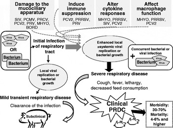
The interactions between different pathogens play an important role in the development and outcome of PRDC. Although different pathogens are capable of inducing disease independently, simultaneous infection with two or more viral pathogens commonly results in more severe clinical signs and lesions (Allan et al., Reference Allan, McNeilly, Ellis, Krakowka, Meehan, McNair, Walker and Kennedy2000; Harms et al., Reference Harms, Sorden, Halbur, Bolin, Lager, Morozov and Paul2001). Some known mechanisms by which pig pathogens interact include damaging of the mucociliary apparatus in respiratory tissues to allow bacterial colonization (Pol et al., Reference Pol, van Leengoed, Stockhofe, Kok and Wensvoort1997; Loving et al., Reference Loving, Brockmeier, Vincent, Palmer, Sacco and Nicholson2010), inducing immune suppression (Renukaradhya et al., Reference Renukaradhya, Alekseev, Jung, Fang and Saif2010), altering cytokine responses (Thanawongnuwech et al., Reference Thanawongnuwech, Thacker, Halbur and Thacker2004), or affecting macrophage function (Chiou et al., Reference Chiou, Jeng, Chueh, Cheng and Pang2000) (Fig. 2). In addition, the sequence of infection is likely also of great importance and should be considered when evaluating outcomes of experimental studies (Table 3). The following paragraphs summarize some of the experimental co-infection models using different combinations of bacteria and viruses or mixed viral–bacterial infections.
Summary of possible mechanisms and outcomes of viral and bacterial infections of the respiratory tract.

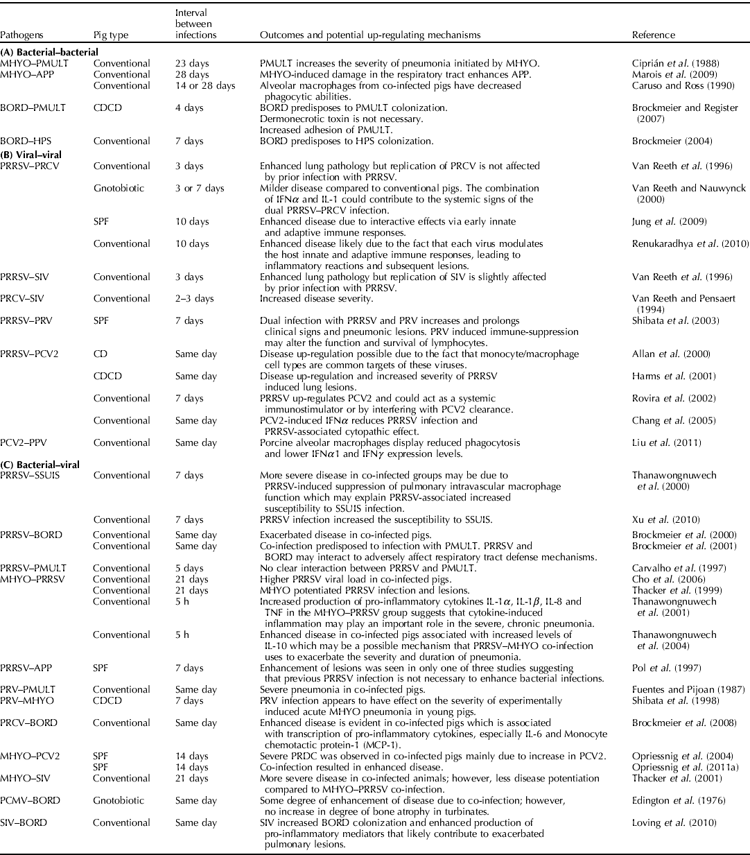
Selected examples of interactions and up-regulating mechanisms in polymicrobial respiratory diseases in pigs. (A) Bacterial–bacterial interactions. (B) Viral–viral interactions. (C) Bacterial–viral interactions. SPF, specific-pathogen-free; CD, colostrum-deprived; CDCD, cesarean-derived, colostrum-deprived pigs
Bacterial–bacterial interactions
A summary of selected study outcomes and potential mechanisms in poly-bacterial respiratory disease is provided in Table 3(A). MHYO and PMULT represent the most commonly identified respiratory bacteria based on case submissions to the ISU-VDL in 2010 (Table 2 and Fig. 1). The combination of these pathogens is frequently identified in the same pig indicating a potential interaction. It is well known and experimentally confirmed that inoculation with MHYO alone causes only mild disease and lung lesions (Thacker et al., Reference Thacker, Halbur, Ross, Thanawongnuwech and Thacker1999, Opriessnig et al., Reference Opriessnig, Thacker, Yu, Fenaux, Meng and Halbur2004). Similarly, inoculation with PMULT alone typically causes no disease and the organism is difficult to recover from experimentally infected pigs (Ciprián et al., Reference Ciprián, Pijoan, Cruz, Camacho, Tórtora, Colmenares, López-Revilla and de la Garza1988; Amass et al., Reference Amass, Clark, van Alstine, Bowersock, Murphy, Knox and Albregts1994). However, dual infection with MHYO and PMULT induces disease characterized by elevated rectal temperatures, a severe cough, and more extensive lung lesions from which both organisms can be isolated (Ciprián et al., Reference Ciprián, Pijoan, Cruz, Camacho, Tórtora, Colmenares, López-Revilla and de la Garza1988; Amass et al., Reference Amass, Clark, van Alstine, Bowersock, Murphy, Knox and Albregts1994). In these experiments, if pigs were MHYO vaccinated before MHYO–PMULT challenge or if the pigs were allowed to recover from MHYO challenge, they were not susceptible to subsequent PMULT challenge (Amass et al., Reference Amass, Clark, van Alstine, Bowersock, Murphy, Knox and Albregts1994). It is thought that the compromised respiratory defense mechanism in this model results from the MHYO-associated ciliostasis that decreases clearance of microorganisms and mucus.
Similar to what was observed with MHYO and PMULT, lesions in pigs co-infected with MHYO and APP were more severe than with single infection or what would be additive of individual infections (Marois et al., Reference Marois, Gottschalk, Morvan, Fablet, Madec and Kobisch2009). In fact, pigs infected with the APP isolate used in that study remained healthy and lung lesions were only seen with concurrent MHYO infection (Marois et al., Reference Marois, Gottschalk, Morvan, Fablet, Madec and Kobisch2009). Alveolar macrophages collected from pigs infected with either MHYO or APP had increased phagocytosis, whereas alveolar macrophages collected from pigs co-infected with both MHYO and APP had decreased phagocytic abilities (Caruso and Ross, Reference Caruso and Ross1990). The data suggested that the suppression was due to MHYO rather than APP and that decreased macrophage functions in a pig infected with MHYO may predispose the pig to secondary infections (Caruso and Ross, Reference Caruso and Ross1990). Combined challenge of pigs with PMULT and the cytotoxins of APP caused more severe lung lesions than did inoculation of either factor alone (Chung et al., Reference Chung, Backstrom and Collins1994), further confirming a relationship between these pathogens.
BORD is another pathogen that by itself may only cause mild disease, but its interaction with other bacteria, especially toxigenic PMULT, has been well documented (Sakano et al., Reference Sakano, Okada, Taneda, Ono and Sato1992, Reference Sakano, Okada, Taneda, Mukai and Sato1997). It has been shown that BORD predisposes the upper respiratory tract to colonization with non-toxigenic PMULT type A (Chanter et al., Reference Chanter, Magyar and Rutter1989; Brockmeier and Register, Reference Brockmeier and Register2007). In addition, pretreatment of tracheal rings with BORD has been shown to increase adhesion of PMULT (Dugal et al., Reference Dugal, Girard and Jacques1990, Reference Dugal, Belanger and Jacques1992; Brockmeier and Register, Reference Brockmeier and Register2007). Interestingly, the expression of the dermonecrotic toxin by BORD appears unnecessary for predisposing to PMULT infection (Brockmeier and Register, Reference Brockmeier and Register2007). BORD has also been shown to predispose the nasal cavity to increasing colonization with HPS (Brockmeier, Reference Brockmeier2004). Possible mechanisms by which BORD predisposes to infection with other bacteria include use of adhesins secreted by BORD (Nicholson et al., Reference Nicholson, Brockmeier and Loving2009), toxin-induced turbinate atrophy (Brockmeier et al., Reference Brockmeier, Register, Magyar, Lax, Pullinger and Kunkle2002), damage to ciliated epithelium (Nakai et al., Reference Nakai, Kume, Yoshikawa, Oyamada and Yoshikawa1988) and others.
Viral–viral interactions
A summary of selected study outcomes and potential mechanisms in poly-viral respiratory disease is provided in Table 3(B). In many pig-producing regions of the world, including the USA (Table 2), PRRSV is the most important viral pathogen in the pig population causing high economic losses (Neumann et al., Reference Neumann, Kliebenstein, Johnson, Mabry, Bush, Seitzinger, Green and Zimmerman2005). When dual infections with PRRSV and PRCV or PRRSV and SIV were examined in conventional pigs, disease was more pronounced in dually infected animals (Van Reeth et al., Reference Van Reeth, Nauwynck and Pensaert1996). When these experiments were repeated with gnotobiotic pigs, the clinical outcome was different. In gnotobiotic pigs, the clinical signs were milder with decreased morbidity and the effect of the co-infection appeared additive rather than synergistic (Van Reeth and Nauwynck, Reference Van Reeth and Nauwynck2000). More recent studies demonstrate that PRRSV modifies the innate immunity, induces immunosuppression and alters disease outcome in pigs subsequently infected with PRCV, thereby exacerbating the inflammatory response to PRCV in pigs (Jung et al., Reference Jung, Renukaradhya, Alekseev, Fang, Tang and Saif2009; Renukaradhya et al., Reference Renukaradhya, Alekseev, Jung, Fang and Saif2010). Exposure to PRCV decreased replication of SIV in the respiratory tract of feeder pigs and disease appeared more severe in co-infected animals than in pigs infected with SIV alone (Van Reeth and Pensaert, Reference Van Reeth and Pensaert1994). It was speculated that the high IFNα levels induced by PRCV were responsible for inhibition of SIV replication (Van Reeth and Pensaert, Reference Van Reeth and Pensaert1994). Dual infection of specific pathogen-free pigs with PRRSV and PRV resulted in increased clinical signs and pneumonic lesions in pigs infected with both viruses, as compared to pigs infected with PRRSV or PRV only (Shibata et al., Reference Shibata, Yazawa, Ono and Okuda2003). However, initial infection of 3-week-old pigs with PRRSV followed by SIV 1 week later did not influence clinical disease outcome associated with SIV (Pol et al., Reference Pol, van Leengoed, Stockhofe, Kok and Wensvoort1997).
Until there was widespread implementation of vaccine protocols, the different disease manifestations of PCV2 in pigs including respiratory disease were associated with high morbidity and mortality rates in pigs (Opriessnig et al., Reference Opriessnig, Meng and Halbur2007). It has been determined that PCV2 by itself is rarely, and only under certain circumstances, associated with disease in conventional pigs. However, it is well known and accepted that PCV2 is up-regulated by a variety of infectious and non-infectious factors including other viruses (Opriessnig and Halbur, Reference Opriessnig and Halbur2011). Infection with a combination of PCV2 and PRRSV has been shown to potentiate replication and distribution of PCV2, and results in a more severe clinical outcome than with either virus by itself (Allan et al., Reference Allan, McNeilly, Ellis, Krakowka, Meehan, McNair, Walker and Kennedy2000; Harms et al., Reference Harms, Sorden, Halbur, Bolin, Lager, Morozov and Paul2001; Rovira et al., Reference Rovira, Balasch, Segalés, García, Plana-Durán, Rosell, Ellerbrok, Mankertz and Domingo2002; Chang et al., Reference Chang, Jeng, Liu, Lin, Chang, Chia, Tsai and Pang2005). In this context, it has been speculated that the activation of macrophages in lymphoid tissues may lead to potentiation of PCV2 replication and development of clinical disease (Allan et al., Reference Allan, McNeilly, Ellis, Krakowka, Meehan, McNair, Walker and Kennedy2000). Additionally, PCV2-infection increased the severity of PRRSV-induced interstitial pneumonia in cesarean-derived, colostrum-deprived pigs (Harms et al., Reference Harms, Sorden, Halbur, Bolin, Lager, Morozov and Paul2001). A longer duration of PRRSV viremia and a higher proportion of PCV2 viremic pigs were identified in the co-infected pigs compared to singularly PRRSV-infected pigs (Rovira et al., Reference Rovira, Balasch, Segalés, García, Plana-Durán, Rosell, Ellerbrok, Mankertz and Domingo2002). In an in vitro study using swine alveolar macrophages, it was determined that PCV2-induced IFNα reduces PRRS infection and the PRRSV-associated cytopathic effect (Chang et al., Reference Chang, Jeng, Liu, Lin, Chang, Chia, Tsai and Pang2005). There is experimental evidence that synergism occurs between PCV2 and PPV, and inoculation of pigs with these viruses commonly results in systemic disease (Allan et al., Reference Allan, Kennedy, McNeilly, Foster, Ellis, Krakowka, Meehan and Adair1999; Krakowka et al., Reference Krakowka, Ellis, Meehan, Kennedy, McNeilly and Allan2000). In a recent study, the effect of PPV- and PCV2-co-infection on phagocytosis and interferon mRNA expression level of porcine alveolar macrophages was investigated and concurrent PCV2 and PPV infection strongly weakened phagocytosis and constantly lowered IFNα1 and IFNγ mRNA expression levels (Liu et al., Reference Liu, Chen, Song, Yang, Li, Zuo, Jiao and Wang2011). The pathogenesis of PCV2 and SIV co-infection remains unclear. Recently, it was determined that co-infection with SIV did not increase the number of PCV2 genomic copies in serum or target tissues or the severity of microscopic lesions associated with PCV2 in lung or lymph node and the humoral response to PCV2 did not differ between PCV2-SIV- and PCV2-infected groups (Wei et al., Reference Wei, Lenz, Van Alstine, Stevenson, Langohr and Pogranichniy2010).
Bacterial−viral interactions
A summary of selected study outcomes and potential mechanisms in respiratory disease due to combined bacterial−viral infection is provided in Table 3(C). Experimental studies have confirmed that PRRSV-infected pigs have increased pulmonary infections associated with SSUIS (Galina et al., Reference Galina, Pijoan, Sitjar, Christianson, Rossow and Collins1994; Thanawongnuwech et al., Reference Thanawongnuwech, Brown, Halbur, Roth, Royer and Thacker2000; Xu et al., Reference Xu, Wang, Li, Lei, Liu, Shi, Wu, Li, Rong, Xu, Sun, Xiang and Cai2010) or BORD (Brockmeier et al., Reference Brockmeier, Palmer and Bolin2000, Reference Brockmeier, Palmer, Bolin and Rimler2001). Moreover, primary infection of pigs with both PRRSV and BORD predisposes pigs to secondary pulmonary infection with PMULT (Brockmeier et al., Reference Brockmeier, Palmer, Bolin and Rimler2001). It is speculated that the mode of action seen with PRRSV- and BORD-co-infection is based on an interaction of these pathogens that adversely affects respiratory tract defense mechanisms leaving pigs especially vulnerable to infection with secondary agents such as PMULT (Brockmeier et al., Reference Brockmeier, Palmer, Bolin and Rimler2001). On the other hand, infection of pigs with PRRSV and PRV or PRRSV alone followed by subsequent inoculation with PMULT resulted only in mild disease leading to the conclusion that there is no direct interaction between PRRSV and PMULT (Carvalho et al., Reference Carvalho, Segalés and Pijoan1997), further supporting the findings with concurrent PRRSV–BORD infection.
It has been demonstrated that pulmonary alveolar macrophages from pigs infected with PRRSV have a marked decrease in their ability to kill bacteria (Solano et al., Reference Solano, Bautista, Molitor, Segalés and Pijoan1998). In contrast, others have demonstrated that PRRSV induces activation of pulmonary alveolar macrophages (Segalés et al., Reference Segalés, Domingo, Balasch, Solano and Pijoan1998, Reference Segalés, Domingo, Solano and Pijoan1999). Co-infection of pigs with PRRSV and MHYO results in a synergistic effect increasing the severity and duration of respiratory disease and lesions (Thacker et al., Reference Thacker, Halbur, Ross, Thanawongnuwech and Thacker1999; Thanawongnuwech et al., Reference Thanawongnuwech, Young, Thacker and Thacker2001, Reference Thanawongnuwech, Thacker, Halbur and Thacker2004; Cho et al., Reference Cho, Dee, Deen, Trincado, Fano, Jiang, Faaberg, Murtaugh, Guedes, Collins and Joo2006). The relatively severe potentiation and prolongation of PRRSV-induced pneumonia by MHYO may be due to the inflammatory responses elicited in the course of both mycoplasmal and PRRSV pneumonia (Thanawongnuwech et al., Reference Thanawongnuwech, Young, Thacker and Thacker2001). It is possible that the MHYO-induced infiltration of macrophages and lymphocytes into the lung parenchyma provides a steady supply of susceptible cells for PRRSV to infect, thus potentiating and prolonging the viral pneumonia. In addition, the inflammatory response induced by both infections may combine to further modulate the local immune response so that clearance of PRRSV is reduced (Thanawongnuwech et al., Reference Thanawongnuwech, Young, Thacker and Thacker2001). Interestingly, PRRSV vaccination in combination with MHYO-infection reduces PRRSV vaccine efficacy and the amount of pneumonia induced by PRRSV is increased (Thacker et al., Reference Thacker, Thacker, Young and Halbur2000b). When the influence of PRRSV on APP-infection in the acute stage (PRRSV-infection 2 days after APP) or the more chronic stage (PRRSV-infection 2 weeks after APP) was investigated, PRRSV enhanced APP-associated disease in the acute stage of APP infection, but this finding was not reproducible in subsequent trials (Pol et al., Reference Pol, van Leengoed, Stockhofe, Kok and Wensvoort1997). The authors concluded that previous PRRSV infection does not necessarily enhance the severity of secondary infections of the respiratory tract.
An alternative, reproducible model to study the pathogenesis of PRRSV-induced multi-factorial respiratory disease may be based on bacterial endotoxins (Van Gucht et al., Reference Van Gucht, Labarque and Van Reeth2004). It has been demonstrated that pigs infected with PRRSV and exposed to bacterial lipopolysaccharide (LPS) had more severe respiratory disease compared to pigs given either PRRSV or LPS alone (Labarque et al., Reference Labarque, Van Reeth, Van Gucht, Nauwynck and Pensaert2002). A synergistic effect between PRRSV and LPS was further confirmed using Chinese PRRSV strains with increased cell surface CD14 expression, resulting in increased cytokine secretion in co-treated pigs (Qiao et al., Reference Qiao, Feng, Bao, Guo, Wan, Xiao, Yang and Zhang2011).
In order to determine whether single or combined infections in 6- to 7-week-old pigs results in pneumonia, pigs were inoculated with different combinations of PRV and PMULT (Fuentes and Pijoan, Reference Fuentes and Pijoan1987). Co-infected pigs developed severe pneumonia with decreased average daily gain, whereas pigs given PRV only did not develop pneumonia and use of a modified-live PRV vaccination had no effect on the study outcome (Fuentes and Pijoan, Reference Fuentes and Pijoan1987). In another study using cesarean-derived, colostrum-deprived pigs co-infected with PRV and MHYO, PRV infection enhanced the severity of experimentally induced acute mycoplasmal pneumonia (Shibata et al., Reference Shibata, Okada, Urono, Samegai, Ono, Sakano and Sato1998).
The effect of PRCV on bacterial diseases was investigated in 2008 using BORD (Brockmeier et al., Reference Brockmeier, Loving, Nicholson and Palmer2008). A synergistic effect between both pathogens with regard to pro-inflammatory cytokine transcription was observed and gross and histopathological lung lesions were found to be more severe in the co-infected group as compared to the groups infected with BORD or PRCV alone (Brockmeier et al., Reference Brockmeier, Loving, Nicholson and Palmer2008). Furthermore, experimental exposure of pigs to bacterial LPS following PRCV-infection led to more severe respiratory disease and increased TNFα and IL-1 levels than exposure to either LPS or PRCV alone (Van Reeth et al., Reference Van Reeth, Nauwynck and Pensaert2000). Thus, infection with PRCV and Gram-negative bacteria may result in more severe disease. Recently it was determined that blocking of TNFα did not reduce disease in the PRCV–LPS model using cesarean-derived, colostrum-deprived pigs (Atanasova et al., Reference Atanasova, Van Gucht and Van Reeth2010).
When the effect of MHYO- and PCV2-co-infection was investigated in growing pigs, it was demonstrated that MHYO potentiated the severity of PCV2-associated lung and lymphoid lesions, increased the amount and prolonged the presence of PCV2 antigen, and increased the incidence of respiratory disease in pigs (Opriessnig et al., Reference Opriessnig, Thacker, Yu, Fenaux, Meng and Halbur2004). Similar results were also observed in mature boars (Opriessnig et al., Reference Opriessnig, Madson, Schalk, Brockmeier, Shen, Beach, Meng, Baker, Zanella and Halbur2011a). The results of a subsequent study, which focused on the role of cytokines in the MHYO-PCV2 model, indicated that MHYO potentiated PCV2 infection by increasing IFNγ and IL-10 mRNA expression levels (Zhang et al., Reference Zhang, Lunney, Baker and Opriessnig2011). The increase of IFNγ and chemokines, and decrease of IFNα in pigs singularly infected with PCV2 or dual-infected was correlated with increased severity of lymphoid lesions and presence of PCV2 antigen. This implies that increased severity of lesions in dual-infected pigs is associated with the presence of PCV2 antigen and alterations of cytokine mRNA expression profiles (Zhang et al., Reference Zhang, Lunney, Baker and Opriessnig2011).
Additionally, the interaction between MHYO and SIV in the induction of pneumonia in susceptible swine was investigated in the pig model (Thacker et al., Reference Thacker, Thacker and Janke2001). While pigs infected with both agents exhibited more severe clinical disease, the relationship between the two pathogens lacked the profound potentiation observed with dual infection with MHYO and PRRSV (Thacker et al., Reference Thacker, Halbur, Ross, Thanawongnuwech and Thacker1999) or dual infection with MHYO and PCV2 (Opriessnig et al., Reference Opriessnig, Thacker, Yu, Fenaux, Meng and Halbur2004). In contrast, the relationship between MHYO and SIV appeared independent of each other, additive and transitory in nature, with the pneumonia induced by each pathogen following a normal course over time and with minimal interaction between the two pathogens (Thacker et al., Reference Thacker, Thacker and Janke2001). Differences between PRRSV, PCV2 and SIV include differences in cell tropism and differences in duration of infection and induced lesions. SIV has a tropism for airway epithelial cells and SIV infection and the lesions are relatively transitory, suggesting that an effective immune response is mounted quickly. These findings demonstrate that the effect of mycoplasmas on viral infections may vary with the viral agent. MHYO decreases or disrupts the function of the mucociliary apparatus (DeBey and Ross, Reference DeBey and Ross1994) and infection with SIV has similar consequences which would potentially lead to increased secondary infections from opportunistic organisms under field conditions. Further characterization of the differences in the interaction between mycoplasmas and viruses will ultimately improve our understanding of the mechanisms that result in the severe pneumonia observed in PRDC cases.
The relationship of PCMV and BORD was investigated, and both PCMV and BORD produced rhinitis and pneumonia when inoculated intranasally into young gnotobiotic pigs (Edington et al., Reference Edington, Smith, Plowright and Watt1976). Some exacerbation of the lesions in the nasal mucosa of pigs given both agents was observed, but the degree of bone atrophy was not increased. It is thought that PCMV may predispose pigs to colonization by BORD (Edington et al., Reference Edington, Smith, Plowright and Watt1976).
A recent study showed that concurrent infection of pigs with SIV and BORD enhanced bacterial colonization and host responses exacerbating pulmonary lesions (Loving et al., Reference Loving, Brockmeier, Vincent, Palmer, Sacco and Nicholson2010). When pigs were inoculated with SIV alone, BORD alone, or both agents, SIV clearance was not altered by BORD infection, but BORD colonization was enhanced. Pulmonary lesions in the co-infected pigs were more severe when compared to singly infected groups, which was associated with an elevated cytokine response leading to the conclusion that SIV-infection increased BORD colonization and enhanced production of pro-inflammatory mediators that likely contribute to exacerbated pulmonary lesions (Loving et al., Reference Loving, Brockmeier, Vincent, Palmer, Sacco and Nicholson2010).
Recently, the effect of lipoteichoic acid (LTA) produced by Staphylococcus aureus to enhance respiratory disease in pigs infected with PRCV was investigated by inoculating pigs with PRCV followed by S. aureus producing LTA 24 h later (Atanasova et al., Reference Atanasova, Van Gucht, Barbé, Duchateau and Van Reeth2011). Specifically, levels of IL-6, IL-12/IL-23 and IFNγ were higher in co-infected groups compared to pigs infected with PRCV alone (Atanasova et al., Reference Atanasova, Van Gucht, Barbé, Duchateau and Van Reeth2011).
Summary and conclusions
Polymicrobial respiratory disease continues to be a major economic threat to the global swine industry. Substantial advancements have been made in the quality, timeliness and breadth of diagnostic and analytic tools available to better understand which pathogens are present over time in different farms and production systems as well as which tissue responses are attributable to specific pathogens individually and in co-infections. Experimental models continue to be developed and analyzed, and field trials conducted to establish best practices for controlling this complex disease syndrome.