Introduction
The tapeworm family Gryporhynchidae Spassky & Spasskaya, Reference Spassky and Spasskaya1973 was initially erected as a subfamily (Spassky & Spasskaya, Reference Spassky and Spasskaya1973) to include those genera of the Dilepididae Railliet and Henry, 1909 that have a three-host life cycle involving crustacean and teleost intermediate hosts and a piscivorous bird definitive host. It was later raised to family status (Spassky, Reference Spassky1995). At first, some workers were cautious about recognizing the new family (Scholz & Salgado-Maldonado, Reference Scholz and Salgado-Maldonado2001; Scholz et al., Reference Scholz, Kuchta and Salgado-Maldonado2002; Georgiev & Vaucher, Reference Georgiev and Vaucher2004). However, the validity of the Gryporhynchidae has been strengthened by morphological (Hoberg et al., Reference Hoberg, Jones and Bray1999) and molecular (Mariaux, Reference Mariaux1998) phylogenetic studies, and the family has recently become accepted in the literature (Beveridge, Reference Beveridge2001; Chervy, Reference Chervy2002; Scholz et al., Reference Scholz, Bray, Kuchta and Repova2004; Ortega-Olivares et al., Reference Ortega-Olivares, Barrera-Guzmán, Haasová, Salgado-Maldonado, Guillén-Hernández and Scholz2008; Scholz et al., Reference Scholz, Boane and Saraiva2008; Yoneva et al., Reference Yoneva, Świderski, Georgieva, Nikolov, Mizinska and Georgiev2008; Marigo et al., Reference Marigo, Bâ and Miquel2010). The family includes 13 genera: Amirthalingamia Bray, Reference Bray1974, Ascodilepis Guildal, 1960, Bancroftiella Johnston, 1911, Clelandia Johnston, 1909, Cyclorchida Fuhrmann, 1907, Cyclustera Fuhrmann, 1901, Dendrouterina Fuhrmann, 1912, Glossocercus Chandler, 1935, Neogryporhynchus Baer & Bona, Reference Baer and Bona1960, Paradilepis Hsü, 1935, Parvitaenia Burt, 1940, Proorchida Fuhrmann, 1908, and Valipora Linton, 1927 (e.g. Spassky & Spasskaya, Reference Spassky and Spasskaya1973; Scholz et al., Reference Scholz, Bray, Kuchta and Repova2004; Ortega-Olivares et al., Reference Ortega-Olivares, Barrera-Guzmán, Haasová, Salgado-Maldonado, Guillén-Hernández and Scholz2008). Additionally, on the basis of the generic diagnosis of Baerbonaia Deblock, 1966 (in Bona, Reference Bona1994), it seems likely that this genus should be included within the Gryporhynchidae (Bona, Reference Bona1994). The geographical distribution of gryporhynchids encompasses Africa, Asia, Europe, North and South America, and Australia, but none has previously been reported from New Zealand.
Adult cestodes of the genus Paradilepis are almost entirely confined to pelicaniform birds, in many cases cormorants (Freeman, Reference Freeman1954; Mahon, Reference Mahon1955; Clark, Reference Clark1957). Species have also been reported from other pelicaniform (spoonbills, pelicans, ibis, herons) and accipitriform (kites and osprey) hosts (Mayhew, Reference Mayhew1925; Freeman, Reference Freeman1954; Mahon, Reference Mahon1955; Khalil, Reference Khalil1961; McLaughlin, Reference McLaughlin1974; Ryzhikov et al., Reference Ryzhikov, Rysavy, Khokhlova, Tolkatcheva and Kornyushin1985; Ortega-Olivares et al., Reference Ortega-Olivares, Barrera-Guzmán, Haasová, Salgado-Maldonado, Guillén-Hernández and Scholz2008). Metacestodes of Paradilepis inhabit a wide range of fish orders as second intermediate hosts: Acipenseriformes, Antheriniformes, Cypriniformes, Esociformes, Gasterosteiformes, Perciformes, Salmoniformes, Scorpaeniformes and Siluriformes (Ching, Reference Ching1982; Ryzhikov et al., Reference Ryzhikov, Rysavy, Khokhlova, Tolkatcheva and Kornyushin1985; Scholz & Salgado-Maldonado, Reference Scholz and Salgado-Maldonado2001; Scholz et al., Reference Scholz, Bray, Kuchta and Repova2004). Although adults of the genus have been described from North America, Europe, Asia, Africa and Australia, metacestodes in fish are known from only North America and Eurasia. However, immature forms of P. minima (Goss, Reference Goss1940) and P. scolecina (Rudolphi, 1819) have been described from the intestine of a cormorant from Australia that ‘had not advanced much from the cysticercoid stage’ (Clark, Reference Clark1957, p. 126).
Cysts containing an unknown metacestode are frequently found in the body cavity of the common bully (Gobiomorphus cotidianus McDowall) (Perciformes: Eleotridae), collected from Sullivan's Dam and Lake Waihola in Otago, South Island, New Zealand. The bully is a small, endemic fish found in fresh to brackish water throughout New Zealand (McDowall, Reference McDowall1990). The objectives of the present study were to: (1) characterize morphologically and molecularly these metacestodes; and (2) grow metacestodes in vitro, thus attempting a description of adult worms in the absence of the definitive host. According to the published literature, 9 of the 13 species recognized by us have more or fewer rostellar hooks than the New Zealand species. The remaining four have 28 hooks, in common with our specimens, but in two species (P. longivaginosus Mayhew, Reference Mayhew1925 and P. simoni Rausch, 1949) the shape and size of the hooks differ significantly, and the third species (P. kempi (Southwell, 1921)) differs in total size by an order of magnitude. Herein, we compare this species to P. minima, from Australia, the closest species, both geographically and morphologically. Our specimens can be distinguished from P. minima on the basis of the size of the scolex and suckers, and width of the strobila. However, these measurements were taken from in vitro-grown specimens, which possibly differ from live worms in their dimensions. Although maturation and degradation of the male reproductive organs appeared to progress normally in our in vitro-grown specimens, the female organs were not discernable. For these latter two reasons, we refrain from naming these specimens as a new species and refer to them here as Paradilepis cf. minima, until such time as adults are available from the definitive host. Our collections of P. cf. minima from the common bully represent the first reports of a gryporhynchid cestode from New Zealand and from eleotrid fish.
Materials and methods
Collection and examination of fish
A total of 195 Gobiomorphus cotidianus specimens were collected from Sullivan's Dam and Lake Waihola in Otago, South Island, New Zealand using a seine net, or push nets, on six occasions during 2009–2010. Fish were killed with an overdose of tricaine methanesulphonate (MS222) (according to the University of Otago Animal Ethics Committee protocol #15/08) and, in some cases, frozen for future dissection. Metacestodes, extracted from the body cavity and mesenteries, were fixed in either 95% ethanol for molecular analyses or 4% buffered formalin for whole mounts.
In vitro technique
Live metacestodes, gathered from the body cavity of bullies, were manually removed from their cysts, washed twice in fish saline and subsequently pipetted into 1.5-ml Eppendorf tubes containing 400 μl trypsin solution (Irwin et al., Reference Irwin, McKerr, Judge and Moran1984) and maintained at 40°C in an incubator (Sanyo, Tokyo). Although designed for the excystment of trematode metacercariae, this medium approximates the requirement for trypsin that has been shown to aid scolex evagination in cestodes (Osuna-Carrillo & Mascaro-Lazcano, Reference Osuna-Carrillo and Mascaró-Lazcano1982; Arme, Reference Arme1987; Markoski et al., Reference Markoski, Bizarro, Farias, Espinoza, Galanti, Zaha and Ferreira2003). Incubation temperature was based upon the body temperature of the assumed bird definitive host. After 3 h, half of the trypsin solution was removed and replaced with the culture medium of NCTC 109 (Sigma, Auckland, New Zealand) supplemented with 20% chicken serum (inactivated at 56°C for 30 min). A mixture of penicillin (120 μg/ml), streptomycin (100 μg/ml) and fish fungicide (0.2 mg/ml) was added to the medium to prevent bacterial and fungal contamination. In addition, 1% w/v d-glucose powder provided a food source (see Smyth, Reference Smyth1952). On the second day, the mixture was replaced entirely with culture medium, which was subsequently changed daily. Two batches of worms were raised successfully. From the first batch (n = 28), three worms were culled on each of days 10, 12, 14, 17 and 21, in order to observe ontogenetic changes, and fixed in hot 4% buffered formalin. Those remaining lived to 23 days and were similarly fixed. The second batch of worms (n = 40) was allowed to grow in the medium for as long as they appeared healthy, and again, by 23 days, those remaining were mostly moribund. They were fixed, where possible, before death to prevent deterioration.
Morphological data
In vitro cultured worms were stained using acetic acid alum carmine stain or Gill's haematoxylin, dehydrated through a gradient series of ethanols, cleared in clove oil and mounted in Canada balsam. Specimens prepared for histological sectioning were dehydrated in a graded ethanol series, cleared in xylene, embedded in paraffin, and 6 μm sections were cut using a rotary microtome, affixed on sodium silicate-coated glass slides placed on a slide warmer, de-waxed in xylene, hydrated through a ethanol gradient series, stained in Gill's haematoxylin, differentiated in Scott's solution, counterstained in eosin, dehydrated in a graded ethanol series, cleared in xylene and mounted in Canada balsam. Hook measurements were taken from unstained metacestodes and in vitro cultured specimens, softened with sodium dodecyl sulphate (SDS) (Wong et al., Reference Wong, Tan and Lim2006) or Hoyer's solution, and squashed gently under a coverslip. Drawings were made using an Olympus drawing tube mounted on an Olympus compound microscope. Measurements of whole mounts were made using Olympus DP2-BSW application software on an Olympus BX51 compound microscope mounted with DP25 camera attachment (Olympus, Tokyo). For scanning electron microscopy the worms were washed overnight in distilled water and post-fixed in 1% osmium tetraoxide for 2 h prior to being dehydrated through a gradient series of ethanols, critical point dried in a CPD030 Bal-Tec critical-point dryer (BalTec AG, Balzers, Liechtenstein) using carbon dioxide, mounted on aluminium stubs using double-sided adhesive carbon tape, and sputter coated with gold/palladium (60:40) to a thickness of 12 nm in an Emitech K575X Peltier-cooled high-resolution sputter coater (EM Technologies, Ashford, Kent, UK). The specimens were viewed with a field emission scanning electron microscope fitted with JEOL 2300F EDS system (JEOL Ltd, Tokyo, Japan) at the Otago Centre for Electron Microscopy (OCEM, University of Otago, New Zealand).
Molecular analysis
Partial sequences were obtained for the small (18S) subunit ribosomal DNA (SSU) from three metacestodes. Genomic DNA was extracted from entire specimens using standard techniques (Devlin et al., Reference Devlin, Diamond and Saunders2004). The SSU fragment was amplified with primers Worm-A and Worm-B (Littlewood et al., Reference Littlewood, Rohde and Clough1999) using BioLine DNA polymerase (Total Lab Systems Ltd., Auckland, New Zealand) in 25 μl reaction mixtures. Cycling parameters were: initial 5 min denaturation phase; 30 cycles of denaturation (1 min at 94°C), primer annealing (1 min at 50°C) and extension (1 min at 72°C); and a 7-min final extension at 72°C. Polymerase chain reaction (PCR) products were cleaned prior to sequencing using ExoSap PCR pre-sequencing purification kit (GE Healthcare, Auckland, New Zealand). Cycle sequencing reactions using the ABI PRISM®BigDye™ Terminator Cycle Sequencing Ready Reaction Kit v.3.1 were performed using the PCR primers in addition to internal primers 930F (Littlewood & Olson, Reference Littlewood and Olson2001) and 18S-A27 (Olson & Caira, Reference Olson and Caira1999), on a 3730XL DNA Analyser (Applied Biosystems, Foster City, California, USA). Sequence data were edited in Bioedit v.7 (Hall, Reference Hall2005) and aligned in MacClade 4.07 (Maddison & Maddison, Reference Maddison and Maddison2005). Sequences were aligned against 23 other taxa from nine families currently included in the Cyclophyllidea (sensu Khalil et al., 1994 but including Gryporhynchidae), and three outgroups from the Tetrabothriidea, Nippotaeniidea and Proteocephalidea. These orders were shown in previous studies to be sister groups basal to the derived cyclophyllideans (Mariaux, Reference Mariaux1998; Olson et al., Reference Olson, Littlewood, Bray and Mariaux2001). Sequences from this study are available from GenBank under accession numbers JQ042915-7. From the original alignment of 2256 bp, regions missing from Mariaux's data and ambiguous regions were excluded, leaving a length of 1032 bp. Modeltest 3.7 (Posada & Crandall, Reference Posada and Crandall1998; Posada & Buckley, Reference Posada and Buckley2004) was used to determine the best nucleotide-substitution model for the data. The generalized time-reversible (GTR) model with a proportion of invariable sites (I) and gamma distribution (G) was determined to provide the best fit to the data based on the Akaike information criterion (AIC). Bayesian inference was performed with MrBayes v3.1.2 (Huelsenbeck & Ronquist, Reference Huelsenbeck and Ronquist2001) using the covarion option according to a GTR+I+G nucleotide substitution model with no initial values assigned and with empirical nucleotide frequencies. Four separate Markov chains were used to estimate posterior probabilities over 5 × 10⁶ generations, sampling the Markov chains at intervals of 100 generations. The first 10,000 trees were discarded as ‘burn-in’ and a 50% majority-rule tree was constructed from the subsequent trees. Nodal support was estimated as the mean posterior probabilities (Huelsenbeck et al., Reference Huelsenbeck, Ronquist, Nielson and Bollback2001) using the sumt command.
Results
Paradilepis cf. minima
Host. Gobiomorphus cotidianus McDowall (second intermediate host).
Definitive host. Unknown. Probably shags, Phalacrocorax carbo novaehollandiae Stephens [Kawau] and/or P. melanoleucos brevirostris Gould [Kawaupaka].
Locality. Sullivan's Dam, Otago, New Zealand (45°48′14″S, 170°31′07″E, freshwater, elevation 318 m).
Other localities. Lake Waihola, Otago, New Zealand (46°01′12″S, 170°05′43″E, brackish, sea level).
Sequence. GenBank accession numbers: JQ042915, JQ042916 and JQ042917, SSU. Vouchers deposited in the Natural History Museum (NHM), London, UK (NHMUK 2011.10.20.5-10), New Brunswick Museum (NBM), Saint John, NB, Canada (NBM 010231.1) and Otago Museum (OM), Dunedin, New Zealand (IV38993).
Site of metacestode infection. Body cavity and mesenteries; multiple encystment observed.
Prevalence of metacestode. 50.8% (31 of 61 from Sullivan's Dam); 34.9% (15 of 43 from Lake Waihola).
Mean intensity of metacestode. 2.9 per infected G. cotidianus, Sullivan's Dam (range 1–10); 1.5 per infected G. cotidianus, Lake Waihola (range 1–4).
Mounted material. Voucher specimens are deposited with the NHM (NHMUK 2011.10.20.5-10 (metacestodes); NHMUK 2011.10.20.11-15 (in vitro-grown adults)), NBM (NBM 010231.2-8, NBM 010232) and OM (IV38985-92). Cross- and longitudinal-sections of 25 vouchers deposited with the NHM (NHMUK 2011.10.20. 1-4).
Other material examined. Whole-mounts and sections of P. minima and Paradilepis sp. described by Clark (Reference Clark1957) deposited with the South Australia Museum under accession numbers AHC 20 594, AHC 20 600 (P. minima), AHC 20 592 and AHC 20 593 (Paradilepis sp.).
Description of in vitro-grown worms
Based on 20 individuals, 14–23 days old (grown in vitro); number of measurements indicated by ‘n’; all measurements are in micrometres unless otherwise stated. Measurements are represented by the range and (mean ± standard deviation).
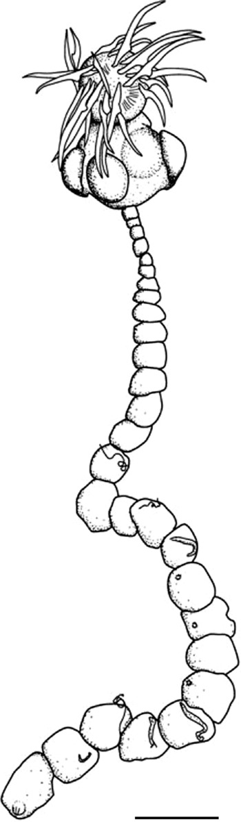
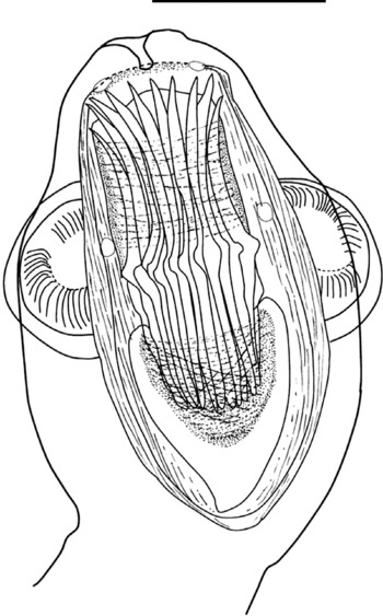
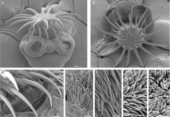
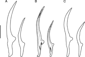
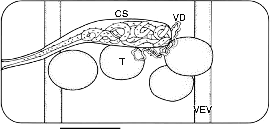
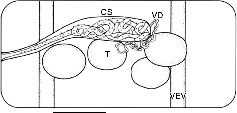
Worms (figs 1–5) very small, 0.96–2.97 (1.80 ± 0.59) mm in length; maximum width 92–203 (124 ± 31) anterior to terminal proglottis. Strobila with 7–35 (16 ± 8) acraspedote proglottids (n = 18), including 2–11 (7 ± 4) immature proglottids (n = 8), and 7–20 (12 ± 4) protandric proglottids (with developed male reproductive organs) (n = 10). Scolex (figs 2 and ) sub-spherical, 239–401 (318 ± 43) long (n = 17), 191–339 (232 ± 35) wide (n = 20). Everted rostellum sub-spherical, thick-walled, muscular, 156–213 (185 ± 25) long (n = 5), 140–159 (148 ± 8) wide (n = 5), flattened on top with capilliform filitriches (fig. 3D) and covered on the sides in coniform spinitriches (fig. 3E). Rostellar apparatus cyclusteroid (see Bona, Reference Bona1994, p. 470), rostellar sheath sac-like, muscular, usually reaching well beyond posterior margin of suckers. Rostellum with two superficial muscular layers, the outer of which consists of slightly diagonal, spiral fibres. Robust rostellar hook pad with thin outer layer of transverse circular muscle fibres, and inner layer of spiral fibres. Sleeve of circular fibres in the wall of rostellar pouch slightly above level of suckers forming sphincter above hooks when rostellum invaginated, and beneath hooks when evaginated (fig. 2). Rostellum armed with double crown of 28 rostellar hooks of urceus pattern (Bona, Reference Bona1994); anterior hooks 157–186 (173 ± 5) long (n = 114), 15–25 (20 ± 2) wide at guard (n = 94); posterior hooks 111–128 (122 ± 3) long (n = 100), 10–19 (16 ± 2) wide at guard (n = 92). Blade around twice length of handle; anterior hooks blade:handle ratio 1.9–2.5 (2.2 ± 0.1), posterior hooks blade:handle ratio 1.7–2.6 (1.9 ± 0.2). Blade gently curved; handle slightly recurved at tip. Very fine (1 μm thick) epiphyseal thickenings between tip of handle and guard on posterior edge of handle (fig. 4). Four circular to sub-circular suckers 81–158 (102 ± 19) long (n = 42), 73–132 (90 ± 17) wide (n = 36), muscular, without spines, covered with scolopate spinitriches (fig. 3F) interspersed with capilliform filitriches (not shown). Proglottids, covered with scolopate spinitriches (fig. 3G); wider than long in anterior one-third; sub-square to circular in central one-third; posterior proglottids longer than wide; terminal proglottis considerably longer than wide (length:width = 1.3–9.9), in several cases an indistinct division giving the impression of terminal two proglottids having ‘fused’. Terminal 2–3 proglottids large and lack testes but eggs not discernible. Terminal proglottis in most specimens has weakly staining oval area that appears to be site of excretory pore where the four longitudinal excretory vessels conjoin (see fig. 1). Genital pores unilateral, left. Testes circular to oval 23–39 (30 ± 5) in diameter (n = 29), and arranged with one poral, one median and two aporal. Cirrus-sac strongly staining, oval, 56–63 (60 ± 3) long, 14–16 (16 ± 0.6) wide (n = 6), with short cylindrical poral region and rounded antiporal region, almost reaching level of aporal excretory vessels; passing dorsally to both ventral and dorsal excretory vessels (fig. 5). Evaginated cirrus long (c. 250) cylindrical, armed proximally with rosethorn-shaped spines 8–9 (8.8 ± 0.4) base length, 5–7 (6.2 ± 0.8) height; distal region appears tapered and less heavily armed, although this is difficult to confirm due to contortions of the cirrus. Vas deferens around antiporal end of cirrus sac; long and highly convoluted.
Paradilepis cf. minima: entire worm, fixed and stained specimen showing haphazard arrangement of hooks on this specimen. Scale bar: 100 μm.

Cyclusteroid rostellar apparatus in a slightly everted specimen. The small circles in the pouch wall are contracted bundles of longitudinal muscles from the neck that penetrate the pouch. The lateral muscle fibres across the blade of the hooks form a sphincter behind the hooks when fully everted, and in front of the hooks when fully inverted. Scale bar: 100 μm.

Electron micrographs of Paradilepis cf. minima: (A) scolex showing everted rostellum; (B) rostellum showing the double crown of hooks; (C) rostellum and scolex showing the transition between the different types of microtriches; (D) apex of the rostellum showing capilliform filitriches; (E) rostellum showing coniform spinitriches; (F) sucker surface showing scolopate spinitriches; (G) strobila showing scolopate spinitriches. Scale bars: A–C, 10 μm; D–G, 1 μm.

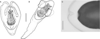
Rostellar hooks, large and small of (A) P. minima, (B) P. cf. minima, (C) P. sp. of Clark. Note the fine epiphyseal thickenings on the large hook of P. cf. minima. Scale bar: 50 μm.

Dorsal view of mature male proglottid: VEV, ventral excretory vessel; T, testis; CS, cirrus sac; VD, vas deferens. Scale bar: 50 μm.
Description of metacestode
Based on nine specimens from G. cotidianus McDowall. Hook measurements based on three unmounted specimens.
Metacestodes (fig. 6A–C) lacking primary lacuna, with scolex invaginated, without cercomer; ‘merocercoid’ of Chervy (Reference Chervy2002), or ‘cercoscolex’ of Jarecka (Reference Jarecka1970). Metacestodes with scolex invaginated 456–523 (489 ± 47) long, 240–261 (250 ± 15) at widest part of body (n = 2), heart-shaped. With scolex evaginated 713–1189 (984 ± 207) long, 313–457 (359 ± 67) wide (n = 5), elongated. With rostellum everted 723–779 (751 ± 40) long, 257–278 (267 ± 15) wide (n = 2). Widest at level of suckers. Suckers circular to oval 107–121 (116 ± 8) long, 81–109 (92 ± 15) wide (n = 3). Rostellum bearing 28 hooks in two circles. Anterior hooks (n = 27) 160–180 (167 ± 5.6) long; width at guard 19–25 (21 ± 1.5); blade 111–125 (116 ± 3.2) long; handle 48–60 (54 ± 3.6) long; blade:handle ratio 1.9–2.5 (2.2 ± 0.14). Posterior hooks (n = 19) 115–130 long (121 ± 3.3); width at guard 14–19 (17 ± 1.2); blade 75–90 (79 ± 3.6) long; handle 34–47 (43 ± 2.8) long; blade:handle ratio 1.7–2.6 (1.9 ± 0.20). Metacestodes enveloped in a close-fitting cyst, which is shed when worms first evert their scolex. This transparent sheath connected to the excretory pore at posterior extremity of the worm, which can clearly be seen as an invagination in the sheath (fig. 6C). Multiple encystment (1–10 metacestodes per cyst) observed.
Paradilepis cf. minima metacestode. (A) Metacestode with scolex inverted. (B) Metacestode with scolex everted. (C) Posterior of a newly everted metacestode, shedding its close-fitting cyst, showing the path of the excretory pore. Scale bars: A, B, 100 μm; C, 100 μm.

Remarks
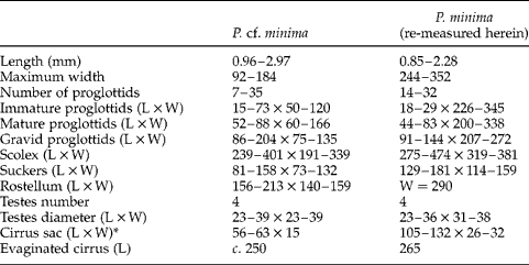
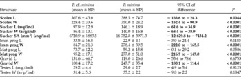
The genus Paradilepis has a complex taxonomic history. In the literature's most comprehensive list Schmidt (Reference Schmidt1986) listed 20 species under the genus. Of these, seven had previously been synonymized or re-assigned (Joyeux & Baer, Reference Joyeux and Baer1950; Spassky, Reference Spassky1961, Reference Spassky1963; Bray, Reference Bray1974) and a further one was re-assigned subsequently (Murai & Georgiev, Reference Murai and Georgiev1987). Paradilepis phalacrocoracis Ukoli, 1968 is a species inquirenda, as the diagnosis was based upon a specimen without a scolex. On the basis of descriptions, we recognize the remaining 12 species from Schmidt's list as members of Paradilepis: P. caballeroi Rysavy & Macko, 1973, P. delachauxi (Fuhrmann, 1909), P. diminuta Huey & Dronen, 1981, P. kempi, P. longivaginosus, P. maleki Khalil, Reference Khalil1961, P. minima, P. patriciae Baer & Bona, Reference Baer and Bona1960, P. rugovaginosus Freeman, Reference Freeman1954, P. scolecina, P. simoni Rausch, 1949, and P. urceus (Wedl, 1855). We also recognize P. urceina Bona, 1975, which was not mentioned by Schmidt. For the purposes of identifying the new specimens from New Zealand, the following species can be eliminated because of the number of rostellar hooks: P. caballeroi (24 hooks), P. delachauxi (20–22), P. diminuta (20), P. maleki (20), P. patriciae (20), P. rugovaginosus (32), P. scolecina (20), P. urceus (20) and P. urceina (20). The remaining four species possess 28 hooks. Of these, the rostellar hooks of P. longivaginosus and P. simoni are not comparable in shape, and are considerably smaller than those of P. cf. minima. Paradilepis kempi has hooks that resemble those of the new specimens in both size and shape, but the entire worm is considerably longer in adult size (50–130 mm) than P. cf. minima (0.96–2.97 mm). Even allowing for the fact that our worms may not be fully developed in terms of final length, this is an order of magnitude difference and a length that is highly unlikely to be attained by these worms. This leaves P. minima as a possible conspecific for the new specimens. Since Goss's original description (Goss, Reference Goss1940) confused specimens of P. minima and P. scolecina and is therefore unreliable (see Clark, Reference Clark1957), we rely on Clark's redescription and our own measurements of her specimens for comparison. Paradilepis minima has 28 rostellar hooks of a similar size and shape (fig. 4), and bears the closest resemblance to P. cf. minima (table 1). However, a comparison of measurements of the scolex and proglottids shows significant differences between P. minima and P. cf. minima (referred to as morphs from hereon) (table 2). For this study, we re-measured Clark's (1957) specimens (n = 7), and in table 2 measurements are compared between the morphs using two-tail unpaired t-tests with uneven variance, with 95% confidence intervals, of the difference between morphs, bound away from 0 when differences are significant. In P. minima the scolex is larger in both length (P = 0.0044) and width (P < 0.0001), and the suckers are significantly larger (P < 0.0001) (table 2). The width of all proglottids is greater in P. minima (P < 0.0001) (table 2), which is manifest in the visual difference of typically rectangular proglottids in P. minima, compared to almost square or circular proglottids in P. cf. minima. Along with her redescription of P. minima, Clark (Reference Clark1957) included a brief description of an unidentified species of Paradilepis, which is around 10 mm long, but incomplete. We have examined some slides of these specimens (although not the entire scolex) and conclude that, although the number, shape and size of the hooks are consistent with P. cf. minima (fig. 4), nonetheless Clark's Paradilepis sp. is too large to be conspecific with P. cf. minima.
Morphometric and meristic data comparing Paradilepis cf. minima with P. minima specimens of Clark (Reference Clark1957). All measurements in micrometres unless indicated otherwise and represented by the range.
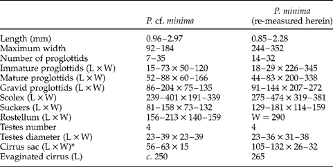
* Cirrus sac measurement not used for formal comparison with P. minima; see text. L, length; W, width.
Comparison of selected measurements (micrometres unless specified otherwise) between Paradilepis cf. minima and Clark's (Reference Clark1957) P. minima using two-tail unpaired t-test with unequal variance. Statistical significance set at P<0.05 and highlighted in bold.

Avg/ind, average per individual; Imm, immature; L, length; Mat, mature; prog, proglottids; SA, surface area; SD, standard deviation; W, width.
Molecular analysis
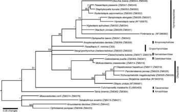
The tree resulting from the SSU sequences is somewhat equivocal, placing Paradilepis in a polytomy. Neither Paradilepis nor Neogryporhynchus are included in the Dilepididae clade, confirming the Gryporhynchidae as a family separate from the dilepidids (fig. 7). The fact that these two genera do not form a clade between themselves suggests considerable unresolved variation between them or possible long-branch attraction. Furthermore, consistent with Mariaux (Reference Mariaux1998), our tree suggests the exclusion of the genus Mesocestoides Vaillant, 1863 from the Cyclophyllidea (fig. 7).
Bayesian inference tree (50% majority-rule tree) based on DNA sequence data (1032 bp) of the SSU gene showing the equivocal position of Paradilepis cf. minima and Neogryporhynchus within the Cyclophyllidea. Vertical bars indicate families. Nodal support expressed as posterior probabilities with * indicating 100% posterior probability.

Discussion
The genus Paradilepis is characterized by a rostellum with a double row of hooks, of urceus or scolecina pattern; unilateral genital pores; genital ducts dorsal to excretory vessels; genital organs without accessory structures; vagina ventral to cirrus sac; uterus sacciform; four testes; and cirrus with rosethorn spines (Bona, Reference Bona1994). The specimens collected as metacestodes from the bully Gobiomorphus cotidianus, and grown in vitro, possess these characteristics where discernable, and were therefore assigned to this genus. On the basis of their length, size and number of hooks, scolex and sucker size, and width of proglottids, these specimens are considered probably distinct from previously described species. However, because the measurements are taken from in vitro cultured specimens, which may not be comparable to worms from the natural host, we refrain from naming them as a new species and describe them herein as P. cf. minima.
Species of the genus Paradilepis show a strong host affinity for piscivorous bird species of the Phalacrocoracidae Reichenbach, 1850, and it is almost certain that P. cf. minima will be found as an adult in the intestines of one or both of New Zealand's inland shags; the black shag (Phalacrocorax carbo novaehollandiae Stephens) and the little pied shag (P. melanoleucos brevirostris Gould) (shag taxonomy follows the findings of Kennedy et al., Reference Kennedy, Gray and Spencer2000, Reference Kennedy, Valle and Spencer2009). These species are found frequently in fresh and brackish bodies of water, often well away from the coast, and subsist on a varied diet of fish and invertebrates, including bullies and other native fish. In studies of shag diets, little pied shags preferred Gobiomorphus species at Lake Taupo (Falla & Stokell, Reference Falla and Stokell1945) and bullies made up the main part of the diet in black shags at Lake Ellesmere and Taupo district (Falla & Stokell, Reference Falla and Stokell1945; Dickinson, Reference Dickinson1951; Boud & Eldon, Reference Boud and Eldon1960). Shag species in New Zealand have full protection so it has not been possible to cull any individuals to prospect for adult worms.
Although cestodes have been cultured in vitro for decades, such methods have principally been used for exploring physiological and biochemical aspects of certain model species, usually important as human pathogens (e.g. Barber & Scharsack, Reference Barber and Scharsack2010; Hemphill, Reference Hemphill2010; Hoole et al., Reference Hoole, Carter and Dufour2010; Willms & Zurabian, Reference Willms and Zurabian2010). Only a few workers have used growth in an artificial medium as a way to identify species discovered as larval stages (Hamilton & Byram, Reference Hamilton and Byram1974; Campbell & Carvajal, Reference Campbell and Carvajal1979; Carvajal et al., Reference Carvajal, Barros and Santander1982; Chambers et al., Reference Chambers, Cribb and Jones2000; Holland et al., Reference Holland, Campbell, Garey, Holland and Wilson2009). Those studies that have been able to grow larval stages into adult cestodes have all concentrated on tetraphyllidean species, and this is the first study, that- we are aware of, to use in vitro cultured worms to aid in identification of a non-tetraphyllidean. Provided that our metacestodes received an initial trypsin ‘shock’, our simple growth medium was sufficient for metacestodes to evaginate, grow proglottids and mature into protandric worms. However, some sub-adult worms, while remaining alive, possibly reacted to the unnatural medium by developing ‘ballooned’ proglottids, suggesting, perhaps, that the osmotic balance of the proglottis had been disrupted. The organs within these proglottids may have been displaced by the ‘ballooning’ effect, but the proglottids became transparent and the maturing male components more visible. However, the square to circular shape of most mature proglottids appears to be real (Clark, Reference Clark1957, mentions that mature segments of P. minima resemble a ‘string of beads’), and the distinctive elongated terminal proglottis is found in every specimen from 14 days old, and even in many newly excysted juveniles, and is therefore likely to be real. If worms cultured in artificial media indeed grow naturally so as to be comparable to those existing in nature, there is much to be learned in terms of ontogeny. In particular, we examined preliminarily the size progression of rostellar hooks and suckers from metacestode to 23-day-old ‘adults’. Although we only found a statistically significant positive correlation between small hook length and age (slope 0.37 ± 0.10; r = 0.747; P = 0.0021), our sample size was small (n = 14) and it would be of interest, with a larger dataset, to compare the allometry of small hooks, large hooks and suckers. Such data could prove a useful tool for ecological studies where identification of metacestodes is based on hook length alone.
Being able to grow live metacestodes to maturity should bypass the difficulty of species determination. Metacestodes have little internal morphology with which to identify them, and identification has always rested on the number, size and shape of the rostellar hooks (Ching, Reference Ching1982; Scholz & Salgado-Maldonado, Reference Scholz and Salgado-Maldonado2001; Scholz et al., Reference Scholz, Bray, Kuchta and Repova2004, Reference Scholz, Boane and Saraiva2008). In addition, hooks can only satisfactorily be observed when the entire worm is squashed, so preserved and whole-mounted metacestodes are almost impossible to use for taxonomic purposes. Until true adults are found in the definitive host, we retain some caution about interpreting ‘adults’ grown in vitro, because we cannot, at this stage, know if or how the artificial conditions alter the development of the worm. For instance, our preserved, in vitro-grown worms appear to be lacking female reproductive organs and eggs. The cirrus sac length appears to differ significantly between P. cf. minima and P. minima (table 1), but we have not used this as a potential diagnostic character, because of the difficulty of measuring the sac in specimens that were not perfectly stained and mounted completely flat. Measuring the cirrus sac can be made difficult by a number of factors: (1) the cirrus sac can appear to be twisted and bent in some specimens; (2) it is rarely seen neatly dorso-ventrally flattened; and (3) the size varies according to the maturity of the proglottis and also according to how far the cirrus is everted. More complete data will emerge if and when adults are found in the definitive host, when it should be possible to erect a new species formally.
The presence of epiphyseal thickenings in this case is notable because they are rarely reported for Paradilepis; indeed, Ryzhikov et al. (Reference Ryzhikov, Rysavy, Khokhlova, Tolkatcheva and Kornyushin1985) state as part of their generic diagnosis, ‘two rows of hooks of parvoid type (without secondary growths of handle and guard)’. While epiphyseal thickenings are clearly a distinctive feature of some gryporhynchid genera (e.g. Cyclustera and Glossocercus; Scholz & Salgado-Maldonado, Reference Scholz and Salgado-Maldonado2001; Scholz et al., Reference Scholz, Kuchta and Salgado-Maldonado2002) they are illustrated only sporadically in the literature for Paradilepis (Baer & Bona, Reference Baer and Bona1960; McLaughlin, Reference McLaughlin1974; Scholz & Salgado-Maldonado, Reference Scholz and Salgado-Maldonado2001; Scholz et al., Reference Scholz, Kuchta and Salgado-Maldonado2002) and mentioned in the text on only one occasion (the ‘flattened, rounded appendages’ of Mahon, Reference Mahon1955). This is undoubtedly because, in this genus, the thickenings are very fine and difficult to observe.
A number of works have investigated the microtriches of cyclophyllidean taxa (for a comprehensive review see Chervy, Reference Chervy2009), but this is the first study to show these features in any species of Gryporhynchidae. Scanning electron micrographs show the rostellum to be covered with both capilliform filitriches (sensu Chervy, Reference Chervy2009), most dense in the centre and radiating outward in between the bases of the rostellar hooks (fig. 3B–D), and dense coniform spinitriches on the rostellar walls (fig. 3E). The scolex is covered with scolopate spinitriches (fig. 3F) interspersed with capilliform filitriches (not shown), whereas the strobila is covered only with scolopate spinitriches (fig. 3G). Further comparison with additional gryporhynchid taxa is necessary to determine the universality of these structures within the family.
An interesting characteristic of P. cf. minima is multiple encystment. Frequently, more than one metacestode is found inside a shared capsule within the mesenteries of the fish. The soft, loosely fitting capsule in which they are found is mostly or entirely of host origin – certainly small blood vessels and melanophores are often incorporated into the outer wall (B. Presswell, pers. obs.). When this is broken open, from one to ten metacestodes can be discovered inside. Where a fish has multiple infections, metacestodes are more likely to be found in a shared capsule than in individual capsules. This phenomenon is observed frequently in Sullivan's Dam specimens, where both prevalence and intensity of infection are higher than at Lake Waihola. Without evidence of asexual proliferation (there is no indication of budding on the inside of the capsule, for instance) we assume that the metacestodes are preferentially, or coincidentally, migrating to the same position in the fish host, where they are encapsulated together by the host tissue.
Our phylogenetic analyses support the contention that, if Mesocestoides is included in the Cyclophyllidea, the Order is paraphyletic, with Mesocestoides basal to Cyclophyllidea+Tetrabothriidea (Mariaux, Reference Mariaux1998). This finding was supported by morphological (Hoberg et al., Reference Hoberg, Jones and Bray1999) and spermatological (Justine, Reference Justine2001) characters, and earlier authors suggested promoting the Mesocestoididae to ordinal level. Our results endorse this position. A total evidence approach, however, found the Mesocestoididae to be a basal member of the Cyclophyllidea (Hoberg et al., Reference Hoberg, Mariaux and Brooks2001). The relationship between Paradilepis and Neogryporhynchus was equivocal, and more samples of gryporhynchids are needed to produce a clearer picture of relationships within the family. In particular, DNA from the Australian P. minima should show the relationship between that species and P. cf. minima, thus shedding light on the biogeography of the genus in Australasia.
Despite this being the first record of a gryporhynchid cestode from New Zealand, it seems likely that the lack of species reported from this country owes more to limited study than a dearth of suitable hosts (Poulin, Reference Poulin2004). There are relatively few species of freshwater fish native to New Zealand, but 87% of these are endemic, including all seven of the known Gobiomorphus species (McDowall, Reference McDowall2010). The New Zealand species are closely related to two congeneric species in Australia, and it seems likely that the genus arrived in New Zealand by dispersal (four species of bully are diadromous) (McDowall, Reference McDowall2010), thus providing a home for the larval stages of parasites carried by the shag species, which are found on both sides of the Tasman Sea. We think it possible that further gryporhynchid cestodes remain to be discovered in other bully species, or indeed, considering the broad range of fish families in which they occur (see above), in the other families of native fish such as the Galaxiidae.
Acknowledgements
Special thanks are due to Dr Leslie Chisholm of the South Australia Museum for lending material from Clark's collection and to Ms Liz Girvan of the OCEM for assistance with navigating the scanning electron microscope. Thank you also to Anya Gonchar for Russian translation. This work has been supported indirectly by the Marsden Fund (Royal Society of New Zealand) and a Zoology Department Performance-Based Research Fund (PBRF) Research Enhancement grant. The New Brunswick Museum provided funds to purchase Canada balsam. Finally, comments from the Parasitology Research Group at the University of Otago contributed to improving a previous version of this manuscript.