In the United States of America, around 1% (8.14 out of 1000) of patients are diagnosed with CHD in the first year of life, and over 90% of these patients are expected to reach adulthood. Reference Marelli, Ionescu-Ittu and Mackie1 In recent years, the prevalence of adults with CHD accounts for more than 50% of the total CHD population at any given time in developed countries. Reference Van der Bom, Bouma and Meijboom2,Reference Warnes, Liberthson and Danielson3 Adults with CHD have comorbidities such as hypertension, diabetes, obesity, and chronic kidney disease, which may increase their operative risk. Reference Agarwal, Sud and Menon4 This is especially important as an increasing number of adults with CHD undergo surgeries as reported by the Society of Thoracic Surgeons Congenital Heart Surgery Database Reference Jacobs, Mavroudis and Quintessenza5 . Zomer et al reported that during a 15-year period, one-fifth of patients living with CHD required surgery for CHD in adulthood and 40% of these surgeries were reoperations. Reference Zomer, Verheugt and Vaartjes6 Prognostic scoring indices that can predict outcomes in adults undergoing surgery for CHD may allow timely intervention and allow better communication in the early post-operative period. Reference Gaies, Gurney and Yen7,Reference Gaies, Jeffries and Niebler8 A few studies have shown that peak vasoactive-inotropic score (VIS) during the first 24 hours had a strong correlation with post-operative morbidity and mortality in infants and adolescents (12–17 years). Reference Gaies, Jeffries and Niebler8,Reference Garcia, Walters and Delius9 Vasoactive-ventilation-renal (VVR) score, a marker of cardiovascular, pulmonary, and renal function, has been reported to predict perioperative outcomes in children. Reference Miletic, Spiering and Delius10–Reference Scherer, Moser and Brown12 However, there is a lack of data validating the prognostic role of the VVR score in adults following surgery for CHD. Therefore, we aim to evaluate the prognostic utility of VVR in adults with CHD who underwent surgery involving cardiopulmonary bypass.
Materials and methods
This is a single-centre retrospective chart review including patients ≥ 18 years of age with CHD who underwent cardiac surgery using cardiopulmonary bypass from December, 2008 to January, 2017. Patients who required extracorporeal membrane oxygenation, renal replacement therapy, required reoperation, or died within the first 48 post-operative hours were excluded. Patients admitted following heart transplantation, or those who had incomplete medical records were also excluded from the study. Heart transplant patients were excluded from the study since this is not a surgical repair or palliation for CHD. The Institutional Review Board at Wayne State University and the Detroit Medical Center approved this study.
Data collection
Demographic and clinical data included age, gender, race, weight, height, number of prior surgeries, and cardiac surgical procedures performed during the admission. Surgical procedures were categorised using the Society of Thoracic Surgery and European Association for Cardiothoracic risk stratification system (STAT categories), with category 1 having the lowest risk and 5 having the highest risk for mortality. Reference O’Brien, Clarke and Jacobs13
Several perioperative variables were also collected, including cardiopulmonary bypass duration, aortic cross-clamp duration, length of hospitalisation, duration of intubation, and inotropic support. Arterial blood gases and lactate measurements were obtained at the discretion of the cardiac intensive care team. Also, clinical management and use of inotropes and ventilation were at the discretion of cardiac intensive care team. Data including the doses of various vasoactive agents, ventilator settings, and serum creatinine during the first 48 post-operative hours were collected. Various ventilatory parameters were collected at the time of recording of arterial blood gases and included respiratory rate, fraction of inspired oxygen, partial pressure of carbon dioxide, peak inspiratory pressure, positive end-expiratory pressure, and mean airway pressure. The doses of each inotropic and vasopressor were recorded immediately on arrival to ICU, and then every 6 hours for the first 48 hours.
Our primary outcome variable was prolonged hospital length of stay and was defined as hospital length of stay greater than 75th percentile for the cohort. Our secondary outcomes were duration of mechanical ventilation and vasoactive drug requirement.
Calculation of prognostic scoring indices
The VIS was calculated for every patient, first on arrival to cardiac ICU, and then every 6 hours for the first 48 hours post-operation.
The VIS was calculated in the following manner:
 $$\eqalign{ {\rm{VIS}} = {\rm{dopamine}}\ {\rm{dose}}\ \left( {{\rm{ug}}/{\rm{kg}}/{\rm{minute}}} \right) \cr \ \ \ \ \ \ \ \ + {\rm{dobutamine}}\ {\rm{dose}}\left( {{\rm{ug}}/{\rm{kg}}/{\rm{minute}}} \right) \cr \ \ \ \ \ \ \ \ + {\rm{1}}00 \times {\rm{epinephrine}}\ {\rm{dose}}\ \left( {{\rm{ug}}/{\rm{kg}}/{\rm{minute}}} \right) \cr \ \ \ \ \ \ \ \ + {\rm{1}}0 \times {\rm{milrinone}}\ {\rm{dose}}\ \left( {{\rm{ug}}/{\rm{kg}}/{\rm{minute}}} \right) \cr \ \ \ \ \ \ \ \ + {\rm{1}}0\ 000 \times {\rm{vasopressin}}\ {\rm{dose}}\ \left( {{\rm{U}}/{\rm{kg}}/{\rm{minute}}} \right) \cr \ \ \ \ \ \ \ \ + \\ {\rm{1}}00 \times {\rm{norepinephrine}}\ {\rm{dose}}\ \left( {{\rm{ug}}/{\rm{kg}}/{\rm{minute}}} \right){\hbox {\hbox {\hbox {\hbox .}}}} \cr} $$
For patients with no vasoactive support at any time point, the VIS equaled zero.
The Ventilation Index was calculated for every VIS measurement, and are as follows:
 $$\eqalign{ {\rm{Ventilation}}\ {\rm{Index}} = \!{\rm{Respiratory}}\ {\rm{rate}} \times \!({\rm{Peak}}\ \!{\rm{inspiratory}}\ \!{\rm{pressure}} \cr \ \ \ \ \ \ \ \ \ \ \ \ \ \ \ \ \ \ \ \ \ \ \ \ \ -{\rm{Positive}}\ {\rm{end}} - {\rm{expiratory}}\ {\rm{pressure}}) \cr \ \ \ \ \ \ \ \ \ \ \ \ \ \ \ \ \ \ \ \ \ \ \ \ \ \times {\rm{Partial}}\ {\rm{pressure}}\ {\rm{of}}\ {\rm{carbon}}\ {\rm{dioxide}}/{\rm{1}}000{\hbox {\hbox {\hbox {\hbox .}}}} \cr} $$
For patients who were not intubated, ventilation index equaled zero at the respective time point.
Pre and post-operative measurements of serum creatinine with every vasoactive-inotropic score were also recorded. For patients whose post-operative serum creatinine values were less than baseline, we assumed ΔCr = 0.
The VVR score was calculated for all the time points: Reference Miletic, Spiering and Delius10
 $${\rm{VVR score = VIS}}\ {\rm{ + }}\ {\rm{Ventilatory\ Index + }}\left( {{{\rm{\Delta} CrX10}}} \right){\rm{{\hbox {\hbox {\hbox {\hbox {\hbox .}}}}}}}$$
We defined VVR at zero hours as the score calculated at the time of patient arrival to cardiac ICU. Peak VVR score during the first day was defined as the highest score obtained any time during the first 24 hours in cardiac ICU, which did not include the score taken at zero hours but included the measurements taken at 6 and until 24 hours after arrival to cardiac ICU. Peak VVR during the second day was defined as the highest score obtained between 30 and 48 hours after arrival to cardiac ICU.
Statistical analysis
All statistical analysis was performed using SPSS 25 software for PC (IBM Inc.) in the Division of Pediatric Cardiology. Continuous variables were expressed as mean with interquartile range or median (range), and the categorical variables were expressed in absolute counts with percentage. The entire cohort was divided into two groups with and without prolonged length of stay. The Student’s t-test and the Chi-square test were used as appropriate for statistical comparisons between the groups with and without prolonged length of stay. The receiver operator curves were generated for each significant score at various time points. Paired comparison of the ROC curves by the method of DeLong et al was performed. Reference DeLong, DeLong and Clarke-Pearson14 Logistic regression analysis was used to evaluate the association between the VVR score and all other statistically significant variables in relationship to our primary outcome and prolonged length of stay. Significance was taken as a p-value of < 0.05.
Results
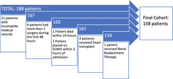
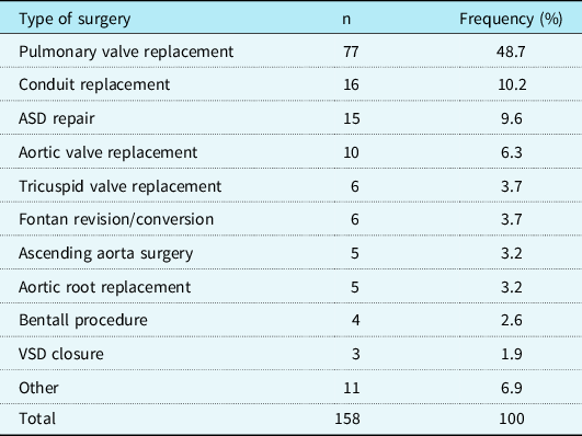
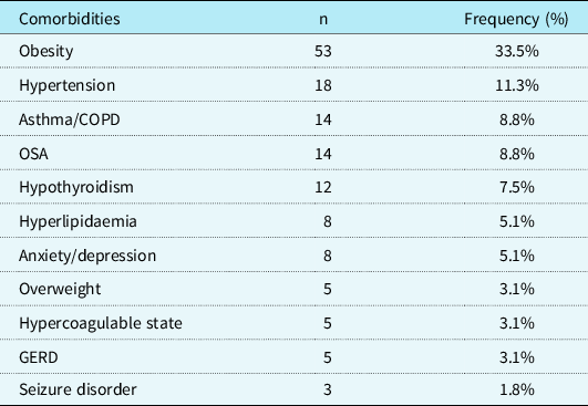
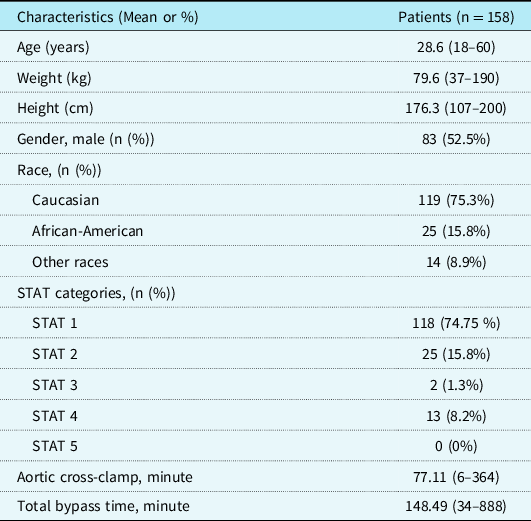
A total of 188 charts were reviewed, of whom 30 patients were excluded after applying the exclusion criteria (Fig 1). Our study cohort included 158 patients, with a median age of 28.6 years (range 18–60.2 years), and 83 (52.5%) were males. The cohort included 119 Caucasian patients (75.3%), 25 African-American patients (15.8%), and the remainder 14 patients of other races (8.9%). There were 87 (55%) patients who had 2 or less surgeries and 71 (45%) patients had more than 2 surgeries in the past. There were 19 (12%) patients who had no prior surgery. Table 1 depicts the most common index surgeries, from which VVR scores were calculated. The most frequent comorbidities in our cohort were obesity and hypertension. Sixty-five (41%) patients had 2 or more comorbidities and 38 (24%) patients had no comorbidities (Table 2). According to the STAT cardiothoracic risk stratification system, 118 (74.7%) patients were in category 1, 25 (15.8%) patients in category 2, 2 (1.3%) patients in category 3, 13 (8.2%) in category 4, and there were no patients in category 5. The baseline demographic and clinic characteristics of the entire cohort are depicted in Table 3.
Exclusion criteria.

Type of surgical procedures.

Most frequent comorbidities.

COPD=Chronic obstructive pulmonary disease; GERD=Gastroesophageal reflux disease; OSA=Obstructive sleep apnea.
Baseline demographic characteristics.

Data are presented as n (%) for categorical variables and mean (25th–75th percentiles) for continuous variables.
STAT=Society of Thoracic Surgery and European Association for Cardiothoracic risk stratification.
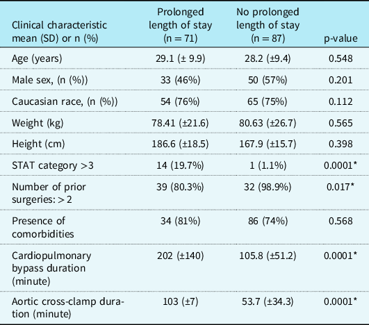
Seventy-one patients had prolonged length of stay, defined as hospital stay equal to or more than 8 days (median length of stay: 5, IQR: 4–8). The groups with and without prolonged length of stay were not different in their baseline characteristics including age, gender, and height and weight (Table 4). There was no difference between the groups with and without comorbidities in regards to prolonged length of stay (p = 0.568). However, the group with prolonged length of stay had a significantly higher number of prior surgeries (>2 prior surgeries, 39 versus 32; p = 0.017) and higher baseline risk for mortality by the STAT cardiothoracic risk stratification system (STAT category >3, 14 versus 1; p < 0.001). Similarly, in the group with prolonged length of stay, the cardiopulmonary bypass duration (202 ± 140 minutes versus 106 ± 51 minutes) and aortic cross-clamp duration (103 ± 7 minutes versus 54 ± 34 minutes) were significantly (p = 0.005) longer compared to the group without prolonged length of stay.
Patients with and without prolonged length of stay.

SD=Standard deviation; STAT=Society of Thoracic Surgery and European Association for Cardiothoracic risk stratification.
*means statistical significant.
Prognostic indices
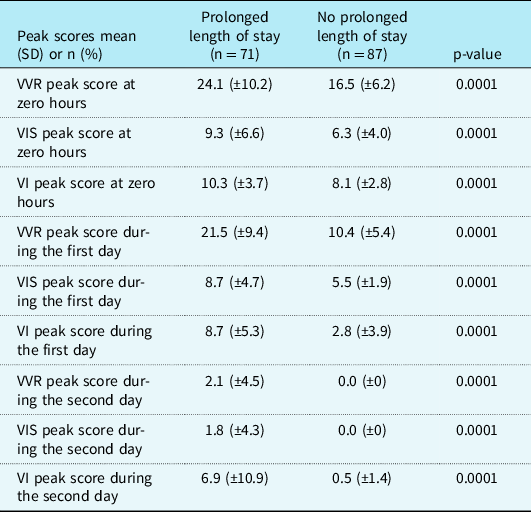
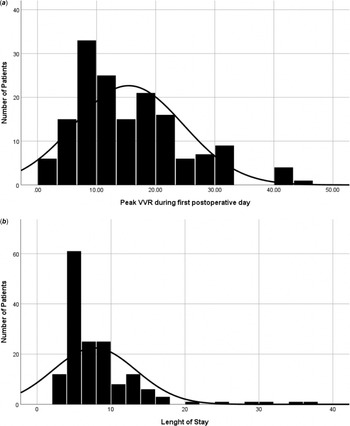
The peak VVR score, peak VIS, and peak ventilation index scores were significantly higher in the group with prolonged length of stay at all time points during the first 48 hours (Table 5). Figure 2a and b depict the distribution of the VVR values and length of stay in our cohort.
Prognostic indices between groups with and without prolonged length of stay.

SD=Standard deviation, VI=Ventilator index; VIS=Vasoactive inotropic support; VVR=Vasoactive-ventilator-renal.
(a and b ) Vasoactive-ventilation-renal score. Length of stay.

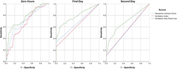
At zero hours of arrival to the cardiac ICU, the area under the curve for peak ventilation index was 0.674 (p = 0.0001), for peak VIS was 0.710 (p = 0.0001), and for peak VVR score was 0.766 (p = 0.0001). During the first day, the area under the curve for peak ventilation index was 0.815 (p = 0.0001), for peak VIS was 0.735 (p = 0.0001), and for peak VVR score was 0.847 (p < 0.0001). For the peak score during the second day, the area under the curve for ventilation index was 0.556 (p = 0.224), for VIS was 0.549 (p = 0.287), and for the VVR score was 0.692 (p = 0.0001). The biggest area under the curve within all the prognostic scoring indices calculated was for peak VVR during the first day by paired comparison of the ROC curves. The ROC curves are illustrated in Figure 3 and Table 5.
Receiver operative curves for peak prognostic scoring indices.

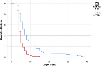
The first post-operative day peak VVR score ≥13 had a sensitivity of 81% and a specificity of 75% for predicting prolonged length of stay. We then divided the cohort into two; patients with a peak VVR score during the first-day score ≥13 and the rest. The group with peak VVR score during the first day ≥ 13 had an increased risk of prolonged time of intubation (OR: 34.8; 95% CI; 8, 151; p = 0.0001) and prolonged use of inotropic support (OR: 40.38; 95% CI; 9.28, 175.71; p = 0.0001).
Kaplan–Meier analysis was performed for patients with peak VVR score during the first day ≥ 13 as indicated in Figure 4 (logrank: p < 0.0001). A logistic regression was performed including the following variables: number of prior surgeries, Society of Thoracic Surgery and European Association for Cardiothoracic risk stratification system categories, aortic cross-clamp duration, peak VVR at zero hours, and peak VVR score during the first day. Only the peak VVR score during the first day was independently associated with our main outcome and prolonged length of stay (p < 0.0001).
Kaplan–Meier length of stay for VVR first day peak ≥ 13.

Discussion
Several perioperative prognostic score indices have been reported to predict morbidity and mortality in children who underwent cardiac surgery for CHD. Reference Garcia, Walters and Delius9–Reference Scherer, Moser and Brown12 However, the data regarding the use of these prognostic indices in adults with CHD during perioperative period is limited. Reference Yamazaki, Oba and Matsui15 Our data show that peak VVR score during the first day was a good predictor of prolonged length of stay in our study cohort. This strong association was the only one that remained valid after multivariate regression analysis, despite multiple other variables being significant in our initial univariate analysis. Peak VVR score during the first day ≥ 13 had strong sensitivity and specificity in predicting prolonged length of stay. Miletic et al reported an association of high VVR scores with poor outcomes in 222 infants with a median age of 107 days. Their results showed that VVR score at 48 hours (median, 38.3) was the best predictor of adverse outcomes in infants (duration of mechanical ventilation, duration of vasoactive agents, hospital length of stay, pediatric ICU length of stay, and duration of chest tubes). Reference Miletic, Spiering and Delius10 Scherer et al also reported that high VVR scores predicted length of stay of 168 children and adults with CHD who underwent surgery. VVR score at 12 hours post-operatively (median, 31) was a reliable predictor for prolonged length of stay. Reference Scherer, Moser and Brown12 Our cohort found that a lower cutoff peak VVR score value during the first day was associated with prolonged length of stay. This could be related to early extubation, less complex surgical interventions, and judicious use of inotropic support in the adult population. There has been a variable cutoff value for VVR scores in previous studies in order to predict adverse outcomes. Reference Miletic, Spiering and Delius10,Reference Scherer, Moser and Brown12,Reference Alam, Akunuri, Jain, Mazahir and Hegde16,Reference Mahan, Gupta, Aronoff, Bruni, Stevens and Moulick17 Miletic et al established the cutoff value of peak VVR score to be 22.5 at 48 hours in their pediatric population. Reference Miletic, Spiering and Delius10,Reference Miletic, Delius and Walters11 While Scherer et al reported a cutoff value of 25 for the peak VVR score at 12 hours in their population that included both children and adults with CHD, Garcia et al described a lower VIS cutoff value in an adolescent population with CHD. Reference Garcia, Walters and Delius9,Reference Scherer, Moser and Brown12 Similarly, a recent study by Yamazaki et al reported a lower cutoff value for VIS score in adults after surgery for CHD correlated with a prediction of adverse outcomes. Reference Yamazaki, Oba and Matsui15
The difference in the cutoff values compared to previous studies could be due to different age groups as our study cohort included only adult patients. There are few other possible reasons for the finding of higher peak VIS and VVR scores in the neonatal population. Reference Kumar, Sharma and Sethi18 First, there is a relatively higher complexity in the operative procedures performed during neonatal period compared to adults. Second, neonates and infants have reduced beta-adrenoceptor receptors due to downregulation induced by an elevated sympathetic tone and younger patients will need higher vasopressors doses, which may ultimately affect the VIS and VVR. Third, many adults are extubated in the operating room or early in the ICU. Reference Yamazaki, Oba and Matsui15,Reference Kozlik-Feldmann, Kramer and Wicht19,Reference Shahin, DeVarennes and Tse20
Regarding clinical applications of the VVR score, Osturk et al proposed that the VVR can be a strong early predictor of clinical results after surgery for CHD. Reference Ozturk, Tanidir and Güneş21 Similarly, we postulate that the VVR is relatively easy to calculate at bedside and it could provide the clinical team with a reliable estimate of length of stay. This will allow the ICU team to communicate and counsel families in a timely manner. In addition, this score is useful during patient handoff, providing a good summary to the incoming care team about the critical state of the patient. Moreover, it can help in classifying the patients by severity of the condition for future clinical research.
Limitations
This study has limitations including being a single-centre retrospective study with a small number of patients and most of them having a STAT category less than 3. In our institution, there is no set protocol to wean the inotropic support or extubate, which could affect the data. However, this study was performed in an institution with seven cardiac intensivists and consistent management practices during the study period. In addition, data collection and statistical analysis were limited to data already recorded in the medical records.
Conclusion
This study is, to our knowledge, the first one validating the VVR score exclusively in adult patients with CHD. Our study validated the role of VVR as a good clinical tool and predictor of prolonged length of stay in adults undergoing surgery for CHD. A peak VVR score during the first day ≥ 13 after surgery demonstrated the best prognostic utility to predict prolonged length of stay. This information can help clinicians in their communication with the patients and their families. Further prospective studies are needed to evaluate the role of the bedside VVR score during the transition of care, and if it can serve to classify patient severity in this population.
Acknowledgements
None.
Financial support
This research received no specific grant from any funding agency, commercial, or not-for-profit sectors.
Conflicts of interest
None.
Ethical standards
The authors assert that all procedures contributing to this work comply with the ethical standards of the relevant national guidelines on human medical regulations and with the Helsinki Declaration of 1975, as revised in 2008, and has been approved by the ethical committee of Children's Hospital of Michigan.