Introduction
Species of Oesophagostomum, commonly known as nodule worms, are parasitic nematodes in pigs and small ruminants, causing major economic impacts worldwide. (Pfukenyi & Mukaratirwa, Reference Pfukenyi and Mukaratirwa2013). More importantly, some Oesophagostomum spp. can also infect humans, causing oesophagostomiasis which is a significant public health problem in some countries (Legesse & Erko, Reference Legesse and Erko2004; Krief et al., Reference Krief, Vermeulen, Lafosse, Kasenene, Nieguitsila, Berthelemy, L'Hostis, Bain and Guillot2010). Although O. columbianun and O. venulosum were the most prevalent Oesophagostomum species in small ruminants in many countries (Jacquiet et al., Reference Jacquiet, Colas, Cabaret, Dia, Cheikh and Thiam1995; Sissay et al., Reference Sissay, Uggla and Waller2007), O. asperum is considered to be the predominant species in small ruminants in China, and causes significant economic losses to the small-ruminant industry (Chen et al., Reference Chen, Yang, Wang, Wang, Tian and Wen2010; Meng et al., Reference Meng, Gao, Wen, Du, Wang, Wang, Meng, Su, Hu, Jin and Yu2012).
The accurate analysis of genetic variation among nodule worm populations has important implications for studying molecular epidemiology and genetic structure of nodule worms, as well as for effective control of oesophagostomiasis. Currently, identification and differentiation of Oesophagostomum species have been based traditionally on morphological features of the adult worms (Bowman, Reference Bowman2009). However, these criteria are often insufficient for specific identification and differentiation, particularly at larval and/or egg stages (Dobson et al., Reference Dobson, Barnes, Birclijin and Gill1992; McManus & Bowles, Reference McManus and Bowles1996). DNA sequencing and analysing are relatively cheap and efficient for studies of genetic variations and population genetics. Mitochondrial (mt) DNA has been shown to provide useful genetic markers for molecular identification and differentiation of many nematodes (Li et al., Reference Li, Liu, Wang, Song, Lin, Zou, Liu, Xu and Zhu2013; Lin et al., Reference Lin, Liu, D'Amelio, Zhang, Song, Weng, Zou and Zhu2013; Zhao et al., Reference Zhao, Liu, Zhao, Cai, Zhu and Qian2013). In addition, the internal transcribed spacer of nuclear ribosomal DNA (ITS rDNA) is also an excellent tool for quickly distinguishing closely related nematode species (Blouin, Reference Blouin2002; Liu et al, Reference Liu, Zhou, Nisbet, Xu, Zhou, Zhao, Wang, Song, Lin and Zhu2014; Pekmezci et al., Reference Pekmezci, Onuk, Bolukbas, Yardimci, Gurler, Acici and Umur2014). Although mt cox3, nad5 and atp6 genes and ITS rDNA have been studied in O. asperum (Jia et al., Reference Jia, Zhao, Hu, Cheng, Du, Bian, Ma and Yu2014; Zhao et al., Reference Zhao, Hu, Song, Jia, Li, Wang, Lin, Xu, Yu and Deng2014), there is a paucity of information on the sequence variability in other mt genes of O. asperum.
The objectives of the present study were to examine sequence variability in mt cox1 and nad1 genes and ITS rDNA regions among O. asperum isolates from different endemic regions in Hunan Province, China. Based on the combined sequences of the pcox1 and pnad1, phylogenetic relationships of O. asperum with other Oesophagostomum species were also re-constructed.
Materials and methods
Extraction of DNA from adult worms
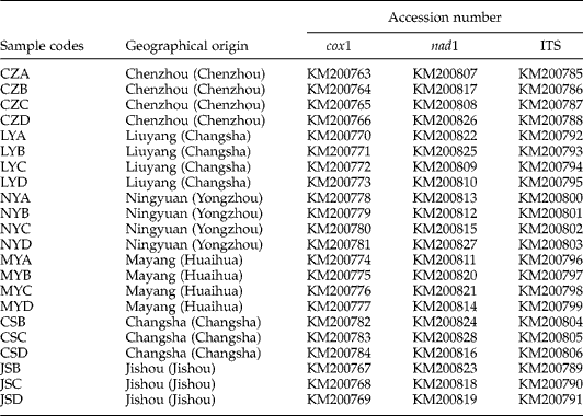
All adult worms of O. asperum (n= 22) were obtained from naturally infected goats from different localities in Hunan Province, China. These samples were washed separately in physiological saline, fixed in 70% (v/v) ethanol and stored at − 20°C until use. Total genomic DNA was extracted from individual samples using sodium dodecyl sulphate/proteinase K treatment, followed by spin column purification (WizardTM DNA Clean-Up, Promega, Madison, Wisconsin, USA) and eluted into 50 μl water according to the manufacturer's recommendations. The sample codes, geographical origins and GenBank accession numbers are listed in table 1.
Sample codes, geographical origins and GenBank accession numbers of Oesophagostomum asperum samples used in the present study.

Enzymatic amplification
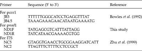
A portion of cox1 (pcox1), nad1 (pnad1) genes and ITS rDNA were amplified using primers listed in table 2. These primers were synthesized on a Biosearch Model 8700 DNA synthesizer (Shanghai, China). Polymerase chain reactions (PCR) (25 μl) were performed in 10 mm Tris–HCl (pH 8.4), 50 mm KCl, 4 mm MgCl2, 200 μm of each deoxynucleoside triphosphate (dNTP), 50 pmol of each primer and 2 U Taq polymerase (Takara Biotechnology, Dalian, China) in a thermocycler (Biometra, Göttingen, Germany) under the following conditions: an initial denaturation at 94°C for 5 min then 94°C for 30 s (denaturation); 55°C for 30 s (annealing); 72°C for 30 s (extension) for 37 cycles, followed by a final extension at 72°C for 5 min (final extension). These optimized amplification conditions for the specific and efficient amplification of individual DNA fragments were obtained after varying annealing and extension temperatures. One microlitre (5–10 ng) of genomic DNA was added to each PCR reaction. Samples without genomic DNA (no-DNA controls) were included in each amplification run. A 5 μl sample of each amplicon was examined by 0.8% (w/v) agarose gel electrophoresis to validate amplification efficiency. PCR products were sent to Sangon Company (Shanghai, China) for sequencing from both directions.
Sequences of primers used to amplify a portion of the cytochrome c oxidase subunit 1 gene (pcox1), NADH dehydrogenase subunit 4 (pnad1) and the nuclear internal transcribed spacers (ITS) from Oesophagostomum asperum.

Sequence analysis and phylogenetic reconstruction
Sequences of three mt genes were separately aligned using the software Clustal X 1.83 (Thompson et al., Reference Thompson, Gibson, Plewniak, Jeanmougin and Higgins1997). Pairwise comparisons were made of the level of sequence differences (D) among O. asperum isolates using the formula D= 1 − (M/L), where M is the number of alignment positions at which the two sequences have a base in common, and L is the total number of alignment positions over which the two sequences are compared (Chilton et al., Reference Chilton, Gasser and Beveridge1995).
The haplotypes, nucleotide diversity (Pi) and haplotype diversity (Hd) of each gene were determined using the DnaSP 5.0 program (Librado & Rozas, Reference Librado and Rozas2009). To study phylogenetic relationships with other Oesophagostomum species, isolates of O. dentatum (FM161882), O. quadrispinulatum (NC_014181), O. columbianum (KC715827) and O. asperum Shaanxi isolate (KC715826) were considered in the present study, with Cylicocyclus insignis (NC_013808) as the outgroup. The pcox1 and pnad1 sequences were aligned singly using Clustal X 1.83 and then combined, and the ambiguously aligned regions were excluded using Gblocks online server (http://molevol.cmima.csic.es/castresana/Gblocks_server. html) with the default parameters (Talavera & Castresana, Reference Talavera and Castresana2007) using the options for a less stringent selection. Three inference methods, namely Bayesian inference (BI), maximum likelihood (ML) and maximum parsimony (MP), were used for phylogenetic reconstructions. The GTR+I+G model of nucleotide evolution was selected as the most suitable model of evolution by JModeltest (Posada, 2008) based on the Akaike information criterion (AIC). BI was conducted with four independent Markov chains run for 1,000,000 metropolis-coupled MCMC generations, sampling a tree every 100 generations in MrBayes 3.1.1 (Ronquist & Huelsenbeck, Reference Ronquist and Huelsenbeck2003). The first 2500 trees were omitted as burn-in and the remaining trees were used to calculate Bayesian posterior probabilities (Bpp). ML analysis was performed with PhyML 3.0 (Guindon & Gascuel, Reference Guindon and Gascuel2003) using the subtree pruning and regrafting (SPR) method with a BioNJ starting tree, and the MtArt model of amino acid substitution with proportion of invariant sites (I) and gamma distribution (G) parameters estimated from the data with four discretized substitution rate classes, the middle of which was estimated using the median. Bootstrap frequency (Bf) was calculated using 100 bootstrap replicates. MP analysis was performed using PAUP* 4.0b10 (Swofford, Reference Swofford2002), with indels treated as missing character states. A total of 1000 random addition searches using tree bisection–reconnection were performed for each MP analysis. Bootstrap frequency (Bf) was calculated from 1000 bootstrap replicates with 10 random additions per replicate in PAUP. Phylograms were drawn using the TREEVIEW program version 1.65 (Page, Reference Page1996).
Results and discussion
Genomic DNA was isolated from individual nematodes from 22 different goats in Hunan Province, China (table 1). The pcox1, pnad1 and ITS rDNA were amplified individually from 22 O. asperum samples and subjected to agarose gel electrophoresis. The PCR products were approximately 450 bp, 750 bp and 850 bp (not shown) in size, respectively. For each DNA region, no size variation was detected on agarose gel among any of the amplicons examined, and no products were amplified from host (goat) DNA and no-DNA controls. The lengths of pcox1, pnad1 and ITS rDNA were 366 bp, 681 bp and 785 bp, respectively. The A+T contents of gene sequences were 71.5–72% (pcox1), 73.7–74.2% (pnad1) and 58–58.8% (ITS rDNA), consistent with previous reports of the A+T contents for parasitic nematodes (Li et al., Reference Li, Lin, Song, Sani, Wu and Zhu2008; Makouloutou et al., Reference Makouloutou, Setsuda, Yokoyama, Tsuji, Saita, Torii, Kaneshiro, Sasaki, Maeda, Une, Hasegawa and Sato2013; Wang et al., Reference Wang, Liu, Li, Xu, Ye, Zhou, Song, Lin and Zhu2013; Jia et al., Reference Jia, Zhao, Hu, Cheng, Du, Bian, Ma and Yu2014).
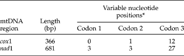
Intra-specific sequence variations within O. asperum were 0–1.6% for pcox1, 0–1.9% for pnad1 and 0–1.7% for ITS rDNA, while inter-specific sequence differences among members of the Oesophagostomum genus were significantly higher, being 11.1–12.5%, 13.3–17.7% and 8.5–18.6% for pcox1, pnad1 and ITS rDNA, respectively. Similarly, sequence variation has also been detected in other nematodes (Li et al., Reference Li, Liu, Wang, Song, Lin, Zou, Liu, Xu and Zhu2013; Lin et al., Reference Lin, Liu, D'Amelio, Zhang, Song, Weng, Zou and Zhu2013; Zhao et al., Reference Zhao, Liu, Zhao, Cai, Zhu and Qian2013; Liu et al, Reference Liu, Zhou, Nisbet, Xu, Zhou, Zhao, Wang, Song, Lin and Zhu2014; Pekmezci et al., Reference Pekmezci, Onuk, Bolukbas, Yardimci, Gurler, Acici and Umur2014). However, previous studies have indicated that the intra-specific sequence variations among O. asperum samples were 0–2.11%, 0–1.84% and 0–1.48% for pnad5, patp6 and pcox3, respectively, while inter-specific sequence differences among Oesophagostomum species in pig and small ruminants were 18–21.3% for pnad5, 18.3–24.5% for patp6 and 10.6–13.7% for pcox3 (Lin et al., Reference Lin, Liu, Song, Zhang, Li, Zou, Yuan, Weng and Zhu2012; Jia et al., Reference Jia, Zhao, Hu, Cheng, Du, Bian, Ma and Yu2014). For pcox1 and pnad1, intra-specific nucleotide variations were related mainly to changes at third codon positions, while fewer changes were detected at first and second codon positions (table 3), consistent with the results of other helminths, including cestodes, trematodes and nematodes (Li et al., Reference Li, Lin, Song, Sani, Wu and Zhu2008; Zhao et al., Reference Zhao, Mo, Zou, Weng, Lin, Xia and Zhu2009; Dai et al., 2012). Intra-specific nucleotide variations represented transitions (A ↔ G or C ↔ T; n= 12 for pcox1 and n= 31 for pnad1) and transversions (A ↔ C, A ↔ T and/or T ↔ G; n= 1 for pcox1 and n= 2 for pnad1). Nine haplotypes, nucleotide diversity (Pi = 0.009) and haplotype diversity (Hd = 0.81), were determined in all pcox1 sequences. Fifteen haplotypes, nucleotide diversity (Pi = 0.008) and haplotype diversity (Hd = 0.939), were determined in all pnad1 sequences. Haplotype diversity was higher in the nad1 gene than that in the cox1 gene. To our knowledge, this is first time that descriptions of Hd and Pi in O. asperum have been reported.
Number and position of codon variations in a portion of the mitochondrial cytochrome c oxidase subnit 1 (pcox1) and NADH dehydrogenase subunit 1 (pnad1) genes among Oesophagostomum asperum isolates in Hunan Province, China.
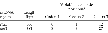
* Determination of the first codon position of each sequence relative to the complete mitochondrial DNA sequence of Oesophagostomum asperum (accession number: KC715826).
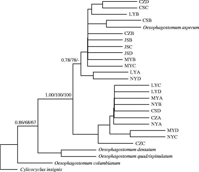
Phylogenetic analyses based on combined sequences of pcox1 and pnad1 among 22 individual O. asperum isolates from China, and four other nematodes including C. insignis as an outgroup, using Bayes, ML and MP analyses, are shown in fig. 1. Topologies of all trees inferred by different methods (Bayes, ML and MP) were identical or similar, with only small differences of Bpp and Bf values (fig. 1). In this tree, all O. asperum isolates grouped together, and the relationship of all isolates of O. asperum was closer to O. dentatum +O. quadrispinulatum than to O. columbianum. These results were not consistent with those of a previous study using ITS rDNA sequences (Jia et al., Reference Jia, Zhao, Hu, Cheng, Du, Bian, Ma and Yu2014). Within O. asperum, the samples from the same geographical region did not appear genetically closer than samples from other geographical locations, suggesting that there were no obvious geographical distinctions in O. asperum based on sequences of the two mtDNA regions. Furthermore, all isolates of O. asperum were clustered into two clades, with moderate support in BI and ML analyses and weakly supported in MP analysis (fig. 1). These results indicated that O. asperum samples from Hunan Province, China appeared to belong to different genotypes.
The phylogenetic relationship among Oesophagostomum asperum isolates with other Oesophagostomum species inferred by Bayesian inference (BI), maximum likelihood (ML) and maximum parsimony (MP) analyses using the combined dataset (cox1+nad1), with Cylicocyclus insignis as outgroup. See table 1 for sample codes; numbers along branches indicate bootstrap values in the order BI/ML/MP and all other values are below 50.
Species of Oesophagostomum parasitize the gastrointestinal tract of humans and animals, and can cause serious clinical disease (oesophagostomiasis). It is estimated that at least 1 million people are at risk of infection (Gasser et al., Reference Gasser, de Gruijter and Polderman2006), and approximately 70–99% of animals harbour Oesophagostomum infection (Romstad et al., Reference Romstad, Gasser, Monti, Polderman, Nansen, Pit and Chilton1997; de Gruijter et al., Reference de Gruijter, Blotkamp, Gasser, Amponsah and Polderman2006). The present study examined the sequence variability in pcox1, pnad1 and ITS rDNA, among O. asperum isolates from goats in Hunan province, China. The combined sequence of pcox1 and pnad1 was used effectively to infer the phylogenetic relationships of Oesophagostomum spp. These results provided useful information for studying population genetics of Oesophagostomum spp., and for the diagnosis and control of Oesophagostomum infections in animals and humans.
In conclusion, genetic variability among O. asperum isolates from different endemic regions in Hunan Province, China was revealed by sequences of mitochondrial cox1 and nad1 genes and ITS rDNA, with higher variation in ITS rDNA than in nad1 and cox1. The results of the present study have implications for studying molecular epidemiology and population genetics of O. asperum.
Financial support
Project support was provided in part by grants from the National Natural Science Foundation of China (Grant No. 31000901), and Outstanding Young Scientific Research Project of the Education Department of Hunan Province (Grant No. 14B092).
Conflict of interest
None.