



Early and correct differential diagnosis of cognitive impairment is of therapeutic and prognostic importance. However, post-mortem studies indicate that clinical diagnoses do not accurately reflect the underlying aetiology of cognitive impairment.Reference Beach, Monsell, Phillips and Kukull1 Frequent misdiagnoses of neurodegenerative cognitive impairment/dementia include other mental disorders, with 15–23% of people with Alzheimer’s disease initially receiving another psychiatric diagnosis and, conversely, around 30% of people with late-life depression (LLD) being erroneously diagnosed with Alzheimer’s disease.Reference Ismail, Smith, Geda, Sultzer, Brodaty and Smith2 In contrast to early-onset depression, people with LLD are more likely to present with impaired memory function and attentional difficulties, formerly referred to as ‘pseudodementia’.Reference Kiloh3,Reference Perini, Cotta Ramusino, Sinforiani, Bernini, Petrachi and Costa4 Moreover, evidence exists for LLD as both a prodrome of dementia (with depressive symptoms occurring in 30–50% of patients with Alzheimer’s disease) and risk factor for dementia.Reference Livingston, Huntley, Sommerlad, Ames, Ballard and Banerjee5,Reference Zubenko, Zubenko, McPherson, Spoor, Marin and Farlow6 Underlying mechanisms linking depression and dementia comprise vascular disease, alterations in glucocorticoids, hippocampal atrophy, increased deposition of β-amyloid plaques, apolipoprotein E-ε4 as a genetic link, an acceleration of cerebral ageing through stress-mediated inflammatory processes and deficits of neurotrophins.Reference Byers and Yaffe7,Reference Forero, Lopez-Leon, Gonzalez-Giraldo, Dries, Pereira-Morales and Jimenez8 In sum, the clinical diagnosis is challenging because of the close entanglement of both entities, underscoring the need for reliable non-invasive diagnostic biomarkers.
[18F]FDG PET in cognitive impairment and LLD
Molecular neuroimaging using positron emission tomography (PET) is an established method for diagnosing different forms of neurodegenerative dementia.Reference Chetelat, Arbizu, Barthel, Garibotto, Law and Morbelli9 By assessment of cerebral glucose metabolism, [18F]fluorodeoxyglucose (FDG) PET provides an imaging biomarker of neurodegeneration, which has been included in the consensus criteria of most neurodegenerative dementias (see Supplementary Material available at https://doi.org/10.1192/bjp.2025.67). Accurate diagnosis of dementia subtypes can be achieved by imaging disease-specific patterns of cerebral glucose metabolism with [18F]FDG PET.Reference Chetelat, Arbizu, Barthel, Garibotto, Law and Morbelli9,Reference Bohnen, Djang, Herholz, Anzai and Minoshima10
In contrast, metabolic alterations in depression are much more subtle, if present at all: in major depressive disorder, meta-analyses yielded increases as well as decreases of glucose metabolism, mainly in circumscribed frontolimbic regions.Reference Sacher, Neumann, Funfstuck, Soliman, Villringer and Schroeter11,Reference Su, Cai, Xu, Dutt, Shi and Bramon12 Systematic studies on metabolic alterations in LLD are rare and yielding conflicting results.Reference Kumar, Newberg, Alavi, Berlin, Smith and Reivich13,Reference Smith, Kramer, Ma, Kingsley, Dhawan and Chaly14 Recent PET studies explored the relationship between LLD symptoms and Alzheimer’s disease: patients with LLD with positive cerebrospinal fluid (CSF) Alzheimer’s disease biomarkers exhibited hypometabolism of Alzheimer’s disease-typical temporoparietal regions and cognitive decline during follow-up, whereas patients with negative Alzheimer’s disease biomarkers did neither.Reference Liguori, Pierantozzi, Chiaravalloti, Sancesario, Mercuri and Franchini15 Similarly, memory task-related metabolism was decreased in an Alzheimer’s disease-like parietal region in patients with LLD and amyloid PET-positive mild cognitive impairment (MCI), whereas amyloid-negative patients showed very limited frontal hypometabolism.Reference Lee, Youn, Hyung, Suh, Han and Eo16 The possible predictive value of [18F]FDG PET was further underlined by two recent studies demonstrating that the risk for cognitive decline to MCI or dementia was related to cortical hypometabolism in Alzheimer’s disease-typical regions rather than depression per se.Reference Krell-Roesch, Syrjanen, Vassilaki, Lowe, Vemuri and Mielke17,Reference Mauri, Gobbo, Princiotta Cariddi, Schiorlin and Versino18
In this light, it is surprising that evidence from formal studies for the discrimination of depressive pseudodementia is entirely lacking, as stated by European Association of Nuclear Medicine and European Academy of Neurology (EANM-EAN) task force for the use of [18F]FDG PET in neurodegenerative cognitive impairment/dementia.Reference Nestor, Altomare, Festari, Drzezga, Rivolta and Walker19 Still, experts voted unanimously for its use and encouraged further research on this topic. In line with aforementioned literature, they stated that a normal scan (aside from subtle frontal hypometabolism) offers strong evidence for pseudodementia, whereas a disease-specific neurodegenerative pattern argues against pseudodementia. It is worth noting that they restricted their recommendation to established dementia, and did not include very mild or subjective cognitive impairment.
Aims of the present study
The present study aimed to determine the impact and possible incremental diagnostic value of [18F]FDG PET for the differentiation between neurodegenerative and non-neurodegenerative cognitive impairment in a real-life gerontopsychiatric clinical population. To this end, both clinical and [18F]FDG PET diagnostic paths were reanalysed by independent and mutually blinded clinical and imaging experts. We assessed the rate of change and accuracy of clinical diagnosis after disclosing [18F]FDG PET results, with reference to a biomarker- and follow-up-supported, interdisciplinary consensus diagnosis. In response to the restriction imposed by the EANM-EAN task force (dementia only), we explored a possible effect of severity of cognitive impairment and, in addition, severity of depression and age on diagnostic performance.
Method
Standard protocol approvals, registrations and patient consents
All procedures contributing to this work comply with the ethical standards of the relevant national and institutional committees on human experimentation and with the Helsinki Declaration of 1975, as revised in 2013, and were approved by the ethics committee of the Albert-Ludwigs-University of Freiburg (proposal number 22-1523-S1-retro). Written informed consent was obtained from all patients. The trial was registered with the German Clinical Trials Register, under registration number DRKS00035787.
Patients
Between January 2018 and December 2022, 112 consecutive patients with depressive symptoms and cognitive dysfunction of uncertain aetiology were referred for [18F]FDG PET imaging from a tertiary psychiatric referral centre. A board-certified dementia specialist reviewed all medical charts and clinical information (including depression ratings, neuropsychological assessment, magnetic resonance imaging (MRI), CSF biomarker, amyloid PET, tau PET, dopamine transporter (DAT) single-photon emission computed tomography (SPECT)), except for [18F]FDG PET imaging. Clinical vignettes based on consensus criteriaReference Hellwig, Frings, Bormann, Vach, Buchert and Meyer20 summarised relevant information for clinical ratings.
Inclusion criteria comprised (a) depressive syndrome as ascertained by the Montgomery–Åsberg Depression Rating Scale ((MADRS) cut-off score ≥7); (b) cognitive deficits as assessed by the Mini-Mental State Examination ((MMSE) cut-off score ≤26), Montreal Cognitive Assessment ((MoCA) cut-off score ≤25) or Consortium to Establish a Registry for Alzheimer’s Disease Neuropsychological Assessment Battery ((CERAD-NAB) cut-off z-score ≤–1 s.d. in at least one cognitive domain) and (c) age ≥50 years. At this stage, 14 patients were excluded from analyses because of incomplete data (n = 98 remaining).
Study procedure
The study procedure and statistical analyses are described and illustrated in the Supplementary Material. Three clinical experts filed baseline clinical diagnoses at time of PET, based on case vignettes. First, clinical raters had to establish diagnoses without knowledge of biomarker information. This entailed a dichotomisation into neurodegenerative or non-neurodegenerative dementia (level 1), and diagnostic subgroups of neurodegenerative dementia based on current consensus criteria (level-2; categories: Alzheimer’s disease, Lewy body diseases (LBD), frontotemporal lobar degeneration (FTLD), other). Second, [18F]FDG PET results were disclosed to clinical experts, who were asked to adjust former diagnoses if necessary.
To this end, all PET scans were rated for disease-specific patterns by two experienced readers, after standardised pre-processing in randomised order (categories: normal, Alzheimer’s disease, FTLD, LBD, other). Finally, a consensus diagnoses as reference standard was established by an interdisciplinary board including neurologists/psychiatrists, cognitive psychologists and nuclear medicine physicians. In accordance with consensus criteria, all available information have been taken into account, including the entire clinical follow-up, imaging results (i.e. [18F]FDG PET and MRI in all patients, tau PET in n = 20, amyloid PET in n = 11; DAT SPECT in n = 30), laboratory findings (particularly, CSF biomarkers in n = 79) and patient files.
Results
Patient characteristics
Table 1 summarises demographic and clinical variables of included patients. On level 1, the clinical diagnosis (before disclosure of [18F]FDG PET results) was neurodegenerative disease in 50 cases and non-neurodegenerative disease in 48 cases. On level 2, the clinical diagnosis was Alzheimer’s disease in 21 cases, LBD in 17 cases, FTLD in 12 cases and non-neurodegenerative disease in 48 cases. The final interdisciplinary consensus diagnosis (including all available clinical, biomarker und follow-up data) was Alzheimer’s disease in 23 cases, LBD in eight cases, FTLD in 13 cases, ‘neurodegenerative disease other’ in one case and non-neurodegenerative disease in 53 cases.
Demographic and clinical characteristics of patient groups

MADRS, Montgomery–Åsberg Depression Rating Scale; MMSE, Mini-Mental State Examination; MoCA, Montreal Cognitive Assessment.
Data are given as mean value (s.d.). Classification as neurodegenerative disease or non-neurodegenerative disease refers to final diagnoses. MMSE and MoCA scores were available in 89 (including MMSE scores calculated from MoCA scores in n = 6) and 38 patients, respectively. There was no significant difference in gender, MADRS, MMSE or MoCA between groups.
There was no significant difference in gender or age and MADRS, MMSE or MoCA scores between groups at level 1 (all P > 0.1, chi-squared and Mann–Whitney U-test).
On level 2, there was no significant difference in gender and MADRS, MMSE or MoCA scores between groups (all P > 0.1, chi-squared and Kruskal–Wallis test). However, age showed a significant group effect (P = 0.040, Kruskal–Wallis test), with the FTLD group (62.1±8.1 years) being younger than the Alzheimer’s disease (70.4±10.0 years, P = 0.022), LBD (73.3±6.1 years, P = 0.002) and non-neurodegenerative disease (68.6±10.2 years, P = 0.028) groups, respectively.
Discrimination of neurodegenerative and non-neurodegenerative diseases (level 1)
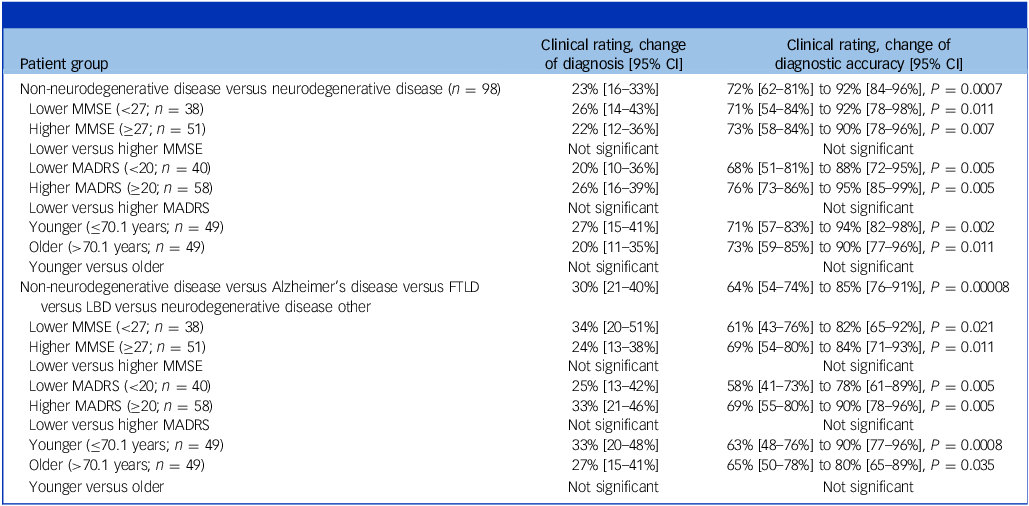
After disclosure of [18F]FDG PET results, level-1 diagnoses were changed in 23 out of 98 (23%; 95% CI 16–33%) patients (Table 2). This led to a significant (P < 0.001, McNemar test) increase in agreement between clinical and [18F]FDG PET diagnoses, from 64% (95% CI 54–74%) (63 out of 98 cases, κ = 0.29) to 88% (95% CI 79–93%) (86 out of 98 cases, κ = 0.75). The accuracy of the level-1 diagnosis at time of PET improved from 72% (95% CI 62–81%) to 92% (95% CI 84–96%) after disclosure of [18F]FDG PET results, with regard to the final interdisciplinary consensus diagnosis (P < 0.001, McNemar test).
Changes of diagnosis and diagnostic accuracy after disclosure of [18F]fluorodeoxyglucose position emission tomography ([18F]FDG PET) results

MMSE, Mini-Mental State Examination; MADRS, Montgomery–Åsberg Depression Rating Scale; FTLD, frontotemporal lobar degeneration; LBD, Lewy body diseases.
Figure 1 summarises the changes of level-1 diagnoses: after disclosure of [18F]FDG PET ratings, the diagnosis was changed from neurodegenerative disease to non-neurodegenerative disease in 26% (13 out of 50; see #1 in Fig. 1), and from non-neurodegenerative disease to neurodegenerative disease in 21% (ten out of 48; see #2 (n = 8) and #2* (n = 2; see below) in Fig. 1) of cases, respectively. Each adjusted level-1 diagnosis, in which the diagnosis was changed from neurodegenerative disease to non-neurodegenerative disease by [18F]FDG PET, was confirmed by the final clinical diagnosis (i.e. exclusion of clinically suspected neurodegenerative disease at the time of PET). In turn, two of the ten changes from non-neurodegenerative disease to neurodegenerative disease were rejected by the final clinical diagnosis (see #2* in Fig. 1). This was motivated by unremarkable additional biomarkers (i.e. normal tau PET, DAT SPECT and CSF biomarkers in one case; normal DAT SPECT in the other) and lack of clinical progression (in both cases). Furthermore, similar fractions of cases with a final diagnosis of neurodegenerative disease and non-neurodegenerative disease in clinically suspected non-neurodegenerative disease (6%, three out of 50) and neurodegenerative disease (6%, three out of 48) at the time of PET, respectively, were missed by the clinical diagnosis also after disclosure of [18F]FDG PET results (see #3 in Fig. 1). All three cases with a final adjustment from non-neurodegenerative disease to neurodegenerative disease showed abnormal Alzheimer’s disease neuropathological biomarkers (i.e. abnormal tau or amyloid PET in n = 2 and abnormal CSF biomarkers in n = 3). The [18F]FDG PET scans of these patients were read to be normal, they showed only MCI with deficits detected in the CERAD battery, and MMSE scores were normal (scores between 27 and 30). In turn, in cases with a final adjustment from neurodegenerative disease to non-neurodegenerative disease, no abnormal biomarkers were detected (as available; normal tau PET, DAT SPECT and CSF markers in n = 1 each), and follow-up showed no progression. Of note, availability of biomarkers did not differ between patients with and without changes in diagnosis from the clinical diagnosis after disclosure of [18F]FDG PET results to the final interdisciplinary consensus diagnosis (e.g. CSF was available in six out of eight patients [75%] and 73 out of 90 patients [81%], respectively; P = 0.675, chi-squared test).
Alluvial diagram of changes of level-1 diagnoses. Depicted is the evolution of individual diagnoses (n = 98) from baseline clinical diagnosis at time of [18F]FDG PET, over the clinical diagnosis after disclosure of [18F]FDG PET results to the final clinical diagnosis (including all available clinical, follow-up and biomarker information). Level-1 diagnosis entails the distinction between a neurodegenerative and a non-neurodegenerative cause of cognitive impairment. #1 to #3 refer to major changes in diagnosis (see text for details). [18F]FDG PET, [18F]fluorodeoxyglucose position emission tomography. #1, cases with a change of diagnosis after disclosure of [18F]FDG PET ratings from neurodegenerative disease to non-neurodegenerative disease; #2, cases with a change of diagnosis after disclosure of [18F]FDG PET ratings from non-neurodegenerative disease to neurodegenerative disease; #2*, subgroup of cases in #2 whose final diagnosis was changed back to non-neurodegenerative disease; #3, cases with a final diagnosis of neurodegenerative disease or non-neurodegenerative disease that were missed by the clinical diagnosis also after disclosure of [18F]FDG PET results.

Discrimination of subtypes of neurodegenerative diseases (level 2)
After disclosure of the [18F]FDG PET results, level-2 diagnoses were changed in 29 out of 98 (30%, 95% CI 21–40%) patients (Table 2). This led to a significant (P < 0.001, McNemar test) increase in agreement between clinical and [18F]FDG PET diagnoses, from 53% (95% CI 43–63%) (52 out of 98 cases, κ = 0.28) to 82% (95% CI 72–88%) (80 out of 98 cases, κ = 0.71). The accuracy of the level-2 diagnosis at time of PET improved from 64% (95% CI 54–74%) to 85% (95% CI 76–91%) after PET, with regard to the final interdisciplinary consensus diagnosis (P < 0.001, McNemar test).
Figure 2 summarises the changes of level-2 diagnoses. Most relevant findings can be summarised as follows (please note that changes from non-neurodegenerative disease to one of the neurodegenerative diseases and vice versa will not be addressed in detail if these have already been addressed above; see level 1): two out of seven patients’ diagnoses that have been changed from non-neurodegenerative disease to Alzheimer’s disease and FTLD after disclosure of [18F]FDG PET were corrected to FTLD (one out of four; one patient with normal tau PET and CSF Alzheimer’s disease biomarkers) and Alzheimer’s disease (one out of three; one patient with abnormal tau PET and CSF Alzheimer’s disease biomarkers), respectively, with the final consensus diagnosis (see #4 in Fig. 2). Interestingly, diagnoses changed from LBD to FTLD or Alzheimer’s disease to FTLD after disclosure of [18F]FDG PET results were confirmed (one out of one and two out of two patients, respectively), whereas one of the two diagnoses changed from LBD to Alzheimer’s disease was rejected (normal CSF Alzheimer’s disease biomarkers; see #5 in Fig. 2). Finally, a substantial fraction of LBD cases according to clinical diagnosis before and after disclosure of [18F]FDG PET results were changed to Alzheimer’s disease by the final consensus diagnosis (three out of 12; normal DAT SPECT in n = 3; see #6 in Fig. 2). Two changes of diagnoses from non-neurodegenerative disease to LBD after disclosure of [18F]FDG PET results were rejected (see above, level 1, normal DAT SPECT) or adjusted to Alzheimer’s disease (normal DAT SPECT) with the final diagnosis (part of #6 in Fig. 2). The level-2 diagnosis ‘neurodegenerative disease other’ was given to one patient after disclosure of [18F]FDG PET results, and was supported by the final consensus diagnosis in this patient (suspected argyrophilic grain disease). Of note, availability of biomarkers did not differ between patients with and without changes in diagnosis from the clinical diagnosis after disclosure of PET to the final interdisciplinary consensus diagnosis (e.g. CSF was available in n = 13/15 (87%) and n = 66/83 (80%), respectively; P = 0.619, chi-squared test).
Alluvial diagram of changes of level-2 diagnoses. Depicted is the evolution of individual diagnoses (n = 98). Level-2 diagnosis entails the additional classification of patients with suspected neurodegenerative diseases into the predefined categories of Alzheimer’s disease, frontotemporal lobar degeneration (FTLD), Lewy body diseases (LBD) and neurodegenerative disease other. #4 to #6 refer to major changes in diagnosis that have not yet been discussed with regard to the changes of level-1 diagnoses (see Fig. 1 and text for details). [18F]FDG PET, [18F]fluorodeoxyglucose position emission tomography. #4, cases with a change of diagnosis from non-neurodegenerative disease to Alzheimer’s disease or FTLD after disclosure of [18F]FDG PET results and a correction with the final diagnosis to FTLD and Alzheimer’s disease, respectively; #5, case whose diagnosis was changed from LBD to Alzheimer’s disease after disclosure of [18F]FDG PET results and changed back with the final diagnosis; #6, cases with a diagnosis of LBD after disclosure of [18F]FDG PET results that was changed to Alzheimer’s disease with the final diagnosis.

Stratified analyses
Stratifying patients by severity of cognitive impairment (51 patients with MMSE score ≥27 points versus 38 patients with an MMSE score <27; not available in nine patients), severity of depression (58 patients with MADRS score ≥20 points versus 40 patients with an MADRS score <20) and age at first visit (i.e. applying a group-split at median age of 70.1 years) yielded the following results (see Table 2).
Changes in clinical level-1 and level-2 diagnoses after disclosure of [18F]FDG PET results occurred with comparable frequencies in the corresponding groups of low versus high MMSE, low versus high MADRS and younger versus older patients (all P > 0.1, chi-squared test).
The diagnostic accuracy concerning level-1 diagnoses did not differ between MMSE, MADRS and age strata before (all P > 0.1) or after disclosure of [18F]FDG PET results (all P > 0.1, chi-squared test), although diagnostic accuracy seems to improve slightly more in the groups with lower MMSE and younger patients. However, these differences did not reach statistical significance (all P > 0.1, chi-squared test).
Concerning level-2 diagnoses, the diagnostic accuracy did not differ significantly between MMSE, MADRS and age strata before (all P > 0.1) or after disclosure of [18F]FDG PET results (all P > 0.1, chi-squared test). Improvement of diagnostic accuracy was again numerically higher in the groups of lower MMSE and younger patients, but this difference was not statistically significant (P > 0.1, chi-squared test).
Discussion
The present study was motivated by the EANM-EAN recommendation to use [18F]FDG PET for differential diagnosis in depressive pseudodementia, which was unanimously positive but also required further research, given the complete lack of systematic studies.Reference Nestor, Altomare, Festari, Drzezga, Rivolta and Walker19 Here, we were able to confirm a high impact and significant incremental diagnostic value of [18F]FDG PET on the clinical diagnosis in a real-life gerontopsychiatric clinical population. It is worth noting that the present study goes beyond the EANM-EAN recommendation by providing additional evidence that the diagnostic value of [18F]FDG PET in this setting is not significantly affected by the severity of cognitive impairment (challenging the dementia-only restriction), severity of depressive symptoms and patient age.
In contrast to disease-specific biomarkers, it has to be emphasised that [18F]FDG PET as a pan-disease biomarker of neurodegeneration fulfils a screening role, which is widely available (e.g. as integral part of oncological imaging), relatively inexpensive (e.g. compared with iterative evaluations) and poses little burden to the patient (e.g. non-invasive, short scan, very low to negligible radiation exposureReference Schiller, Frings, Thurow, Meyer and Mix21).
Concerning discrimination of neurodegenerative disease from non-neurodegenerative disease causes of cognitive impairment (level-1 decisions), we found that additional information of the [18F]FDG PET result led to a change in clinical diagnoses in about every fourth (23%) patient, which is expected to directly impact patient management. The correctness of diagnostic changes imposed by [18F]FDG PET is supported by a strong increase in diagnostic accuracy compared to final, biomarker- and follow-up-supported interdisciplinary consensus diagnosis (from 72 to 92%). In line with the EANM-EAN statement,Reference Nestor, Altomare, Festari, Drzezga, Rivolta and Walker19 [18F]FDG PET was particularly valuable for the exclusion of neurodegenerative dementia (only three out of 16 false-positive neurodegenerative disease cases by clinical diagnosis were still missed after disclosure of PET, and there was no incorrect change from neurodegenerative disease to non-neurodegenerative disease after PET disclosure).
Concerning the discrimination of subtypes of neurodegenerative dementia (level-2 decisions), the clinical diagnosis changed in 30% of cases, increasing its accuracy from 64 to 85%. Reasons for such a low accuracy (64%) before [18F]FDG PET use are a notoriously incorrect differential diagnosis of cognitive impairment based on clinical criteria as revealed by post-mortem studies and our study design with multiple diagnostic groups (compared with a binary decision with an a priori 50/50 chance of correctness). [18F]FDG PET proved particularly valuable for identification of FTLD (five out of six cases missed by the clinical diagnosis were correctly identified after disclosure of PET; Fig. 2). Interestingly, a substantial fraction of Alzheimer’s disease cases were still missed even after disclosing [18F]FDG PET (i.e. eight out of 23). Half of these were assumed to have LBD but were finally corrected to Alzheimer’s disease because of a normal DAT SPECT. LLD was shown to be associated with Lewy body pathology and prodromal depression may precede the diagnosis of LBD by years.Reference Nunes, Suemoto, Rodriguez, Paraizo Leite, Nascimento and Pasqualucci22,Reference Schrag, Horsfall, Walters, Noyce and Petersen23 This knowledge may have provoked a too frequent clinical diagnosis of LBD, which in turn, is the only diagnostic group with a decline in frequency during diagnostic workup (see Fig. 2). In line with literature, this underlines the importance of liberally performing DAT SPECT in suspected LBD, despite the fact that DAT SPECT may be false-negative in a minor fraction of early LBD.Reference Jreige, Kurian, Perriraz, Potheegadoo, Bernasconi and Stampacchia24,Reference Thomas, Attems, Colloby, O’Brien, McKeith and Walker25 The other half of missed Alzheimer’s disease cases were finally correctly identified by CSF biomarkers, which underscores their value in early or questionable Alzheimer’s disease.Reference Grothe, Moscoso, Ashton, Karikari, Lantero-Rodriguez and Snellman26
The impact of [18F]FDG PET for differential diagnosis of dementia has been confirmed by post-mortem studies.Reference Bohnen, Djang, Herholz, Anzai and Minoshima10 Meta-analyses indicated a sensitivity of 87–93% and specificity of 73–81% for discriminating post-mortem verified Alzheimer’s disease from other types of dementia (i.e. FTLD, LBD) and healthy controls.Reference Bohnen, Djang, Herholz, Anzai and Minoshima10,Reference Cure, Abrams, Belger, Dell’agnello and Happich27 A similar overall accuracy was obtained by the present level-2 PET reads (86%, data not shown; 85% in Bohnen et alReference Bohnen, Djang, Herholz, Anzai and Minoshima10). Interestingly, according to one of the post-mortem studies [18F]FDG PET seems to be particularly valuable for differentiating between Alzheimer’s disease and FTLD (accuracy 90%).Reference Foster, Heidebrink, Clark, Jagust, Arnold and Barbas28 This is in line with our results, confirming the strength of [18F]FDG PET in identifying FTLD (see above). Earlier clinical studies showed that the diagnostic value of [18F]FDG PET in dementia (and Alzheimer’s disease in particular) is higher in patients with early onset (≤65 years) compared with those with late onset (>65 years).Reference Hellwig, Frings, Bormann, Vach, Buchert and Meyer20,Reference Matsunari, Samuraki, Chen, Yanase, Takeda and Ono29 The accuracy of the clinical diagnosis increased more and reached a higher level in younger patients compared with older patients after disclosure of [18F]FDG PET in the present study (e.g. level-2 diagnosis: from 63 to 90% v. 65 to 80%), but this did not attain statistical significance between groups. Given that the severity of hypometabolism usually correlates with the severity of cognitive impairment and further cognitive decline,Reference Blazhenets, Ma, Sorensen, Schiller, Rucker and Eidelberg30 it may be expected that the incremental diagnostic value of [18F]FDG PET is higher in patients with more cognitive impairment compared with those with less cognitive impairment. This would support the dementia-only restriction imposed by the EANM-EAN task force for the use of [18F]FDG PET in pseudodementia.Reference Nestor, Altomare, Festari, Drzezga, Rivolta and Walker19 However, we could not find a significant difference between patients with lower and higher MMSE score (<27 v. ≥27 points) (see Table 2). Finally, we stratified our analyses by the severity of depressive symptoms, which also had no significant effect.
Measures of depression and cognition (e.g. MMSE, MADRS) were not correlated with each other in the present study. Still, caution is warranted, since comorbid depression may affect the diagnostic accuracy of cognitive screening instruments, particularly in case of MCI.Reference O’Caoimh and Molloy31 On the other hand, the accuracy of depression case finding tools in elderly patients with dementia is also limited.Reference Goodarzi, Mele, Roberts and Holroyd-Leduc32 We decided to employ the MADRS, since previous studies validated its use for depression rating in an elderly populationReference Heo, Murphy and Meyers33 and in early-onset dementia.Reference Leontjevas, van Hooren and Mulders34 Unlike the most commonly used Hamilton Rating Scale for Depression, the MADRS allows for explicit assessment of cognitive symptoms.Reference Montgomery and Asberg35 Moreover, the item ‘loss of insight’ in the Hamilton Rating Scale for Depression has to be viewed with caution in the context of dementia, since anosognosia is frequent among these patients.Reference de Ruijter, Schoonbrood, van Twillert and Hoff36,Reference Hamilton37
Limitations
The following limitations of the present study have to be acknowledged: because of its retrospective nature, there is an increased risk of bias. We addressed this issue by employing anonymised, highly structured case vignettes, which have already been successful used in an earlier study.Reference Hellwig, Frings, Bormann, Vach, Buchert and Meyer20 We used a study protocol that comprised several steps of decision-making to ensure a complete blinding of the clinical raters against [18F]FDG PET, follow-up and biomarker information. Conversely, the raters of the [18F]FDG PET scans were also blinded against all other data (except age). The majority of patients in our study sample received psychotropic drugs at the time of PET. Although medication effects on cerebral glucose metabolism have been described, we consider the associated risk of bias to be small, given that treatment-induced changes appear to be rather subtle and restricted (in particular, to the anterior cingulate cortex).Reference De Crescenzo, Ciliberto, Menghini, Treglia, Ebmeier and Is38 Inevitably, not all data and information were available in all patients. For example, depression diagnosis was established without using the Structured Clinical Interview for DSM-5 Disorders or similar instruments. Still, the most important parameters were accessible in the majority of patients; for instance, MADRS, MMSE and CSF biomarkers in 98 (100%), 89 (91%) and 79 (81%) patients, respectively. We verified that the rate of diagnostic adjustments by the final interdisciplinary consensus diagnosis did not depend on biomarker availability. Additional SPECT and PET imaging biomarkers (i.e. tau, amyloid and DAT) were frequently acquired according to clinical discretion (n = 52, 53%). We consider this a strength of our study; however, a prospective acquisition of all biomarker scans in all patients would not be permissible, given the accumulating costs and radiation exposure of multiple scans (i.e. [18F]FDG, amyloid and tau PET and DAT SPECT).
We enrolled consecutive geriatric in-patients with depression and cognitive impairment from a tertiary referral centre in Germany. Thus, our results are primarily applicable to patients with similar characteristics (i.e. late-onset, mostly severe and/or treatment-refractory depression with persisting cognitive deficits requiring in-patient treatment) and not necessarily transferable to different settings (e.g. psychiatric or primary care out-patients, different healthy systems). Moreover, we did not address the cost-effectiveness of different diagnostic scenarios. On one hand, ‘sole’ monitoring of a potential antidepressant response on mood and cognition over time may avoid wasteful testing and healthcare spending. On the other hand, an early consideration of [18F]FDG PET in the diagnostic process may allow for more accurate counselling of patients and caregivers, avoiding additional or repeated tests (e.g. MRI, neuropsychology), reducing therapeutic harm (e.g. medication side-effects) and, as a future perspective, using new therapeutic agents (potentially slowing progression). This may delay and reduce healthcare and nursing costs. Therefore, future prospective studies need to explore the applicability of the present findings to other settings and the cost-effectiveness of PET imaging in different scenarios.
Although we enrolled an overall sizeable number of patients, the size of subgroups is relatively small and precludes additional in-depth analyses. Further studies need to address possible determinants of diagnostic performance (e.g. clinical ratings, see EANM-EAN restriction) and possible associations with neuropathological changes. The latter may not only include the biomarkers of neurodegenerative diseases, but also help to unravel the controversial contribution of cerebrovascular disease.Reference Schiller, Frings, Thurow, Meyer and Mix21 Finally, we did not have access to a post-mortem neuropathological diagnosis as reference. To assess the impact and incremental diagnostic value of [18F]FDG PET on the clinical diagnosis, we had to disclose [18F]FDG PET results to the clinical raters. We addressed this very common dilemma by using a multidisciplinary consensus diagnosis as reference, including all available clinical, biomarker and follow-up data. In line with the literature, we consider this reference diagnosis a reasonable substitute of post-mortem data.Reference Gabel, Foster, Heidebrink, Higdon, Aizenstein and Arnold39 The access to [18F]FDG PET results may have led to an overestimation of its diagnostic value. However, it has to be emphasised that the present approach is in line with diagnostic criteria for Alzheimer’s disease, LBD and FTLD, which strongly support the use of [18F]FDG PET.
In conclusion, the present study in a real-life gerontopsychiatric clinical population demonstrates that [18F]FDG PET has a high impact on the clinical diagnosis and provides a significant incremental diagnostic value beyond the clinical diagnosis in depressive cognitive impairment. Thus, [18F]FDG PET should be considered in the diagnostic work-up of patients with psychiatric disorders and cognitive impairment. As a pan-disease biomarker, [18F]FDG PET is particularly valuable for exclusion of neurodegenerative disease and confirmation of FTLD, whereas the diagnosis of early Alzheimer’s disease and suspected LBD are the domains of Alzheimer’s disease neuropathological biomarkers and DAT SPECT, respectively.
Supplementary material
The supplementary material can be found at https://doi.org/10.1192/bjp.2025.67
Data availability
The data that support the findings of this study are available on request from the corresponding author, S.H. The data are not publicly available due to restriction e.g. their containing information that could compromise the privacy of research participants.
Author contributions
S.H. and P.T.M. contributed to the conception and design of the study, acquisition and interpretation of data and drafting the manuscript. L.F. contributed to the acquisition and analysis of data, and drafting and reviewing the manuscript. M.H., N.S., G.B., K.D. and J.B. contributed to data acquisition and reviewing the manuscript. All authors gave final approval of the revised version. All authors agree to be accountable for all aspects of the work ensuring that questions related to the accuracy or integrity of any part of the manuscript are appropriately investigated and resolved.
Funding
This research received no specific grant from any funding agency, commercial or not-for-profit sectors.
Declaration of interest
S.H., L.F., M.H., N.S., G.B. and P.T.M. report no conflicts. K.D. is a member of the Neurotorium Editorial Board, Lundbeck Foundation. The Berta-Ottenstein programme for Advanced Clinician Scientists, Faculty of Medicine, University of Freiburg, supported J.B.






eLetters
No eLetters have been published for this article.