Hospitals are increasingly turning to products with integrated antimicrobials, such as silver and triclosan, to reduce the risk of pathogen transfer from surfaces. Coinciding with the use of silver in healthcare products, an increase in silver and antibiotic resistance has been observed.Reference Finley, Norton, Austin, Mitchell, Zank and Durham1, Reference Kaweeteerawat, Na Ubol, Sangmuang, Aueviriyavit and Maniratanachote2 Although these silver antimicrobial products are being marketed as a tool to decrease the spread of pathogens, they may simultaneously be exerting selective pressures, resulting in a silver- and antibiotic-resistant microbiome.
Cell phones are increasingly integrated into the hospital setting; healthcare workers carry their phones with them as they move between patient’s rooms and use them to receive pages or access medical data through online applications. Even with proper hand cleaning, recontamination of hands with pathogens can occur after handling a cell phone.Reference Jeske, Tiefenthaler, Hohlrieder, Hinterberger and Benzer3 However, the phone has been overlooked reservoir of silver and antibiotic resistance. We performed this study to determine whether silver-coated phone screens on cell phones select for silver and antibiotic resistance genes.
Methods
After informed consent, participants provided samples of the palms of both hands as well as the fronts and backs of their cell phones. After the initial sampling, each phone was sterilized with 70% ethanol and an Antimicrobial Corning Gorilla Glass screen cover (Corning, Corning, NY) was applied.4 Sterile cotton swabs (Puritan, Guilford, ME) were dipped in 1X phosphate buffer solution prior to sample collection. Surfaces were swabbed completely using a rolling motion. Samples were transported to the laboratory within 1 hour of collection and stored at −20°C. DNA extraction was performed within 24 hours of collection. Sampling was repeated on days 7 and 30 after screen application using the same methods.
Total genomic DNA was extracted from swab tips using the MoBio BiOstic Bacteremia isolation kit (MoBio, Carlsbad, CA). One modification was made to the supplier protocol; swab tips were removed using a sterile razor blade and added directly to bead tubes; however, step 1 of the supplier protocol was skipped. DNA was eluted at 25 µL. Isolated DNA was stored at −20°C until further processing. DNA was used as the template for polymerase chain reaction (PCR) amplification of the V3–V4 region (314F and 805R) of the 16S rRNA gene, and 300 paired-end sequencing was performed using Illumina MiSeq (Duke Sequencing Core, Duke University, Durham, NC).
FASTQ sequence files were processed as previously described by Volkoff et al.Reference Volkoff, McCumber, Anderson and Gunsch5 The diversities of the microbes within a sample (α) and between different samples (β) were calculated using vegan software.Reference Oksanen, Blanchet and Kindt6 Analysis of variance (ANOVA), a comparison of the variance between defined groups, was used determine whether there were significant effects by time or collection surface for the diversity metrics. Upon finding these factors were significant, post hoc testing was conducted using Tukey’s honestly significant difference (HSD) to compare the individual samples using the Stats software package in R.7
A novel multiplexed qPCR assay targeting the tetK, mecA, vanA, silE, silP, and silRS genes was developed and utilized for hand samples and targeting combined gDNA from the cell-phone front or screen side (CF) and back (CB) samples. A full description of the methods, primer, and probes used are available in the Supplementary Material online.
Results
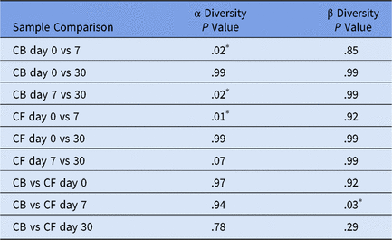
Overall, no major shifts were observed in the overall microbial community structure of personal phones from 26 resident physicians. However, ANOVA revealed significant differences in diversity metrics across time and sample location. The results of Tukey’s HSD post hoc testing of α and β diversities are shown in Table 1. Comparing the β diversity using the Bray-Curtis distance of samples across collection time and sample type, there was one significant time point with a β diversity between the CF and CB swab samples at day 7 (P = .03). Comparisons of the respective sides of the cell phone showed decreased α diversity on day 7 compared to days 0 and 30. The α diversity between day 7 and 30 CF was the only time point that was not significantly different for day 7 (P = .07). while all other diversity sample values showed no difference between days 7 and 30. None of the α or β diversity comparisons between days 0 and 30 were significant.
P Values for Tukey HSD Test Comparison of α and β Diversities for Corresponding Sample Comparisons

Note: HSD, honestly significant difference; CB, back-side cell-phone samples; CF, front-side (screen) samples of cell phone.
* P < 0.05.
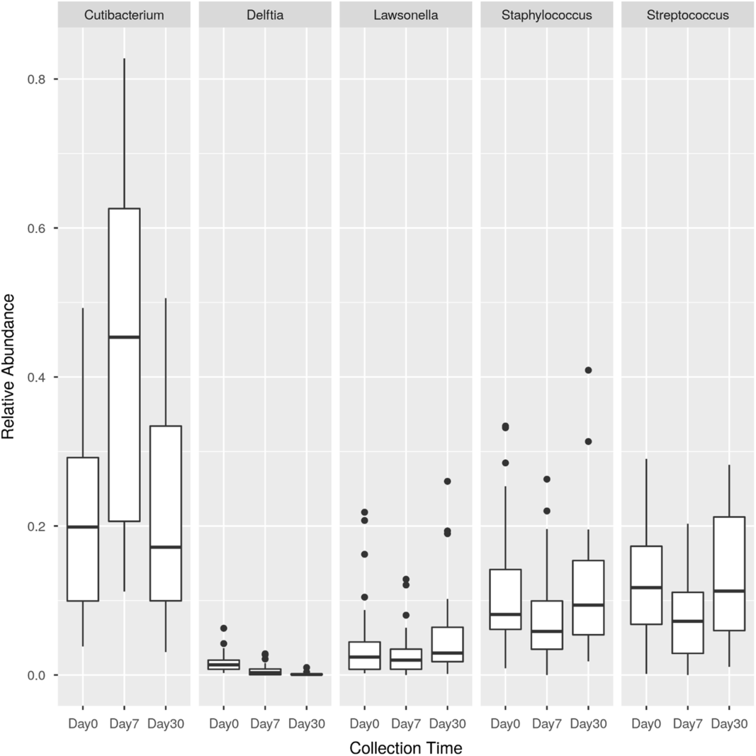
No increase in silver- or antibiotic-resistance genes was observed. The 5 most abundant genera found on the CF samples were Cutibacterium, Delftia, Lawsonella, Staphylococcus, and Streptococcus. Relative abundance for these genera at each sample collection time point is shown in Fig. 1.
Box and whisker plot of the relative abundance of the 5 most abundant genera detected on the front side (screen) of cell phones by sample collection time.

Discussion
To our knowledge, this is the first evaluation of silver-coated phone screens as reservoirs of silver- and antibiotic-resistant genes. The 5 most abundant genera detected on resident cell phones were common commensal skin flora, suggesting that an individual’s hand microbiome influences cell-phone microbiomes, consistent with a previously published study.Reference Grice, Kong, Renaud and Young8, Reference Meadow, Altrichter and Green9 At 30 days postapplication, no significant effect of the silver antimicrobial screen cover on the overall phone microbiome was observed. The similar phone microbiomes of days 0 and 30 suggest the transmission of bacteria from a cell phone via hands to patients would be a similar risk from a cell phone with or without this antimicrobial screen at least over periods of time >30 days. Even though the cell-phone microbiomes were similar, 16S rRNA gene amplicon sequencing is unable to discern between inactive and viable bacteria. Therefore, viability differences would need to be confirmed by culturing. However, silver was ineffective in reducing bacterial viability in a previous culture-based study that revealed no significant difference in colony-forming units of bacteria found on silver-impregnated surgical scrubs compared to cotton scrubs worn by healthcare providers.Reference Anderson, Addison and Lokhnygina10 According to the manufacturer of the screens, the antimicrobial properties are due to leaching of silver ions from the glass.4 Thus, another explanation for the observed lack of difference could be that effective doses of silver were no longer being leached from the glass, thus it lost its antimicrobial properties by day 30. This hypothesis would need to be verified by measuring silver concentrations on the phones.
Although no change was observed in the microbiome over 30 days, the α diversity was significantly lower at day 7 than at days 0 and 30 for both CF and CB, suggesting that the richness of species diversity was lower on day 7. In addition, on days 0 and 30, the β diversity of CF to CB was insignificant; however, the CF and CB on day 7 were significantly different. By treating the CB samples as a control at each time point, at day 7, the screen is potentially still active and causing shifts in the microbial community. Future studies should investigate the longitudinal leaching of silver from the protective cover and samples should be collected at shorter time intervals to capture potentially deterministic or stochastic changes in the cell-phone microbiome. However, the results of this study suggest that cell phones should still be considered contaminated surfaces regardless of the introduction of antimicrobial barriers, and healthcare providers should continue to adhere to proper hand hygiene.
Acknowledgments
Any opinions, findings, conclusions or recommendations expressed in this material are those of the author(s) and do not necessarily reflect the views of the NSF or the EPA. This work has not been subjected to EPA review, and no official endorsement should be inferred. This work was approved by the DUMC IRB Office and operated under the IRB protocol no. Pro0078279.
Financial support
This material is based upon work supported by the National Science Foundation (NSF) and the Environmental Protection Agency (EPA) under NSF Cooperative Agreement EF-0830093, Center for the Environmental Implications of NanoTechnology (CEINT). A. W. McCumber received support from the National Institutes of Health (grant no. T32GM008555). S. J. Volkoff received support from the National Science Foundation (grant no. DGE 1545220).
Conflicts of interest
All authors report no conflicts of interest relevant to this article.
Supplementary material
To view supplementary material for this article, please visit https://doi.org/10.1017/ice.2019.280