Implications
We have demonstrated that melatonin implants inserted around the spring equinox are able to induce reproductive activity in Mediterranean goats that are in daily contact with bucks. This work tries to confirm that those treatments that include exogenous melatonin and that are associated to the male effect could in fact be applied without separation of the sexes. Isolation of males is a practice that implies a management more difficult of the animals and a higher availability of equipment at the farms.
Introduction
Most breeds of sheep and goats from temperate latitudes and some either from or adapted to subtropical latitudes exhibit seasonal variations of sexual activity (Chemineau et al., Reference Chemineau, Daveau, Maurice and Delgadillo1992a; Delgadillo et al., Reference Delgadillo, Canedo, Chemineau, Guillaume and Malpaux1999; Rivera et al., Reference Rivera, Alanis, Chaves, Ferrero and Morello2003). The timing of reproductive seasonality in these small ruminants, as well as in most mammals, is controlled by photoperiod (Chemineau et al., Reference Chemineau, Malpaux, Delgadillo, Guerin, Ravault, Thimonier and Pelletier1992b). Melatonin, the main secretory product from the pineal gland, is the neuroendocrine signal that transduces information about the environmental light received by the retina. This reproductive seasonality is a major limitation in farm animal species (Yeates, Reference Yeates1949).
Various treatments have been proposed to control the phenomenon, including male effect, melatonin, photoperiod or combinations. The seasonal variation in reproductive activity observed under natural conditions is profoundly altered when animals are subjected to alternations of 3 months of long days and 3 months of short days. Under these conditions, the sexual activity of males and females was shown to peak during short days (Chemineau et al., Reference Chemineau, Pelletier, Guérin, Colas, Ravault, Touré, Almeida, Thimonier and Ortavant1988; Delgadillo et al., Reference Delgadillo, Cortez-López, Duarte and Malpaux2000; Delgadillo et al., Reference Delgadillo, Cortez, Duarte, Chemineau and Malpaux2004). Under field conditions, the long-day part of the treatment is easy to apply, as extra illumination can be provided indoors or outdoors.
Exogenously administered melatonin from continuous slow-release implants has been shown to advance the onset of the breeding season in goat males and females by mimicking the stimulatory effect of short days (Chemineau et al., Reference Chemineau, Malpaux, Delgadillo, Guerin, Ravault, Thimonier and Pelletier1992b; Zarazaga et al., Reference Zarazaga, Gatica, Celi, Guzmán and Malpaux2009a). Traditionally, in Mediterranean latitudes, exogenous melatonin treatment is implanted around the spring equinox and accompanied by a prior separation of males and females for 45 days, inducing a male effect to optimise the response during the anoestrous period, obtaining a higher synchronisation at mount. This isolation between males and females has been assumed to be necessary for the male effect to successfully induce ovulation (for details, see review Delgadillo et al., Reference Delgadillo, Gelez, Ungerfeld, Hawken and Martin2009). However, this isolation implies a management more difficult of the animals and a higher availability of equipment at the farms.
This study therefore had a double aim. The first part was to determine whether following a treatment with long days from the second fortnight of November to late February, followed by a melatonin implant, is effective in stimulating reproductive activity in female goats that are in daily contact with bucks during the seasonal anoestrus (short-term effect). The second part was to determine whether this treatment causes a variation in the reactivation of reproductive activity in the subsequent natural breeding season (medium-term effect).
Material and methods
Animals and management
This experiment was performed in accordance with the Spanish Animal Protection Policy RD1201/2005, which conforms to the European Union Directive 86/609 regarding the protection of animals used in scientific experiments.
The study was conducted on an experimental farm (latitude 37°15′) of the University of Huelva, which meets the requirements of the European Community Commission for Scientific Procedure Establishments (1986). Thirty-four adult and non-pregnant female Payoya goats between 1.5 and 4 years old at the beginning of the experiment, which had kidded at least 5 months previously, were used.
The experiment started on 4 November 2005 and finished on 27 October 2006. During the whole experiment, animals were maintained under intensive management and were fed daily with lucerne hay, barley straw ad libitum and commercial concentrate, according to the INRA standards (Morand-Fehr and Sauvant, Reference Morand-Fehr and Sauvant1988), to maintain adult weight or allow growth in younger females throughout the experiment. The concentrate was a commercial mixture of maize (26.3%), bean (20%), oats (14.1%), cottonseed (13.7%), pea (13.4%), lupin (7.3%), barley (0.2%), wheat (0.2%), sunflower seeds (0.2%) and a commercial concentrate as mineral–vitamin complement (4.6%). All animals had free access to water and mineral blocks containing trace elements and vitamins.
Experimental design
Females were distributed at random into groups balanced according to their live weight (LW), body condition score (BCS) and age. The control group (C, n = 17) was kept in a communal yard with an uncovered area and without any supplementary light throughout the experiment. From 14 November 2005 until 20 February 2006, 17 does (LD-M group) were placed in a light-proof building and exposed to long days (16 h light: 8 h dark; lights on 0600 h: lights off 2200 h). The photoperiod was regulated by an electric timer that controlled white fluorescent strip lights providing approximately 200 lux at the goats’ eye level during the light phase, and a ventilator system was used for air renewal.
On 20 February, photoperiodic-treated females (LD-M group) were re-exposed to the natural variations in day length and received one subcutaneous melatonin implant, which contained 18 mg of the hormone (Melovine®, CEVA Salud Animal, Barcelona, Spain) at the base of the left ear. At this moment, the LD-M group was kept in the same communal yard as the C group.
To avoid a possible male effect that could modify the reproductive activity in experimental does, before the onset of the experiment, 12 bucks were allocated close to them in a separate barn. Seven of them were treated similarly to treated females (long days and melatonin) and five were not treated (Zarazaga et al., Reference Zarazaga, Gatica, Celi, Guzmán and Malpaux2010). The photoperiodic treatment of the males was simultaneous to that of the females. For this reason, during the photoperiodic treatment of the LD-M group, it was in contact with the treated males but in a separate barn.
Measurements
Oestrous activity was tested daily using aproned bucks. From 4 November 2005 until the end of the experiment, males were introduced in pairs during at least 30 min, at 0900 h, into the experimental female groups (LD-M and C groups) for three consecutive hours. During the rest of the day, bucks were isolated in their barn but remained close to the females. Females standing at mounting by the male were considered in oestrus. Ovulation rate was evaluated by laparoscopy 7 days after positive identification of the oestrus. The goats received a sedative intramuscular injection (Rompun®, Bayer, Spain) prior to laparoscopy. During laparoscopy, the animals were placed in dorsal recumbency on a cradle at a 45° angle. A 7-mm laparoscope (Karl-Storz, Tuttlingen, Germany) was used for visualisation, and the ovaries were exposed by pulling the fimbria in different directions with atraumatic forceps. In order to determine the occurrence of silent ovulations and to confirm ovarian activity, blood samples were collected weekly from each animal by jugular venipuncture and assayed for progesterone. Immediately after collection, samples were centrifuged and plasma was stored at −20°C until assay. LW and BCS were recorded weekly by the same handler (Hervieu et al., Reference Hervieu, Morand-Fehr, Schmidely, Fedele and Delfa1991).
Plasma progesterone concentrations were assayed by radioimmunoassay, using the technique described by Terqui and Thimonier (Reference Terqui and Thimonier1974). The sensitivity of the assay was 0.125 ng/ml. The intra- and inter-assay coefficients of variation were 7.0% and 1.6%, respectively.
All hormonal analysis was performed at the hormonal analysis laboratory of INRA (Nouzilly, France).
Definitions of reproductive activity
The non-breeding season (seasonal anoestrus) and the natural breeding season were defined according to the results obtained at the same latitude and with a similar method by Zarazaga et al. (Reference Zarazaga, Guzmán, Domínguez, Pérez and Prieto2005) and Zarazaga et al. (Reference Zarazaga, Guzmán, Domínguez, Pérez, Prieto and Sánchez2009b). The reproductive activities measured during the non-breeding season induced by the treatment were: the first ovulatory activity, the first oestrus, the last ovulatory activity, the last oestrus, the length of the cyclic ovulatory activity and the length of the cyclic oestrus activity. The reproductive activities at the reactivation of reproductive activity in the natural breeding season were: the first ovulatory activity and the first oestrus.
Ovulatory activity was confirmed when two or more consecutive plasma samples had progesterone concentrations above baseline (0.5 ng/ml; Zarazaga et al., Reference Zarazaga, Guzmán, Domínguez, Pérez, Prieto and Sánchez2009b) with subsequent cyclicity. For each goat, the date of the last plasma progesterone value below baseline, which was followed by the first extended cyclic pattern, was taken as the onset of ovulatory activity. Cessation of ovulatory activity was considered to have occurred when three or more consecutive plasma samples had concentrations below baseline. The date of the first plasma progesterone value below baseline at the completion of the last ovarian cycle, according to the progesterone pattern, was taken as the end of ovulatory activity.
Sexual activities observed during the experiment were grouped as (i) oestrous cycle, when detected oestrous behaviour was accompanied by a later increase in plasma progesterone levels above 0.5 ng/ml; (ii) ovulatory activity, when an increase in plasma progesterone levels above 0.5 ng/ml in at least two consecutive samples occurred but was not preceded by detected oestrous behaviour (silent ovulation) and (iii) ovulation rate of each detected cycle.
Statistical analysis
The effect of the treatment and time on LW, BCS and progesterone was analysed using an analysis of variance for repeated measures. Daily changes of LW or monthly changes of BCS were calculated for each goat, and differences between groups were analysed by one-way ANOVA. The date of the first detected oestrus or first ovulatory activity induced by the treatment during the seasonal anoestrus, and the date of the first detected oestrus or the first ovulatory activity in the natural breeding season were determined for each goat. The one-way ANOVA was used to evaluate the effect of treatment on these reproductive parameters. The ovulation rate of each detected oestrus in each period (seasonal anoestrus and natural breeding season) was compared using a non-parametric t-test (the Mann–Whitney U-test). The incidence of ovarian activity and oestrus induced after the melatonin implant insertion detected during the seasonal anoestrus or during the natural breeding season was compared between groups, using the χ 2 test.
To study the effect of LW or BCS on ovulation rate at each detected oestrus, females were divided into groups depending on their LW at each oestrus: group 1 (LW ⩽ 50 kg) and group 2 (LW > 50 kg), or depending on their BCS at the same time: group A (BCS ⩽ 2.50) and group B (BCS ⩾ 2.75). They were compared using a non-parametric t-test (the Mann–Whitney U-test).
Differences were considered significant at P < 0.05. Analysis of data was computed using the SPSS package (Statistical Package for the Social Sciences (SPSS), 2006).
Results
Effect of treatment on LW and BCS
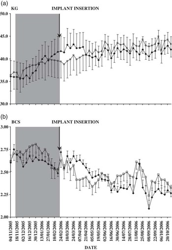
The statistical analysis for repeated measures showed a clear effect of time (P < 0.001) on LW (Figure 1a). Moreover, a significant interaction of time–treatment was observed for LW (P < 0.05). The LD-M group clearly increased their LW during the photoperiodic treatment from November to February, with a mean LW gain of 53.9 ± 7.7 g/day; however, during this period the C group had a mean LW gain of 27.4 ± 8.0 g/day (P < 0.05). In April, the treated group had losses in LW, and the C group continued with a slight increase in LW (−25.5 ±& 11.3 g/day v. 19.9 ± 10.7 g/day, for the LD-M and C groups, respectively, P < 0.01). Finally, until the end of the experiment, both groups had a relatively stable LW.
Weekly means (±s.e.m.) for live weight (a) and body condition score (b) of Mediterranean goat females subjected to natural changes in day length (C group, □) or artificial long days from 14 November 2005 to 20 February 2006, and then treated with one s.c. melatonin implant (LD-M group, ⧫). The shaded area indicates the duration of the long-day treatment.

Similarly, the statistical analysis for repeated measures showed a clear effect of time (P < 0.001) on BCS (Figure 1b), with a marked decrease from the start of the experiment (2.66 ± 0.06) until the end (2.33 ± 0.07). However, no effect of treatment or interaction of time and treatment was observed for this variable.
Effect of treatment on the end of the breeding season
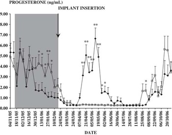
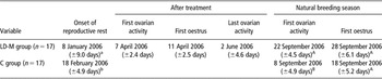
Figure 2 shows the changes in progesterone concentrations during the experiment. A clear effect of time and a treatment–time interaction (P < 0.001) was shown. However, no effect of treatment was observed on this variable. The progesterone concentrations in the LD-M group decreased earlier than in the control group. From 6 January 2006 to 3 February 2006, the progesterone concentrations in the C group were higher than in the LD-M group (with differences of at least P < 0.05). This reveals significant differences between groups at the onset of the reproductive rest (Table 1).
Weekly means (±s.e.m.) of progesterone concentrations (ng/ml) of Mediterranean goat females subjected to natural changes in day length (C group, □) or artificial long days from 14 November 2005 to 20 February 2006, and then treated with one s.c. melatonin implant (LD-M group, ⧫). The shaded area indicates the duration of the long-day treatment. *P < 0.05, **P < 0.01.

Mean (±s.e.m.) date of the onset of the reproductive rest, the first ovarian activity, the first oestrus and last ovarian activity after the treatment and date of the first ovarian activity and the first oestrus of the natural breeding season in Mediterranean goat females subjected to artificial long days from 14 November 2005 to 20 February 2006 and then treated with s.c. melatonin implants (LD-M group) or in goats subjected to natural changes in day length (C group)

Values with different letters in the same column (a, b) are significantly different at P < 0.001.
Values with different letters in the same column (A, B) are significantly different at P < 0.05.
Effect of treatment during the seasonal anoestrus
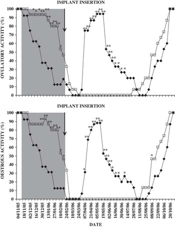
The concentrations of progesterone in the two groups were similar until 14 April 2006, when the concentrations for the LD-M group began to rise, whereas the concentrations of the C group remained at basal levels until 2 June 2006 (with differences of at least P < 0.05; Figure 2). Progesterone concentrations rose above baseline 46.9 ± 2.4 days following implant insertion. The mean duration of the ovarian activity induced by the treatment was 55.1 ± 5.9 days. Any female in the control group showed ovarian activity during this period of the natural seasonal anoestrus. Furthermore, the treated group was characterised by 94.1% of the animals presenting ovarian activity in response to the treatment used (Figure 3).
Percentages of Mediterranean goat females showing reproductive activity: ovarian activity (top) or oestrous activity (bottom) subjected to natural changes in day length (C group, □) or artificial long days from 14 November 2005 to 20 February 2006, and then treated with one s.c. melatonin implant (LD-M group, ⧫). The shaded area indicates the duration of the long-day treatment. *P < 0.05, **P < 0.01.

During this period, no oestrous activity was observed in the C group, despite the animals being in contact with those of the LD-M group and bucks. However, in the LD-M group, 88.2% of the animals showed oestrous activity (Figure 3). The ovulation rate of the first detected oestrus in the LD-M group was 1.93 ± 0.20 corpora lutea.
Effect of treatment on the natural breeding season
The C group resumed the natural breeding season (ovarian activity) at an earlier date than was observed for the LD-M group (P < 0.05; Table 1). During the reactivation of the natural breeding season, only on 1 September were there differences between groups in progesterone concentrations.
No differences between groups were observed on the reactivation of the oestrous activity (Table 1) or the ovulation rate for the first oestrus (1.82 ± 0.3 v. 1.60 ± 0.1 corpora lutea for the LD-M and C groups, respectively). Similarly, no effect or interaction between treatment and the categories of LW or BCS was observed.
Discussion
This study demonstrated that, in Mediterranean goat females, the treatment associating 3 months of long days and exogenous melatonin and daily contact with bucks is able to induce reproductive activity during the natural seasonal anoestrus (short-term effect). During this period, none of the females from the control group was stimulated by contact with the treated females or with males. Finally, this treatment induces a slight retardation in the onset of ovarian activity in the natural breeding season (medium-term effect). In the two cases (short- and medium-term effect), the reproductive activity or the ovulation rate was not influenced by LW or the BCS.
As has been described by our group in other experiments, a pattern of LW changes, associated with reproductive activity, was observed in all groups, despite the fact that the animals were fed to maintain their LW; such variations in LW have been reported previously in this species (Zarazaga et al., Reference Zarazaga, Guzmán, Domínguez, Pérez and Prieto2005). The changes occurred differently between the LD-M and C groups, due to the photoperiodic-melatonin treatment. The increase in LW in the LD-M group following the long-day treatment could be due to a direct effect of the long-day treatment, probably by increasing the food intake (Barenton et al., Reference Barenton, Ravault, Chabanet, Daveau, Pelletier and Ortavant1988). After, the LW decrease in the same group was probably due to the greater activity of animals as a consequence of the sexual activity induced (Walkden-Brown et al., Reference Walkden-Brown, Restall, Scaramuzzi, Martin and Blackberry1997), and/or due to the decrease in food intake mediated by melatonin (Argo et al., Reference Argo, Smith and Kay1999; Marie et al., Reference Marie, Findlay, Thomas and Adam2001).
The treatment with photoperiod establishes that the LD-M group started the induced seasonal reproductive rest (ovarian and oestrous activity) before the C group, indicating that photoperiod controls reproductive activity.
The date of the start of the treatment-induced reproductive activity during the seasonal anoestrous demonstrates the effectiveness of inserting melatonin implants after a photoperiodic treatment of 3 months of long days at Spanish latitudes in goats in daily contact with bucks. Similar results have been observed by our group following the insertion of exogenous melatonin implant around the spring equinox, without a previous long-day treatment (Zarazaga et al., Reference Zarazaga, Gatica, Celi, Guzmán and Malpaux2009a). However, it will be necessary to study the oestrus/ovulatory response to the LD-M treatment in entire females isolated from males and in contact with males to confirm that the presence of males did not modify the response to the LD-M treatment in entire females. Véliz et al. (Reference Véliz, Poindron, Malpaux and Delgadillo2006) suggest that previous isolation of seasonally anovulatory does from bucks to stimulate their reproductive activity by the male effect is unnecessary if sexually active bucks are introduced to them. With regard to the results obtained at Mediterranean latitudes, this assumption might not be applicable, because in our experiment none of the control females showed oestrous activity although the males were sexually active, as they were treated similarly to females (Zarazaga et al., Reference Zarazaga, Gatica, Celi, Guzmán and Malpaux2010). Another factor that can stimulate the reproductive activity in non-treated females is the presence of females in oestrus. In a study by Walkden-Brown et al. (Reference Walkden-Brown, Restall and Henniawati1993), 87% of goats responded to the male effect when oestrous females were present at the time of male introduction v. 72% in their absence. In fact, the presence of only a few oestrous ewes or goats may induce sexual activity in the rest of the females, even in the absence of any male (Bouillon et al., Reference Bouillon, Lajous and Fourcaud1982; Restall et al., Reference Restall, Restall and Walkden-Brown1995). However, in our study, no control female showed oestrous or ovarian activity independent of the fact that they were in contact with males or stimulated females.
Together with the data obtained by our group using a treatment with melatonin implants (Zarazaga et al., Reference Zarazaga, Gatica, Celi, Guzmán and Malpaux2009a), our findings are, as far as we are aware, the first of this type observed in melatonin-implanted goats, because the male effect is a normal practice when melatonin implants are used. The only results in the bibliography, from a similar study of the effectiveness of melatonin implants without separation of the males, are those obtained in ewes (Forcada et al., Reference Forcada, Zarazaga and Abecia1995). The treated ewes showed no reproductive activity during the seasonal anoestrus (the melatonin implants were inserted later than in our experiment); they did, however, exhibit an earlier onset of the natural breeding season.
The reactivation of the ovarian activity, according to the progesterone concentrations, in the natural breeding season in the treated females was later than in the control group. To our knowledge, no data exist about the medium-term effect of this treatment in goats. However, in bucks, the differences between groups are similar to those described by Delgadillo et al. (Reference Delgadillo, Carrillo, Morán, Duarte, Chemineau and Malpaux2001) working with Creole bucks at subtropical latitudes, and by Guzmán et al. (Reference Guzmán, Gatica and Zarazaga2008) working at the same latitude and with a similar treatment to that used in this experiment. The reason for the difference in the onset of reproductive activity between groups treated with melatonin after the long-day treatment or those exposed to natural photoperiod was probably the exhaustion of the melatonin implant, which delayed perception of the natural long days. Indeed, the increasing photoperiod between the winter and the summer solstices has a critical role in the regulation of the onset of the breeding season (Malpaux et al., Reference Malpaux, Robinson, Wayne and Karsch1989).
In conclusion, the results of this experiment indicate first that photoperiodic-melatonin treatment is able to induce reproductive activity during the seasonal anoestrus of goats with daily contact with males; second, the results suggest that at Mediterranean latitudes the use of sexually active males or contact between reproductively active (treated females) and non-active females (control females) does not stimulate reproductive activity in non-treated does; and finally, this treatment induces a slight delay in the onset of the natural breeding season in the treated females, because only the onset of ovarian activity was modified.
Acknowledgements
The authors wish to thank the Assay Laboratory of the Station de Physiologie de la Reproduction et des Comportements (INRA, Nouzilly, France) for carrying out the radioimmunoassays. This work was supported by Grant PETRI 95-0964.OP from C.I.C.Y.T. (Spain). The authors are grateful to CEVA Salud Animal, Barcelona, Spain for providing Melovine® implants. None of the authors of this paper has a financial or personal relationship with other people or organisations that could inappropriately influence or bias the content of the paper.