INTRODUCTION
Nasopharyngeal carcinoma (NPC) is a common disease among head and neck carcinomas. Radiotherapy is one of the most important treatment modalities for NPC patients with or without chemotherapy. Combined chemoradiotherapy and the use of new imaging modalities have shown to be effective.Reference Lagendijk, Leemans Ch, Buter, Berkhof and Slotman1,Reference Ciernik, Dizendirf and Baumert2
Tumour control for NPC is directly related to the dose delivered to the tumour. However, high dose of radiation area is also associated with a significant increase in toxicity that may adversely affect quality of life for the patient. Dose escalation using traditional two-dimensional radiotherapy planning is limited by the tolerance of adjacent critical organs such as spinal cord, brain stem, lenses, optical nerves, parotid glands and mandibles.Reference Zhou, Fei and Wu3 Intensity-modulated radiotherapy (IMRT) delivers a high dose to target volumes including the primer gross tumour volume (GTV) and high risk subclinical region while minimizing dose at organs at risk (OARs). IMRT has therefore become the ideal modality to treat patients with NPC. Many authors have shown that IMRT provided improved target coverage while significantly sparing sensitive normal tissues when compared to three-dimensional conformal radiotherapy.Reference Kristensen, Kjaer-Kristoffersen and Sapru4–Reference Chao, Low, Perez and Purdy8 Previous literature demonstrated that IMRT improves locoregional control, reduces side effects such as xerostomia and improves quality of life.Reference Wang, Schultz and Paul9 Using the inverse approach, the desired dose distribution for IMRT plans by an optimal intensity map is obtained with optimization algorithms. The implementation of IMRT requires the knowledge of setup uncertainties and the delineation of target volumes on the basis of anatomic or molecular imaging modalities and a quality control for the entire procedure.
Accurate delineation of the target volumes is the most important issue in the application of IMRT. Inaccuracy in the delineation of the target volumes by a small amount may result in severe complications as either overdosing of critical organs or underdosing of tumour tissues.
Computed tomography (CT) which gives the electron density information of the structures is a commonly employed imaging modality to delineate target volumes. A disadvantage of CT images is the poor soft tissue contrast, although they provide superior spatial accuracy and accurately show the tumour extension to the bone. Magnetic resonance (MR) imaging using different sequences is an alternative imaging modality to CT for evaluating soft tissue or bone extensions of tumour s and metastatic lymph node involvement in NPC.
Emami et al. published that CT-MR fusion improved the determination of target volumes in NPC.Reference Emami, Sethi and Petruzzelli10 Although MR images provide additional information to increase the accuracy of contouring of the tumour and the normal tissues in NPC, Reference Lee, Xia, Quivet and Sultanem18FlouroDeoxyGlucose-positron emission tomography (Reference Lee, Xia, Quivet and Sultanem18F FDG-PET) has also been used increasingly recently for contouring, staging and prediction of the tumour response. Combining PET imaging with CT imaging accurate anatomical information of anatomy improves staging accuracy compared to the use of CT or MR images alone or combined. By aligning the biological information obtained with PET with the anatomical information provided by CT, combined PET-CT imaging can overcome the limitations associated with PET alone. This has the advantage of avoiding possible false-positive and false-negative results associated with PET alone.Reference Price and Jones11 Increased diagnostic accuracy with PET-CT could also play a role in IMRT planning by reducing inter-observer variability during target delineation, and extension of GTV, the clinical target volume (CTV) and the planning target volume (PTV) for both primer tumour and regional lymph nodes.Reference Guido, Fuccio and Rombi12 Furthermore, IMRT makes it possible to apply a heterogeneous dose to the target inside using assured hypoxia information at the tumour site with PET-CT. Riegel et al. previously noted that the variability of GTV delineation in head and neck cancers using PET-CT fused images decreased among multiple observers.Reference Riegel, Berson and Destian13
NPC patients in our department have been treated with IMRT and/or chemotherapy since 2006. The aim of our study is to investigate the effectiveness of PET-CT for staging before radiotherapy, target delineation including GTV, CTV and PTV for IMRT compared with CT-MR fused images and an early response assessment after IMRT.
METHODS AND MATERIALS
Patients
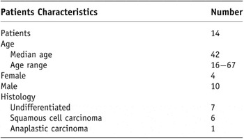
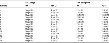
Our current study covers 14 NPC patients who were treated in our department with IMRT combined with chemotherapy between February 2006 and November 2008. Treatment IMRT plans of patients were done using both PET-CT and MR findings. Clinical characteristics of all the patients are presented in Table 1. Median age of our patients, including 10 males and 4 females, was 42 with ranges 16 and 67. All patients received physical examination, blood counts, liver function tests, dental examination, PET-CT, MR imaging and planning CT scan before the initiation of chemoradiotherapy. Patients were staged with MR and PET-CT as defined by the American Joint Committee on Cancer.
Characteristics of the patients

All patients were treated with IMRT explained in detail below and with concurrent chemotherapy. They were seen weekly during IMRT by the radiation oncologist. Patients were seen for follow-up in 2 months and evaluated with MR scans. A PET-CT was obtained in 3 months after treatment and the patients were followed-up with PET-CT every six months afterwards. Any toxicity was noted by the radiation oncologist during weekly visits during the treatment.
Treatment planning CT, MR and PET-CT procedures
All patients had CT simulation, MR and PET-CT scans in supine position, immobilized with head rest. CT simulation and PET-CT images were performed with customized thermoplastic mask extended to the shoulders which allowed precise repositioning of the patient over the course of treatment. Although MR and CT planning images were taken from brain to mid-thorax, PET-CT whole body images were taken. PET-CT scans (Biograph 2, Siemens Medical Solutions, Molvern, PA) were obtained at the Department of Nuclear Medicine. All patients had MR imaging done as well. MR scans were done following a standard protocol which includes different contrast enhancement sequences (such as pre-contrast T1 and T2 weighted). MR images were interpreted experienced diagnostic radiology physicians and PET-CT by nuclear medicine physicians. While evaluating PET-CT scan, a nuclear medicine specialist identified malignancy if the standard uptake value (SUV) was above 2.Reference Schwartz David, Ford and Rajendran14
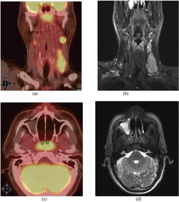
Enrolled patients of this study were treated by IMRT while the definition of primer tumour, involved lymph nodes and critical organs were done using both PET-CT and MR findings. To investigate the effect of adding PET-CT to IMRT planning for NPC patients regarding of staging, tumour volumes and follow-up of patients, all PET-CT and MR-CT sets of patients were evaluated, retrospectively. Initially, PET images were fused with CT data using mutual information registration algorithm which uses image intensity values to match sets. Determination of fusion accuracy was done visually in every case by the radiation oncologist based on the bone structures and spinal cord. Figure 1 shows an example of MR, PET-CT, CT images of a patient. Using PET-CT fused image information, our experienced radiation oncologist delineated GTV, CTV, PTV and critical structures. MR images were fused with simulation CT images and delineated target volumes in the same way as the PET-CT data set.
Initial (a) PET-CT coronal, (b) MR coronal, (c) PET-CT axial and (d) MR axial images of a patient who had T2N2M0 disease by PET-CT and T1N2M0 disease by MR images.

Target volumes delineation and treatment planning
PET-CT and MR images were discussed jointly by the nuclear medicine physicians, radiologist, and radiation oncologist. PET-CT and CT-MR data sets were used to contour GTV, CTV by the same experienced radiation oncologist. The window threshold of PET scan during delineation was set same level for all patients’ images which were suited for tumour volume delineation.
GTV, CTV, PTV and OAR volume definitions were performed according to the guidelines of International Committee of Radiation Unit (ICRU) Report No 62 and Radiation Therapy Oncology Group (RTOG) Nasopharyx Protocol 0225. 15,Reference Lee, Garden, Kramer and Xia16 The primer tumour at the nasopharynx was defined as a GTVNP (nasopharyx gross tumour volume) and the clinically involved lymph nodes as GTVLN (lymph node gross target volume). The CTVNP (nasopharyx clinical target volume) and CTVLN (lymph node clinical target volume) were defined as the GTVNP and GTVLN in addition to a margin for potential microscopic spread and moving variability. The elective target volume included all regional lymphatics (CTV1) was outlined in accordance with the recommendations by Gregorie et al.Reference Gregorie, Levendag and Ang17. PTVs consisted of CTVNP, CTVLP and CTV1 with a 3 mm margin around the CTVs to compensate for the variability of treatment set-up and organ motion. Critical structures included the brainstem, spinal cord, parotid glands, optic nerves, chiasm, eyeballs, lenses, temporal-mandible joint and mandible. 70 Gy, 66–60 Gy and 56–54 Gy were prescribed to the PTVNP (nasopharyx planning target volume), PTVLN (lymph node planning target volume) and PTV1 (elective lymph nodes region) in 35 fractions for both planning sets, respectively. We tried to make sure the CTV doses covered at least 95% of the target while keeping the maximum dose of plans under 110% of 70 Gy. The maximum tolerance dose to the OARs was as follows; Brainstem; 54 Gy, spinal cord; 45 Gy, optical nerves and chiasm; 54 Gy, lens; 6 Gy, mean dose of parotid glands ≤26 Gy and the volume receiving 21 Gy should be <15% of volume, mandible and T-M joints; 60 Gy. Clinically acceptable achieved plans using these doses were selected to compare critical structure doses for both data sets.
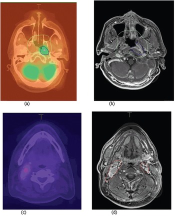
Two separate IMRT plans were obtained for PET-CT based delineation and CT-MR based delineation with the same dose prescriptions. Figure 2 shows delineated PET-CT and CT-MR images of the same patient. A comparison of dose–volume histograms (DVH) was done to evaluate OAR doses at the PET-CT- and CT-MR-based IMRT plans.
(a,b) Delineated fused GTVNP and CTVNP in the PET-CT and CT-MR datasets, respectively. (c,d) GTVLN in the PET-CT and CT-MR data sets, respectively.

DVHs were compared to evaluate volume differences of GTVNP between PET-CT and CT-MR and the involvement of critical structures based on PET-CT and CT-MR images. A statistical analysis could not be done for GTV volume differences given the limited of patients.
Follow-up
All patients completed their planned courses of IMRT and chemotherapy and they were followed-up for 4 to 37 months after completion of IMRT. Median follow-up time was 16 months. During follow-up, MR scan of the head and neck region was taken to evaluate IMRT response of all patients in the 2nd month. All patients also had PET-CT scan in the 3rd month. Patients were later on followed-up with a PET-CT scan every 6 month and MR scan also was performed if clinically necessary.
RESULTS
Staging
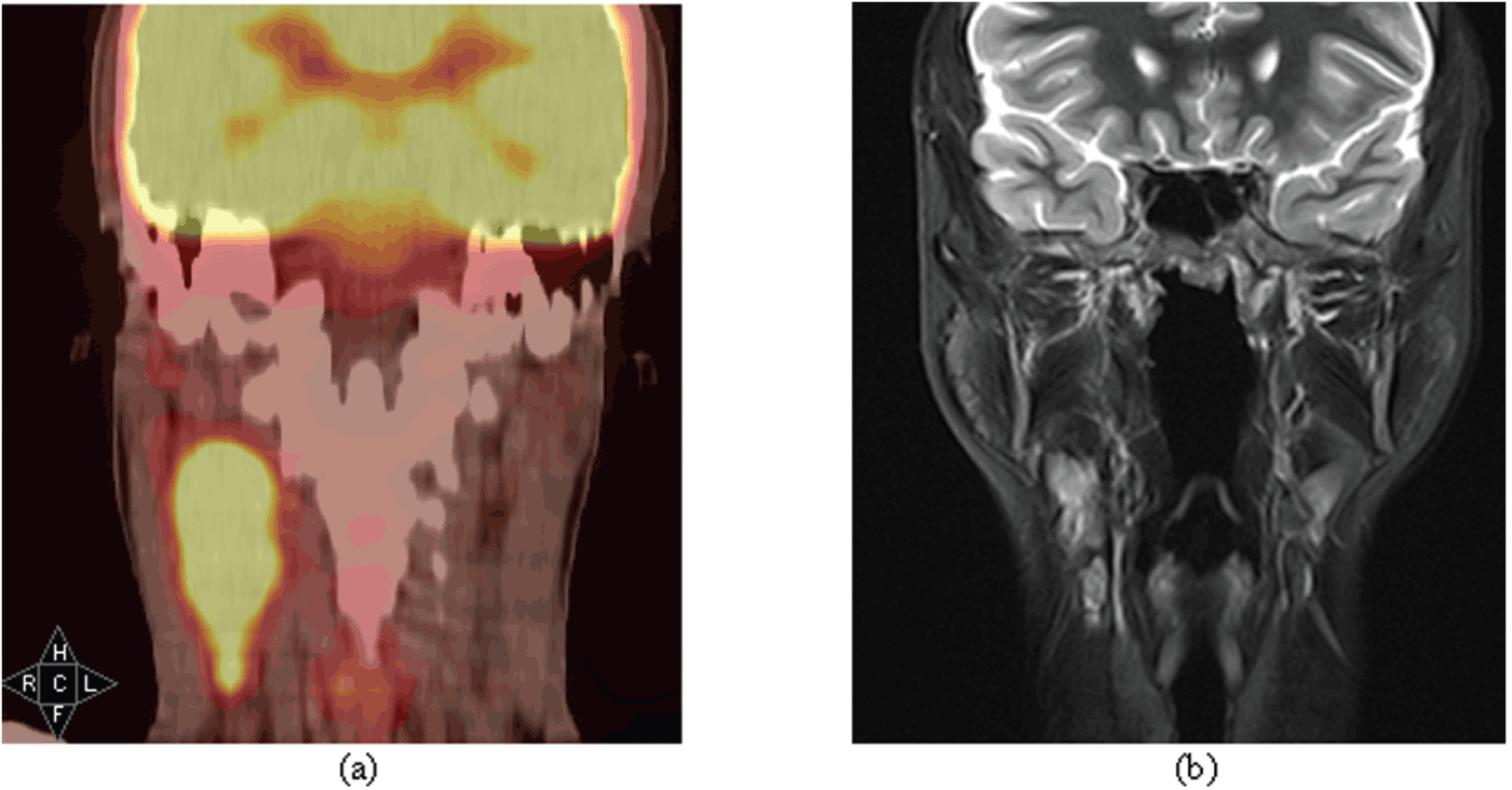
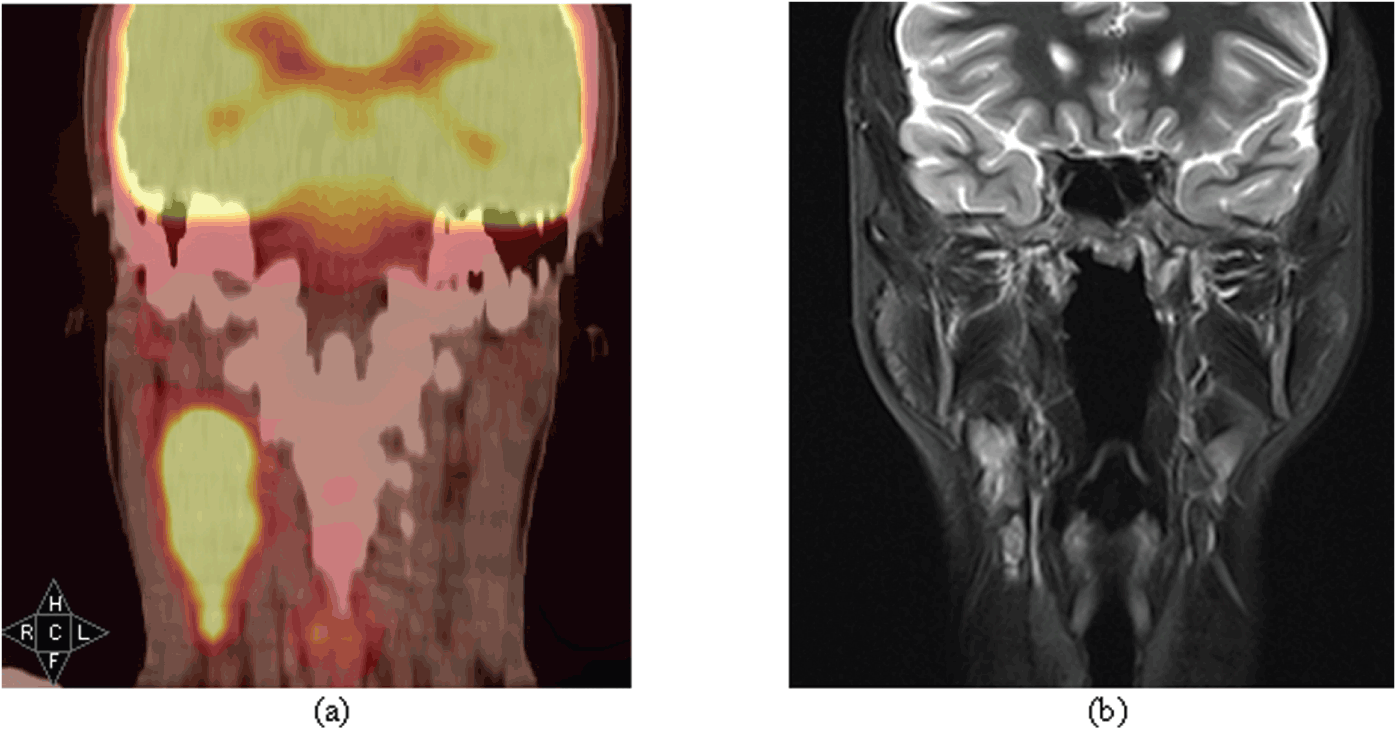
PET-CT imaging changed the MR-based TNM (Tumour Lymph Nodes Metastasis) classification in 11 of 14 patients. The differences were noted in the extension of the primer tumour and the number or the size of the lymph nodes. One patient was upstaged whereas five were downstaged. Table 2 summarized clinical data for the 14 NPC patients. Three cases had intense uptakes of FDG suggestive of nodal involvement. These findings with PET-CT images upstaged the patients from N0 to N2, from N0 to N1 and from N2 to N3a, respectively. Figure 3 shows variations of initial MR and PET-CT images for a patient who had a different N classification at the initial MR and PET-CT images.
(a) Initial PET-CT images of a patient with lymph nodes uptake. (b) Initial MR images of same patient.
Classification and staging of the patients with American Joint Committee on Cancer (AJCC) protocol

One of the patients was found to have distant metastases in both lungs, and its treatment intent was changed from curative to palliative. Three cycles of chemotherapy was initiated. The following PET-CT revealed a good response to chemotherapy while FDG uptake was only in the left jugulo-digastric lymph nodes. He was included in the study and treated with IMRT and chemotherapy as other patients.
GTVNP, GTVLN and CTVNP volumes
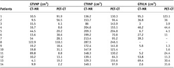
All MR and PET images were fused with simulation CT images obtained subsequent to PET images and using normalized mutual information. The radiation oncologist verified the fused images by visually. The same radiation oncologist delineated all target volumes of CT-MR and PET/CT, separately. Table 3 presents volumes of GTVNP, GTVLN and CTVNP for both image sets.
Volume differences between PET-CT and CT-MR data sets

The median GTVNP was 49.25 cm3 (range of 4.1–122.9 cm3) and 18.8 cm3 (range 2.2–110.1 cm3) for CT-MR and PET-CT sets, respectively. In eight cases, the delineated GTVNP in PET-CT set was smaller than the delineated GTVNP in the CT-MR set. For the remaining six cases, PET-CT presented larger GTVNP volumes than CT-MR sets. In nine cases, the delineated CTVNP which included the nasopharynx in PET-CT was significantly smaller than CTVNP defined on the CT-MR data set. Seven beam IMRT plans with the same prescription were developed and compared, while sparing of the critical structures, to evaluate the effect of volume differences on the IMRT plans.
Evaluation of the critical structures
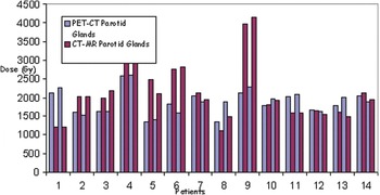
GTVNP, CTVNP and GTVLN were delineated on the CT-MR and PET-CT data sets and IMRT plans were performed to evaluate efficacy of the PET-CT scan in terms of normal tissue sparing. Critical structure doses depend on the target volumes and the distance to them. Parotid glands were well-preserved in six cases which had a smaller GTVNP and CTVNP at the PET-CT data sets. Mean IMRT plan doses for the parotid glands were found to be higher than in the CT-MR data set in one patient although he had a smaller GTVNP and CTVNP in the PET-CT set. Figure 4 shows right and left parotid glands doses for both data sets.
Right and left parotid glands doses for both plans of datasets.

In most of the patient plans, IMRT provided coverage of 95% of the CTVNP volume with 70 Gy, while keeping critical organs doses within their tolerance levels. We tried to obtain 70, 66 and 56 Gy receiving volumes of CTVNP, CTVLN and CTV1 should be 95% for CT-MR and PET-CT data sets in IMRT plans. Right and left parotid gland mean doses were smaller than in the IMRT plan of CT-MR for seven cases that had a smaller CTVNP and GTVNP than the PET-CT set.
Follow-up
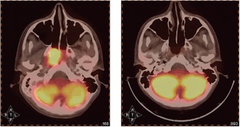
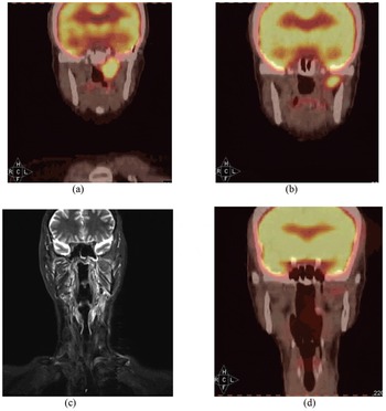
MR and PET/CT scans taken after IMRT were compared to evaluate treatment response and visualize recurrences and residual tumours if any. Figure 5 shows the responses to IMRT and combined chemotherapy for one patient. Only one patient, with T3N1M0 disease at the initial PET-CT, experienced a local recurrence in the CTVNP. An MR scan confirmed the lesion with only T2 trim sequence as a local recurrence inside the CTVNP, treated with a radical dose of 70 Gy. This patient had CyberKnife (Accuray, Sunnyvale, CA) treatment to destroy the residual disease using a total dose of 25 Gy in five fractions. Figure 6 shows control MR and PET/CT images. Another patient, with initially T3N0M0 disease, developed a brain metastasis on PET-CT 3 months following radiotherapy. A third patient scan developed liver metastasis in 6 months and a brain metastasis was detected in a patient with T3N0M0 disease with PET/CT in 3 months after radiotherapy.
PET-CT images of pre and post treatments.

(a) Pre-treatment PET-CT, (b) control PET-CT, (c) control MR and (d) after Cyberknife treatment images of same patient who had a local residual disease after IMRT.

The remaining patients were complete responders to IMRT and chemotherapy. We analyzed the MR and PET-CT scan findings in 10 patients to evaluate the pattern of residual disease or recurrence. MR showed a decrease in the size and number of lymph nodes in four patients while PET-CT showed no uptake in treatment site. MR scan showed a lymph node within the CTV region in one case which did not show any FDG uptake. MR scan reported a 3 mm residual lesion on the wall of nasopharynx while PET-CT scan showed no uptake of FDG at the GTVNP in another case. All patients had positive responses to IMRT and chemotherapy in their second control MR and PET-CT images.
DISCUSSION
IMRT ± chemotherapy is the preferred primary treatment for NPC due to its anatomical location. Recent studies showed that IMRT increased the rate of local control by increasing the dose at the tumour site while sparing adjacent critical organs.Reference Lee, Xia, Quivet and Sultanem18,Reference Cheng, Chao and Low19 The success of IMRT depends on the accurate delineation of the target volumes. The delineation of the target during radiotherapy planning was generally done using only CT or fused MR-CT and PET-CT. PET offers additional advantages over CT and MR for tumour delineation, as it can provide both anatomical and biological tumour information that allows an accurate GTV definition with hypoxia and perfusion. Specific biological information can be obtained using a PET scan, but offers poor spatial resolution in comparison with CT. Combining PET-CT can overcome the limitations associated with PET alone and it presents the advantage of defining GTV in radiotherapy. Daisne et al. showed that PET-CT scan was the most accurate imaging modality to define head and neck region target volumes when compared to CT, MR and PET with surgical specimens.Reference Daisne, Duprez and Weynand20 The comparison of CT and PET-CT planned radiation therapy has shown that inter and intra variations in drawing the GTV were reduced when using PET-CT for delineation.Reference Breen, Publicover, Silva and Pond21 Ciernik et al.Reference Ciernik, Dizendirf and Baumert2 and Paulino et al.Reference Paulino, Koshy and Howell22 also reported that 6 out of 12 patients with head and neck cancer had a significant change in GTV when using PET-CT compared with CT alone. This study contained a relatively small patient number but the results showed efficiency of PET-CT in the IMRT planning.
We included the entire visible tumour at the NPC site as GTVNP as well as involved lymph nodes as seen on CT-MR and PET-CT scan. In most cases, the GTVNP defined by the PET-CT set was smaller than the GTVNP defined by CT-MR. Some tumours could have less metabolically active areas which were not detected with MR and CT images. Our results are similar to those of other studies that delineated GTVNP volumes using PET-CT. A tumour volume decrease when using PET-CT scan also allows us to increase the dose of GTV without increasing the damage on the adjacent critical organs.
The main impact of PET-CT lies in its superior diagnostic accuracy, as compared with MR or CT for involved lymph nodal disease.Reference Schoder, Yeung, Gonen, Kraus and Steven23 CT is often a poor investigation technique for lymph node staging when compared to MR and PET/CT scans. Many lymph nodes that did not meet the criteria of metastasis on MR-CT were diagnosed positive by PET-CT. In our study, the GTVLN volumes increased with by PET-CT in 4 of 14 cases while PET-CT presented different result from MR in terms of lack and presence of involved node in two other cases. However, the sensitivity of PET-CT in the node-negative neck compared with that of examination of a pathological specimen after neck node dissection is only about 70%.Reference Gregorie, Haustermans and Xavier24 Although involved lymph nodes defined by PET-CT scan were included in the GTVLN and treated with a dose of 66 Gy, the limited number of patients included does not allow concluding that PET-CT is more specific than CT or MR.
Several studies have shown that PET-CT scanning is significantly more accurate than CT concerning the definition of the overall TNM staging.Reference Breen, Publicover, Silva and Pond21,Reference Deantonio, Beldi, Gambaro and Loi25 In our study, the implementation of PET-CT changed TNM categories of 11 patients and staging of 5 patients in terms of nodal and/or tumour size. Similar results were reported by Deantonio et al. who found that PET-CT fusion changed TNM categories in 38% and the clinical stage in 14% of 36 patients.Reference Deantonio, Beldi, Gambaro and Loi25 Local tumour extension and staging of regional lymph nodes involvement is best evaluated using PET-CT.
NPC is very radiosensitive, and most NPC tumours regress within 3 months following radiotherapy. Shu-Hang et al. also noted that PET-CT is more specific than MR and CT in detecting residual or recurrent nodal metastasis in head and neck malignancies, with sensitivities ranging from 67% to 100% and specificities from 77% to 100%.Reference Shu-Hang, Tung-Cheig and Sheng-Chieh26 In general, a period of 4 weeks and preferably 6–8 weeks following radiotherapy is reasonable to reduce misinterpretation and yet fulfil clinical requirement.Reference Wong, Chang and Cheng27 In our study, 14 patients had been followed for a median of 16 months by MR scan of head and necks as well as PET-CT scan. We found one nodal recurrence with MR and PET-CT scan, within CTV that was accepted as a local failure. That patient had CyberKnife treatment using a total dose of 25Gy. During the follow-up period, MR and PET-CT images were taken to evaluate the response of IMRT and chemotherapy for 10 patients. Comparison of these images findings showed differences for six patients in term of residual and recurrence findings at the primer tumour and lymph nodes area. However, second control images which were available in eight patients were similar with each other. In conclusion, we found that PET-CT scan is superior to MR scan in investigating treatment response during early follow-up.
Critical organ sparing was achieved with IMRT plans for both MR-CT and PET-CT data sets in our study. We demonstrated with a relatively small number of patients that IMRT plans are capable of providing adequate coverage of PET-CT targets which are smaller than targets defined by MR-CT. Critical organ doses were markedly reduced when GTVNP was delineated using the PET-CT fused images. Ten patients presented response to IMRT and chemotherapy which were treated with IMRT using CT-MR and PET-CT findings. We showed that the definition of the exact GTV and CTV with PET-CT imaging cause to better parotid gland sparing at the nasopharynx IMRT. Although better parotid glands sparing could achieved with smaller GTV and CTV which were delineated using PET-CT imaging, shrunk target volumes in the PET-CT images could be dangerous. It may cause to locoregional recurrence after IMRT in the parotid gland areas. Evaluated patients in this study were treated with IMRT which was obtained using combination of MR and PET-CT images.
However, this study which fused both CT-MR and PET-CT images may introduce the possibility of error due to accuracy of fusion, and which was visually evaluated taking into account bony structures. A second limitation is in the estimation the threshold settings for the PET windows suited for tumour volume delineation.
The aim of our study was to show the effectiveness of the use of PET-CT in IMRT for NPC patients and its contribution to evaluate treatment response. The present study suggests that PET-CT-based IMRT planning is feasible, accurate, and easily integrated into the routine radiation therapy planning to help with target volume definition when scan was performed with same condition simulation CT scan to reduce fusion errors. The clinical outcomes were considered in this study on a relatively small number of patients. The use of combined PET-CT imaging could improve target volume delineation and nodal staging. It enables an increase in dose inside the GTV and CTV which includes uptake of FDG. PET-CT also contributes to the detection of unexpected nodal or distant metastasis. Overall, PET-CT appeared to provide significant information on control scans for most of our patients whose MR imaging studies showed tumour/lymph nodes residual or recurrence.