Introduction
The Mycocaliciales (Mycocaliciomycetidae, Eurotiomycetes) is an order of resinicolous, saprobic and lichenicolous fungi, especially well known for species having pin-like (mycocalicioid) ascomata (Tibell & Wedin Reference Tibell and Wedin2000; Tuovila Reference Tuovila2013). Currently seven genera are accepted within the Mycocaliciales, but recent phylogenetic analysis shows that the genera Chaenothecopsis (Vain.) Oxner, Mycocalicium Vain. and Phaeocalicium A. F. W. Schmidt are polyphyletic and not yet well delimited (Tibell & Vinuesa Reference Tibell and Vinuesa2005; Tuovila Reference Tuovila2013; Thiyagaraja et al. Reference Thiyagaraja, Ertz, Lücking, Wanasinghe, Aptroot, Cáceres, Hyde, Tapingkae and Cheewangkoon2022; Ertz et al. Reference Ertz, Diederich, Lendemer, Komposch, Harris and Huereca2023).
The anamorphs of Mycocaliciales show a remarkable diversity and include both hyphomycetes and coelomycetes, sometimes even within one species (Tibell Reference Tibell1991, Reference Tibell1997; Tibell & Vinuesa Reference Tibell and Vinuesa2005). The pycnidia of the coelomycete anamorphs vary from very simple structures in which the pycnidial wall is not or hardly differentiated from the conidiogenous cells (Asterophoma-anamorphs; Hawksworth Reference Hawksworth1981; Tibell Reference Tibell1991), to other species where pycnidia develop with complex wall structures consisting of multiple layers (Tibell Reference Tibell1995). In some species, for example in Chaenothecopsis viridireagens (Nádv.) A. F. W. Schmidt, multiple pycnidia can be formed on stalks forming tree-like structures (Tibell Reference Tibell1993). Some species have been shown to produce a hyphomycete anamorph in culture, for example Chaenothecopsis schefflerae (Samuels & D. E. Buchanan) Tibell (Samuels & Buchanan Reference Samuels and Buchanan1983).
The conidiogenous cells in most anamorphs of Mycocaliciales are phialidic, often with a more or less distinct collarette, or sometimes showing percurrent proliferations (Samuels & Buchanan Reference Samuels and Buchanan1983; Tibell Reference Tibell1991, Reference Tibell1995; Ertz et al. Reference Ertz, Diederich, Lendemer, Komposch, Harris and Huereca2023). The hyphomycete anamorph of Chaenothecopsis haematopus Tibell produces the conidia in chains (Tibell & Constantinescu Reference Tibell and Constantinescu1991; Tibell Reference Tibell1997). In most species, the conidia are ellipsoidal, aseptate and hyaline or brown (Tibell Reference Tibell1997; Tibell & Vinuesa Reference Tibell and Vinuesa2005). Recently, however, a lichenicolous species of Mycocalicium was described having campylidia-like conidiomata and appendiculate conidia (Ertz et al. Reference Ertz, Diederich, Lendemer, Komposch, Harris and Huereca2023).
In the Netherlands, an unknown sporodochioid lichenicolous fungus was found on Lecanora expallens Ach. in several localities. The characteristics did not fit any described species. DNA analysis revealed that it belonged to the Mycocaliciales. It is described here as new to science and is the first species of Mycocaliciales known to produce sporodochia, further demonstrating the large anamorphic diversity in Mycocaliciales.
Methods
Material collection and morphological study
Specimens of the unknown lichenicolous species were collected over several years in the Netherlands, and stored in BR and the private herbaria of the authors. Many sightings were documented with images via www.waarneming.nl, a website on which volunteers can record their observations of all organisms. We have therefore provided the waarneming.nl observation ID in the specimen information. Specimens were studied with stereomicroscopes (×7–45 magnification) and light microscopes (×40–1000 magnification). Macroscopical images were captured using a mirrorless camera through a Raynox DCR 150 tube lens with a ×4 or ×10 microscope lens. For microscopic observations and measurements, specimens were mounted in water and images were captured with a Canon 1200D camera mounted on a light microscope.
Molecular methods
Two well-preserved herbarium specimens (van der Kolk 3280, 3979) that were two weeks old were used for DNA isolation. A sterile scalpel blade was used to take a large number of conidia from a sporodochium for direct PCR, as described in Ertz et al. (Reference Ertz, Tehler, Irestedt, Frisch, Thor and van den Boom2015). The conidia were placed directly in microtubes with 20 μl H2O. Amplification reactions were prepared for a 50 μl final volume, as detailed in Ertz et al. (Reference Ertz, Sanderson, Łubek and Kukwa2018). The nuITS rDNA (ITS1 + 5.8S + ITS2) was amplified using the primers ITS1F and ITS4 (White et al. Reference White, Bruns, Lee, Taylor, Innis, Gelfand, Sninsky and White1990), and the nuLSU rDNA was amplified using the primers LIC15R (Miadlikowska et al. Reference Miadlikowska, McCune and Lutzoni2002) and LR6 (Vilgalys & Hester Reference Vilgalys and Hester1990). The PCR cycling conditions for the nuITS was as follows: 10 min at 95 °C; 35 cycles of 45 s at 95 °C, 1 min at 52 °C and 75 s at 72 °C; 10 min of final elongation at 72 °C. Those for the nuLSU consisted of: 10 min at 95 °C; 25 cycles of 45 s at 95 °C, 40 s at 52 °C and 150 s at 72 °C; 14 cycles of 45 s at 95 °C, 40 s at 52 °C and 150 s at 72 °C (+ 5 s per cycle); 10 min of final elongation at 72 °C. Both strands were sequenced by Macrogen® using the amplification primers, and with the additional primer LR3 for nuLSU (Vilgalys & Hester Reference Vilgalys and Hester1990). Sequence fragments were assembled with Sequencher v. 5.4.6 (Gene Codes Corporation, Ann Arbor, Michigan, USA). Sequences were subjected to megaBLAST searches to verify their closest relatives and to detect potential contaminations.
Phylogenetic analysis
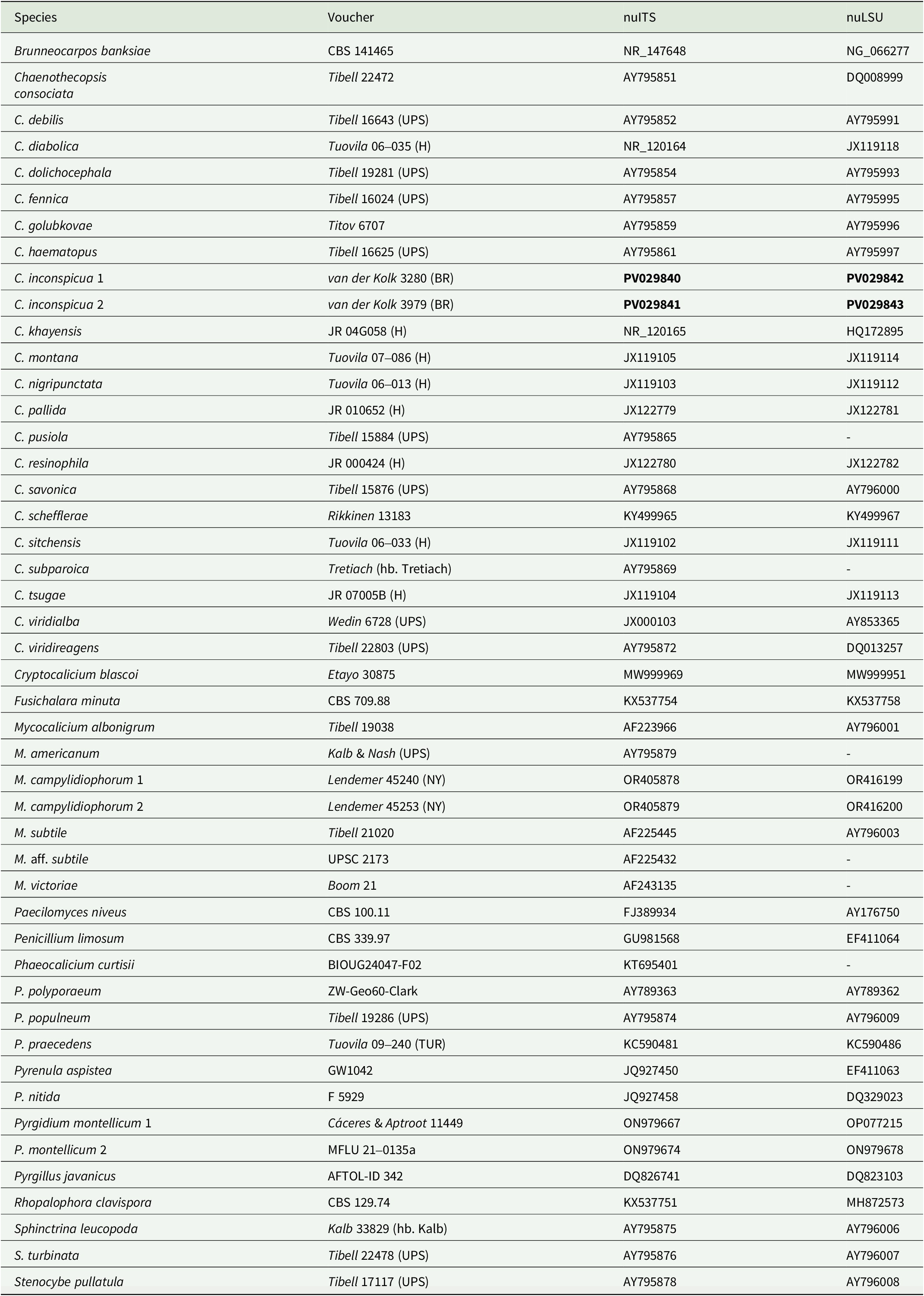
The sequences were included in the two-locus dataset (nuLSU and nuITS) used for the phylogeny of the order Mycocaliciales published in Ertz et al. (Reference Ertz, Diederich, Lendemer, Komposch, Harris and Huereca2023) (Table 1). They were aligned manually using Mesquite v. 3.04 (Maddison & Maddison Reference Maddison and Maddison2015). Terminal ends of sequences, ambiguously aligned regions, and introns were delimited manually and excluded from the datasets.
Species names, voucher specimens and GenBank Accession numbers of Mycocaliciales and outgroup taxa used in the phylogenetic analyses. Newly generated sequences are in bold.

The resulting matrix of Mycocaliciales consisted of 47 terminals and 1469 (1045 for nuLSU and 424 for nuITS) unambiguously aligned sites. Three species of Pyrenulales, viz. Pyrenula aspistea (Ach.) Ach., P. nitida (Weigel) Ach. and Pyrgillus javanicus (Mont. & Bosch) Nyl. were used as the rooting taxa in the Mycocaliciales dataset.
Best-fit evolutionary models were estimated using the Akaike Information Criterion (AIC) as implemented in jModelTest v. 2.1.10 (Darriba et al. Reference Darriba, Taboada, Doallo and Posada2012). The TIM1 + I + G model was selected for the nuLSU dataset and the TIM2ef + I + G model was selected for the nuITS dataset.
Analyses for topological incongruence among loci were carried out. The six taxa for which nuLSU sequences were not available were first removed from the nuITS dataset in order to analyze both datasets with the same 41 terminals. The single-locus datasets were analyzed with a maximum likelihood (ML) approach using the program RAxML v. 8.2.12 (Stamatakis Reference Stamatakis2014) on the CIPRES Web Portal (Miller et al. Reference Miller, Pfeiffer and Schwartz2010) with 1000 ML bootstrap iterations (ML-BS). The GTRGAMMA model was used, and node support was assessed running 1000 bootstrap replicates. We analyzed the two single-locus datasets for topological incongruence by assuming a conflict to be significant when two different relationships (one being monophyletic and the other being non-monophyletic) for the same set of taxa were both supported with bootstrap values ≥ 70% (Mason-Gamer & Kellogg Reference Mason-Gamer and Kellogg1996; Reeb et al. Reference Reeb, Lutzoni and Roux2004). Based on this criterion, no conflict was detected and the nuLSU and nuITS datasets were concatenated.
Bayesian analyses were carried out on the two-locus dataset under the selected models for two partitions (nuLSU, nuITS) and using the Metropolis-coupled Markov chain Monte Carlo method (MCMCMC) in MrBayes v. 3.2.7a (Huelsenbeck & Ronquist Reference Huelsenbeck and Ronquist2001; Ronquist & Huelsenbeck Reference Ronquist and Huelsenbeck2003) on the CIPRES Web Portal (Miller et al. Reference Miller, Pfeiffer and Schwartz2010). Two parallel MCMCMC runs were performed, each using four independent chains and 20 million generations, sampling trees every 1000th generation. Posterior probabilities (PP) were determined by calculating a majority-rule consensus tree generated from the 30 002 post-burn-in trees of the 40 002 trees sampled by the two MCMCMC runs, using the sumt option of MrBayes. Convergence between runs was verified using the PSRF (Potential Scale Reduction Factor), where values were all equal or close to 1.000.
In addition, a maximum likelihood analysis was performed on the two-locus dataset of Mycocaliciales using RAxML v. 8.2.12 (Stamatakis Reference Stamatakis2014) on the CIPRES Web Portal (Miller et al. Reference Miller, Pfeiffer and Schwartz2010), with 1000 ML bootstrap iterations (ML-BS) and the GTRGAMMA model. This two-locus dataset was divided into two partitions (nuLSU and nuITS).
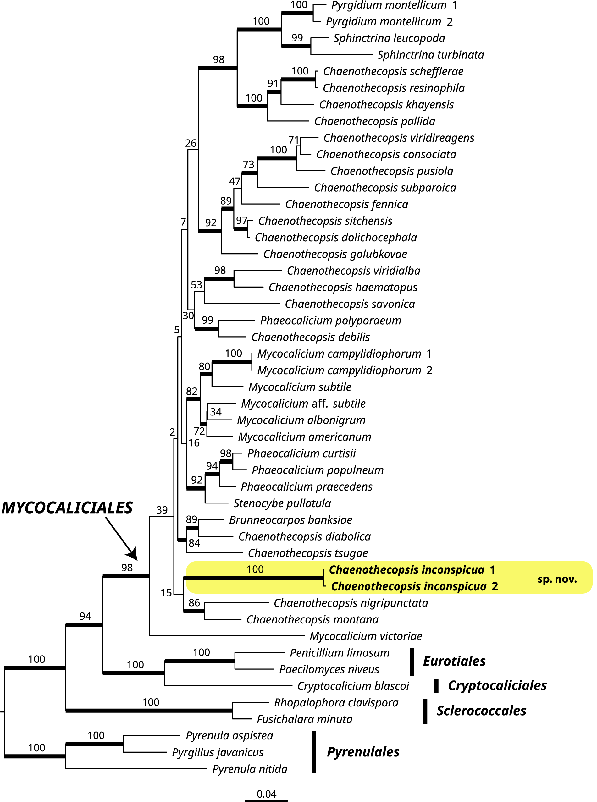
The ML tree did not contradict the Bayesian tree topology for the strongly supported branches. Therefore, only the ML tree is shown, with the ML-BS values added above or near the internal branches. Internodes with ML-BS ≥ 70 and PP ≥ 0.95 were considered to be significant and are represented by thicker lines (Fig. 1). Phylogenetic trees were visualized using FigTree v. 1.4.2 (Rambaut Reference Rambaut2012).
Phylogeny of Mycocaliciales (with eight species of other orders as outgroup) based on a dataset of nuLSU and nuITS sequences that resulted from the RAxML analysis. Maximum likelihood bootstrap values are shown near internal branches. Internal branches considered strongly supported by the Bayesian (PP ≥ 0.95) and RAxML (ML-BS ≥ 70) analyses are represented by thicker lines. The newly sequenced samples of Chaenothecopsis inconspicua are in bold. The lineage corresponding to the new species is highlighted. In colour online.

Results
Four new sequences (two nuLSU and two nuITS) were obtained for this study (Table 1). The RAxML tree obtained from the combined two-locus analysis of the Mycocaliciales dataset is shown in Fig. 1. The main well-supported lineages are in accordance with the results obtained by Tibell & Vinuesa (Reference Tibell and Vinuesa2005), Tuovila et al. (Reference Tuovila, Schmidt, Beimforde, Dörfelt, Grabenhorst and Rikkinen2013) and Ertz et al. (Reference Ertz, Diederich, Lendemer, Komposch, Harris and Huereca2023). The order Mycocaliciales is strongly supported, but the nodes of the backbone of the Mycocaliciales clade are mainly poorly supported. The genera Chaenothecopsis, Mycocalicium and Phaeocalicium are polyphyletic. The new species, represented by two terminals, is sister to the clade of Chaenothecopsis montana Rikkinen and C. nigripunctata Rikkinen.
Taxonomy
Chaenothecopsis inconspicua van der Kolk, Ertz, Hessen & Boers sp. nov.
MycoBank No.: MB 857482
Differing from all other Chaenothecopsis species by the sporodochioid conidiomata and by its ecology, growing on the thallus of Lecanora expallens.
Type: The Netherlands, Drenthe, Ruinen, 52.7632°N, 6.3535°E, on Lecanora expallens on the rain-sheltered side of a roadside oak tree (Quercus robur) in village, 11 November 2023, H. van der Kolk 3979 (BR—holotype; hb. van der Kolk—isotype). GenBank Accession numbers: PV029841 (nuITS), PV029843 (nuLSU).
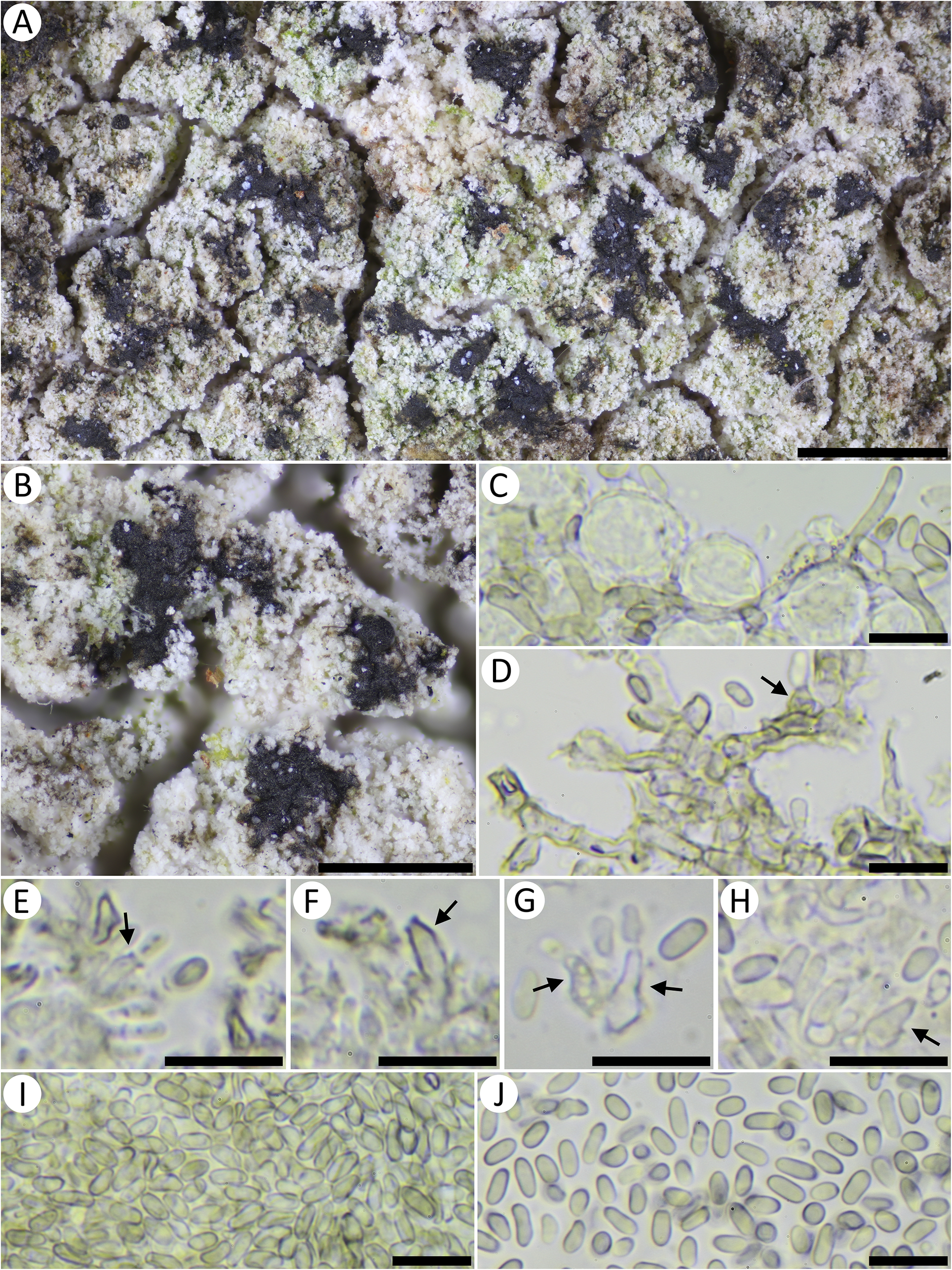
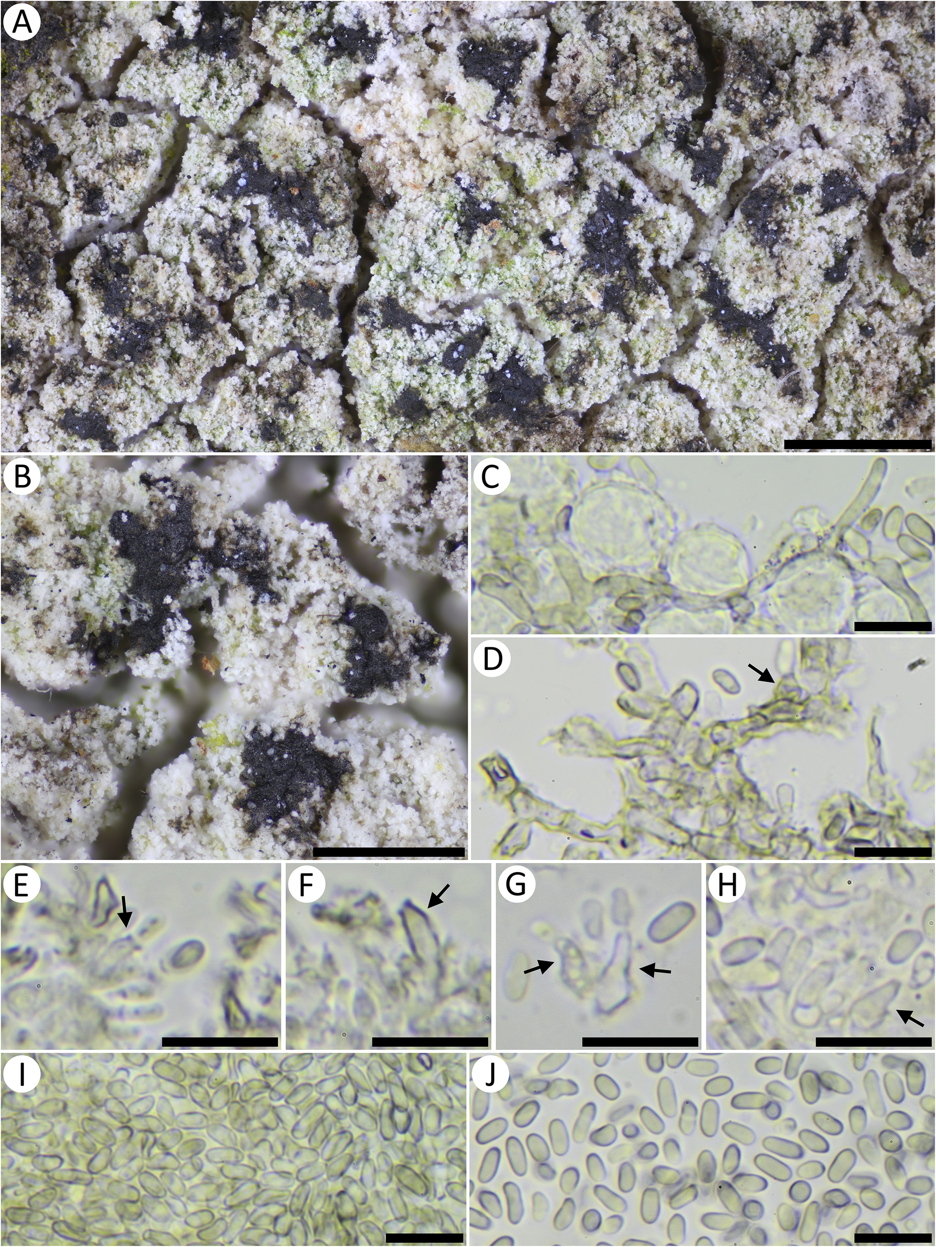
Chaenothecopsis inconspicua (holotype). A & B, habitus on Lecanora expallens. C & D, mycelium. E–H, conidiogenous cells (arrowed). I & J, conidia. C–J, mounted in water. Scales: A = 1 mm; B = 0.5 mm; C–J = 10 μm. In colour online.
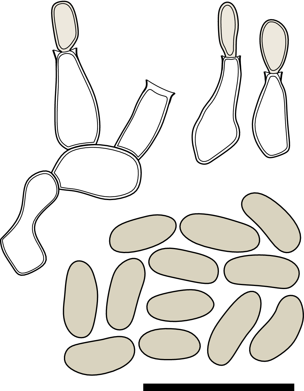
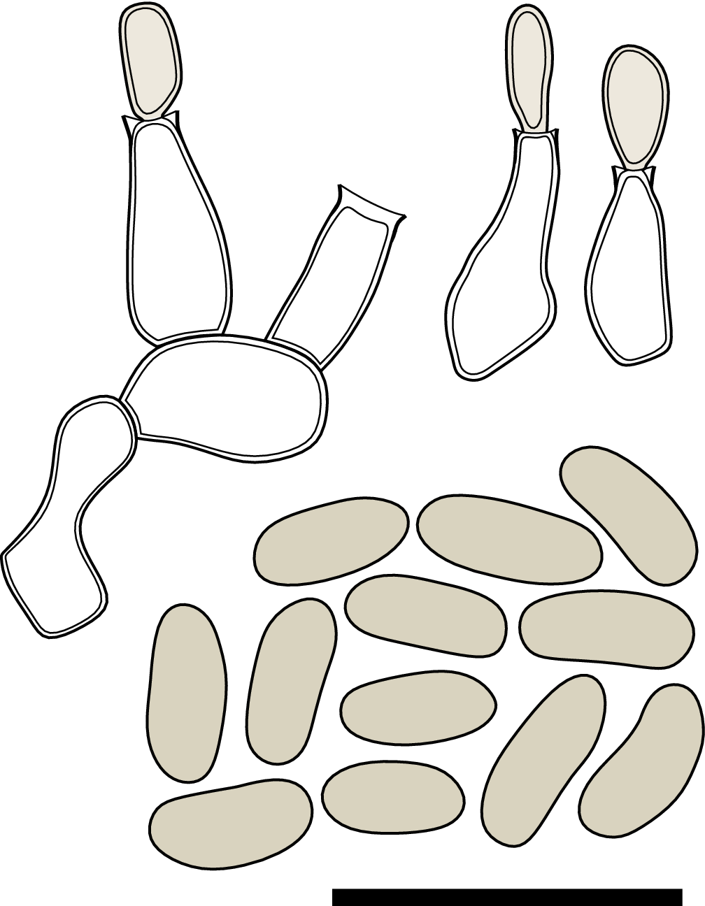
Chaenothecopsis inconspicua, conidiogenous cells and conidia. Scale = 10 μm.
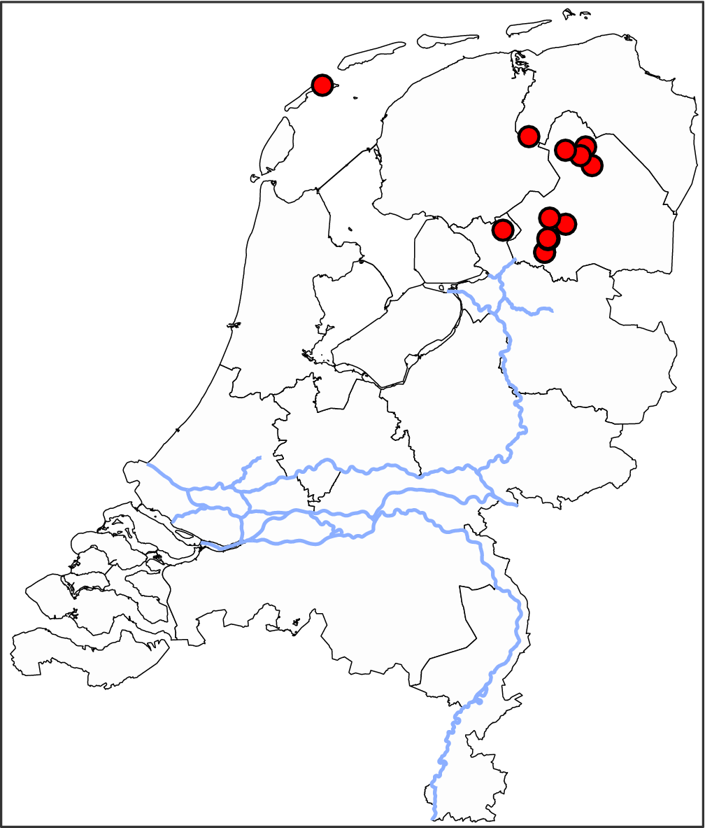
Currently known distribution of Chaenothecopsis inconspicua in the Netherlands. In colour online.

Teleomorph: unknown.
Anamorph: conidiomata sporodochioid, brown-black to black, appearing smooth, first small and irregularly rounded, 75–300 μm diam., later multiple sporodochia can become confluent and form irregularly rounded or ellipsoid clusters up to 700 μm diam. Mycelium immersed in the host thallus, composed of light brown hyphae with an irregular outline, 1.4–3.0 μm wide. Conidiophores reduced to the conidiogenous cell, monoblastic, variable in shape, cylindrical to pyriform, phialidic, often with a slightly thickened collarette, colourless or light brown, 4–8 × 1.5–3 μm. Conidia light brown, simple (very rarely 1-septate), smooth-walled, ellipsoid, sometimes slightly curved, sometimes one end slightly truncated, 4.3–5.9 × 1.9–2.5 μm, l/w ratio = 1.9–3.0.
Etymology
The epithet inconspicua refers to the inconspicuous conidiomata.
Distribution and ecology
Currently known only from the Netherlands, where it seems to be fairly common in the northern half of the country (Fig. 4). Lichenicolous on Lecanora expallens on rain-sheltered side of older deciduous trees, including Quercus robur and Acer pseudoplatanus. Most records are from old roadside oak trees. The fungus often causes a yellow discoloration of the host thallus.
Additional specimens examined
The Netherlands: Drenthe: Dwingelderveld, on Lecanora expallens on oak (Quercus robur) along forest edge, 52.8115°N, 6.4567°E, 23 xii 2020, J. Boers s. n., waarneming.nl ID: 204794711; Dwingeloo, on Lecanora expallens on oak (Quercus robur) in village, 52.8337°N, 6.3657°E, 11 ii 2022, H. van der Kolk 2700 (hb. H. van der Kolk), waarneming.nl ID: 233979129; Koekangerveld, on Lecanora expallens on oak (Quercus robur) in village, 52.7157°N, 6.3347°E, 30 xi 2022, H. van der Kolk 3280 (BR; hb. H. van der Kolk), waarneming.nl ID: 260269713 (GenBank Accession numbers: PV029840 (nuITS), PV029842 (nuLSU)); Ruinen, on Lecanora expallens on oak (Quercus robur) in village, 52.7593°N, 6.3546°E, 30 xi 2022, H. van der Kolk 3283 (hb. H. van der Kolk), waarneming.nl ID: 260269708; ibid., on Lecanora expallens on oak (Quercus robur) in village, 52.7622°N, 6.3614°E, 27 iv 2022, B. Hessen s. n., waarneming.nl ID: 239282138; Loon, on Lecanora expallens on oak (Quercus robur) in village, 53.0126°N, 6.6127°E, 17 iv 2022, B. Hessen s. n. (hb. B. Hessen), waarneming.nl ID: 238128888; Vries, on Lecanora expallens on oak (Quercus robur) in village, 53.0776°N, 6.578°E, 4 vi 2023, B. Hessen s. n., waarneming.nl ID: 274863156; Koekangerveld, on Lecanora expallens on oak (Quercus robur) in village, 52.7158°N, 6.3346°E, 27 vii 2022, B. Hessen s. n., waarneming.nl ID: 250544157; Norg, on Lecanora expallens on oak (Quercus robur) in village, 53.0671°N, 6.4618°E, 22 v 2022, B. Hessen s. n. (hb. B. Hessen), waarneming.nl ID: 242593519; Zeijen, on Lecanora expallens on oak (Quercus robur) in village, 53.0477°N, 6.5468°E, 23 vi 2022, B. Hessen s. n. (hb. B. Hessen), waarneming.nl ID: 246549677. Friesland: Vlieland, on Lecanora expallens on maple (Acer pseudoplatanus) in village, 53.2958°N, 5.0658°E, 18 i 2020, H. van der Kolk s. n., waarneming.nl ID: 293366657. Overijssel: Steenwijk, on Lecanora expallens on oak (Quercus robur) in village, 52.7938°N, 6.0987°E, 1 x 2022, B. Hessen s. n., waarneming.nl ID: 255912387. Groningen: De Wilp, on Lecanora expallens on oak (Quercus rubra) in village, 53.1158°N, 6.2528°E, 9 x 2023, B. Hessen s. n., waarneming.nl ID: 290197211.
Discussion
Chaenothecopsis inconspicua can be recognized by the black sporodochia that contain a dense mass of non-septate, ellipsoid, light brown conidia and by its lichenicolous habit on Lecanora expallens. Chaenothecopsis inconspicua is in many aspects similar to the anamorphs that are known in Mycocaliciales. Specifically, it shares with species of Chaenothecopsis and Mycocalicium, the cylindrical to pyriform, phialidic conidiogenous cells with a thickened collarette and the non-septate, ellipsoid, smooth conidia (Hawksworth Reference Hawksworth1981; Tibell Reference Tibell1990, Reference Tibell1995, Reference Tibell1997). However, as far as we are aware, it is the first species of Mycocaliciales shown to produce sporodochioid conidiomata. So far, the anamorphs of Mycocaliciales are known to produce pycnidia (Tibell Reference Tibell1997; Tibell & Vinuesa Reference Tibell and Vinuesa2005), campylidia-like conidiomata (Ertz et al. Reference Ertz, Diederich, Lendemer, Komposch, Harris and Huereca2023), or in culture sometimes also hyphomycetous anamorphs arising from colonies of hyphae (Tibell Reference Tibell1997). Chaenothecopsis inconspicua can therefore easily be distinguished from all other known Mycocaliciales by its black sporodochioid conidiomata and lichenicolous habit on Lecanora expallens.
The genus Chaenothecopsis is currently polyphyletic and not well delimited (Tibell & Vinuesa Reference Tibell and Vinuesa2005; Tuovila Reference Tuovila2013; Thiyagaraja et al. Reference Thiyagaraja, Ertz, Lücking, Wanasinghe, Aptroot, Cáceres, Hyde, Tapingkae and Cheewangkoon2022; Ertz et al. Reference Ertz, Diederich, Lendemer, Komposch, Harris and Huereca2023). The phylogenetic placement of the type species of the genus, Chaenothecopsis rubescens Vain., is still uncertain. The only sequence of this species in GenBank (Accession OQ717807) was not included in the phylogenetic analyses because in a previous study it proved difficult to align with sequences of all other Sphinctrinaceae, such that confirmation of its identity is needed (Ertz et al. Reference Ertz, Diederich, Lendemer, Komposch, Harris and Huereca2023). Although it is therefore uncertain whether the new species belongs to Chaenothecopsis in the strict sense, we prefer to provisionally place it in this genus, awaiting further studies to properly delimit the genera within the Mycocaliciales. In our phylogenetic analysis, Chaenothecopsis inconspicua forms a weakly supported clade together with its sister clade containing C. montana and C. nigripunctata. Both Chaenothecopsis montana and C. nigripunctata are resinicolous species described from North America and known only by their sexual state as pin-like ascomata (Rikkinen Reference Rikkinen2003; Tuovila et al. Reference Tuovila, Larsson and Rikkinen2012).
Several other anamorphic lichenicolous fungi are known from Lecanora expallens (see identification key). Both Briancoppinsia cytospora (Vouaux) Diederich et al. and Phoma lecanorina Diederich differ from Chaenothecopsis inconspicua in forming pycnidia with hyaline and slightly more elongated conidia (Diederich Reference Diederich1986; Diederich et al. Reference Diederich, Lawrey, Sikaroodi, van den Boom and Ertz2012). Lichenodiplis lecanorae (Vouaux) Dyko & D. Hawksw. also differs from Chaenothecopsis inconspicua in forming pycnidia and having 1-septate conidia (Hawksworth & Dyko Reference Hawksworth and Dyko1979). The macroscopical appearance of C. inconspicua is similar to that of species of Psammina Sacc. & M. Rousseau, several of which also frequently grow on Lecanora expallens. The sporodochia of C. inconspicua especially resemble the smooth sporodochia of Psammina filamentosa Kolk & Earl.-Benn. However, Psammina species are microscopically easily distinguished by their palmate conidia with numerous arms (van der Kolk et al. Reference van der Kolk, Earland-Bennett and Hawksworth2020, Reference van der Kolk, Ertz and Diederich2024). Lastly, Minutoexcipula beaglei Etayo is reported from Lecanora aff. expallens from Chile. It resembles Chaenothecopsis inconspicua in the black sporodochia, but Minutoexcipula beaglei has conidia that are 1-septate (Diederich et al. Reference Diederich, Zhurbenko, Pinault, Berger, Etayo and van den Boom2024a).
Species of Feltgeniomyces Diederich are also similar to the new species in their brown to blackish sporodochia, and in the type of conidiophores and conidia, but they differ in the much more stacked conidiogenous cells (more loosely arranged in Chaenothecopsis inconspicua). Feltgeniomyces lecanorae Zhurb. is the only species of the genus reported from Lecanora, but it differs from C. inconspicua by the paler (dark brown, becoming light brown and translucent when wet) and smaller (up to 120 μm diam.) sporodochia, shorter and broader conidia (3–4 × 2.5–3 μm), and being reported from a different host species (Lecanora chlarotera s. lat.) (Diederich et al. Reference Diederich, Etayo and Zhurbenko2024b). Feltgeniomyces luxemburgensis Diederich is similar to the new species in its black sporodochia, but it has much larger conidia (5–10 × 4–6.5 μm) and is reported from a different host genus (Lecidella). Molecular data are needed to verify the systematic position of the genus Feltgeniomyces, which is currently unknown.
Chaenothecopsis inconspicua has been mostly found on older trees and is probably not rare in the northern half of the Netherlands, where oak trees with large thalli of Lecanora expallens are common. It has most often been found on roadside trees in urban areas, but occasionally also in forested areas. The distribution of the new species could suggest that it is sensitive to SO2 pollution, which in the previous century affected the southern part of the Netherlands more than the northern half (van Dobben & de Bakker Reference van Dobben and de Bakker1996), and to this day has shaped distribution patterns of many lichen species. Other Mycocaliciales species in the Netherlands, including Chaenothecopsis pusilla (A. Massal.) A. F. W. Schmidt and C. savonica (Räsänen) Tibell., are almost exclusively found on the wood of dead trees or, very rarely, in deep crevices of very old oak trees. So far, no other species of Mycocaliciales was found growing on the same tree as C. inconspicua.
Key to the lichenicolous fungi on Lecanora expallens

Author ORCIDs
Henk-Jan van der Kolk, 0000-0002-8023-379X; Damien Ertz, 0000-0001-8746-3187.
Competing Interests
The authors declare none.