Introduction
Transmissible gastroenteritis virus (TGEV) is the aetiological agent of transmissible gastroenteritis (TGE), a condition associated with high morbidity in animals of all ages and high mortality in suckling piglets. It occurs at almost every swine farm and results in severe economic losses. It is therefore important to prevent and control the disease. From previous reports we can see that serodiagnostic assays for TGEV, such as virus neutralization, indirect immunofluorescent antibody, haemagglutinin and enzyme-linked immunosorbent assays (ELISAs), require the large-scale preparation of native antigen (Liu et al., Reference Liu, Kokuho and Kubota2001; Carman et al., Reference Carman, Josephson and McEwen2002; Song et al., Reference Song, Kang and Oh2006; Kim et al., Reference Kim, Kim, Pyo, Tark, Song and Hyun2007). Some researchers intend to replace viral antigens with recombinant protein in the diagnostic ELISA.
TGEV is a member of the Coronaviridae family. It has the largest single-stranded positive-polarity RNA genome among all RNA viruses in nature. The RNA genome is encapsulated in multiple copies of the nucleocapsid (N) protein and is packaged as a helical nucleocapsid in the mature enveloped virion. Numerous studies have shown that the N gene of TGEV is highly conserved, and high levels of N protein-specific antibody are produced at the early stage of TGEV infection (Briton et al., Reference Briton, Carnemes, Page, Garwes and Parra1988). Therefore, the N gene is an ideal candidate antigen for use in the development of TGEV diagnostic techniques.
In this paper, we describe the cloning and expression of the structural N gene, using expressed recombinant protein to develop an ELISA to detect the N serum antibody. This work will enable the detection of N protein antibodies from TGEV-infected pigs.
Materials and methods
Enzymes and reagents
Enzymes (restriction endonucleases, polymerases and ligases) and T4 DNA ligase were purchased from TaKaRa Corp (Dalian, China). The DNA gel recovery kit was obtained from Sangon Corp (Shanghai, China), and the PET-32a(+)vector, TGEV-positive sera were prepared in our laboratory.
The TGEV strain SC-Y was isolated from diarrhoeal piglet faecal samples and blindly subcultured eight times in the swine testicular cell line. Total RNA was extracted using Trizol reagent (Invitrogen, CA, USA) according to the manufacturer's instructions.
Amplification and construction of the prokaryotic expression vector
A pair of primers was designed according to the N sequence of TGEV Purdue strain in GenBank and synthesized by TaKaRa Biotechnology Corp:
P1: 5′-AAC TTC GAA ATG GCC AAC C-3′
P2: 5′AGC TCG AGC ATC TCG TTT AG3′
Reverse transcription was carried out in 20 μl volumes, containing 5 μl of viral RNA, 4 μl of 5×RT buffer, 2 μl of dNTPs (2.5 mmol), 0.5 μl of RNase inhibitor (40 U/μl), 1.0 μl of reverse primer (50 pmol), 2 μl of Avian myeloblastosis virus (AMV) reverse transcriptase (5 U/μl), and 5.5 μl dH2O. Samples were incubated at 42°C for 30 min. The reaction was terminated by heating at 99°C for 5 min.
Polymerase chain reaction (PCR) was performed in 50 μl volumes containing 5 μl of the first-strand cDNA template, 10 μl of 5× PCR buffer, 0.5 μl of each 100 pmol forward and reverse primer, 0.25 μl of Taq DNA polymerase (5 U/μl) and 33.75 μl dH2O. The PCR parameters were 2 min at 94°C, and 30 cycles of 60 s at 94°C, 60 s at 55°C, 90 s at 72°C, and a final extension step of 10 min at 72°C. The PCR amplified products were visualized by gel electrophoresis. The product of the PCR was subcloned into the prokaryotic expression vector PET-32a(+) on corresponding restriction enzyme sites. The recombinant plasmid, namely PET-N, was transformed into competent Escherichia coli BL21 (DE3). Positive clones were identified by Bsp1191 and Xho I.
Inducing expression of N protein in BL21
A transformed colony containing the recombinant vector (PET-N) and negative control vector PET-32a(+) was inoculated into Luria-Bertani (LB) medium containing 100 μg/ml ampicillin, and shaken at 37°C until the OD600 value reached 0.4–0.6. IPTG was then added to a final concentration of 1 mmol/l and incubated for 4 h.
SDS-PAGE and Western blot analysis
The expressed protein was subjected to 10% sodium dodecyl sulphate polyacrylamide gel electrophoresis (SDS-PAGE) and Western blot, performed according to the standard method of Sambrook et al. (Reference Sambrook, Fritsch and Maniatis1989).
Indirect ELISA for N serum antibody
The ELISA plates were coated with the purified protein, and a checkerboard titration was carried out to determine the optimal concentration of N proteins to coat the well of the microtitration plate and the optimal dilution of the test specimens. The cutoff value for positive was set after testing 30 known negative samples. Positives of sera of classical Swine fever virus, Pseudorabies virus, Porcine parvovirus, Japanese encephalitis virus and Porcine reproductive and respiratory syndrome virus were used to evaluate cross-reactivity to the indirect ELISA. ELISA results for 62 clinical samples were compared using the TGEV/PRCV antibody diagnosis kit (Svanova) and its sensitivity and specificity calculated.
Results
Amplification and construction of the recombinant plasmid PET-N
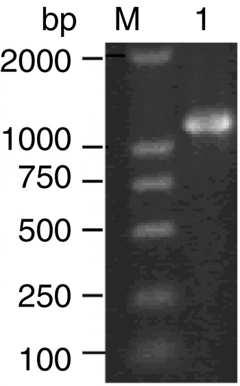
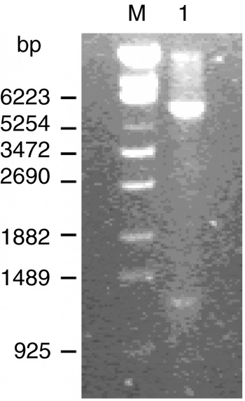
The target fragment of the N gene, with an expected size of 1174 bp, was amplified from a cDNA template of the TGEV strain SC-Y by reverse-transcriptase polymerase chain reaction (RT-PCR; Fig. 1). The PCR products were cut with Bsp1191 and Xho I and subcloned into the PET-32a expression vector. The cloning was confirmed by enzyme restriction (Fig. 2).
PCR results for the N gene. Lanes: M, DL-2000 marker; 1, Product of N gene.

Identification of recombinant plasmid by restriction digestion. Lanes: M, λ-ECOT-14 DNA marker; 1, recombinant PET-N digested by Bsp1191 and Xho I.

Prokaryotic expression of N protein and immunogenicity detection
Expression of N protein in E. coli
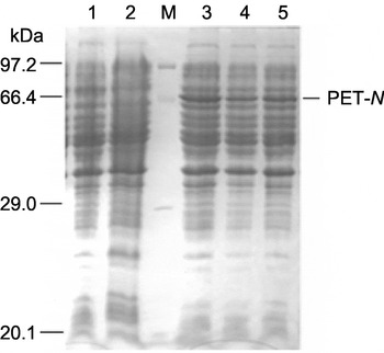
The induced cells were collected by centrifugation, and the supernatant removed and re-suspended in loading buffer. SDS-PAGE of total cell protein with coomassie blue staining showed a target protein of approximately 66 kDa. No typical bands were observed in uninduced cells and cells without PET-N (Fig. 3).
SDS-PAGE of protein expressed from recombinant plasmids in BL21(DE3). Lanes: M, protein molecular weight marker; 1, PET-N (uninduced); 2. PET-32a(+); 3–5, PET-N (expressed products induced by 1 mmol/l IPTG).

Western blot analysis
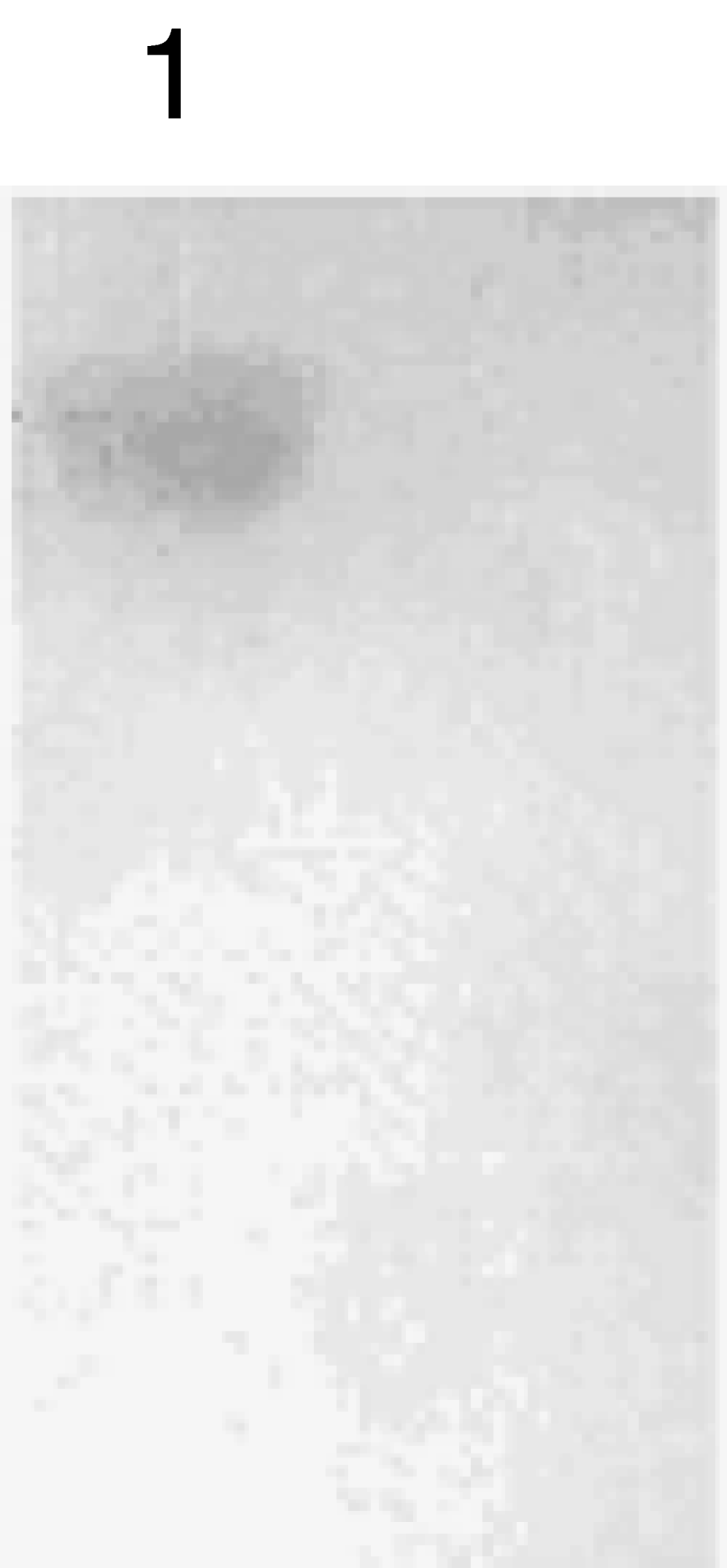
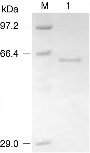
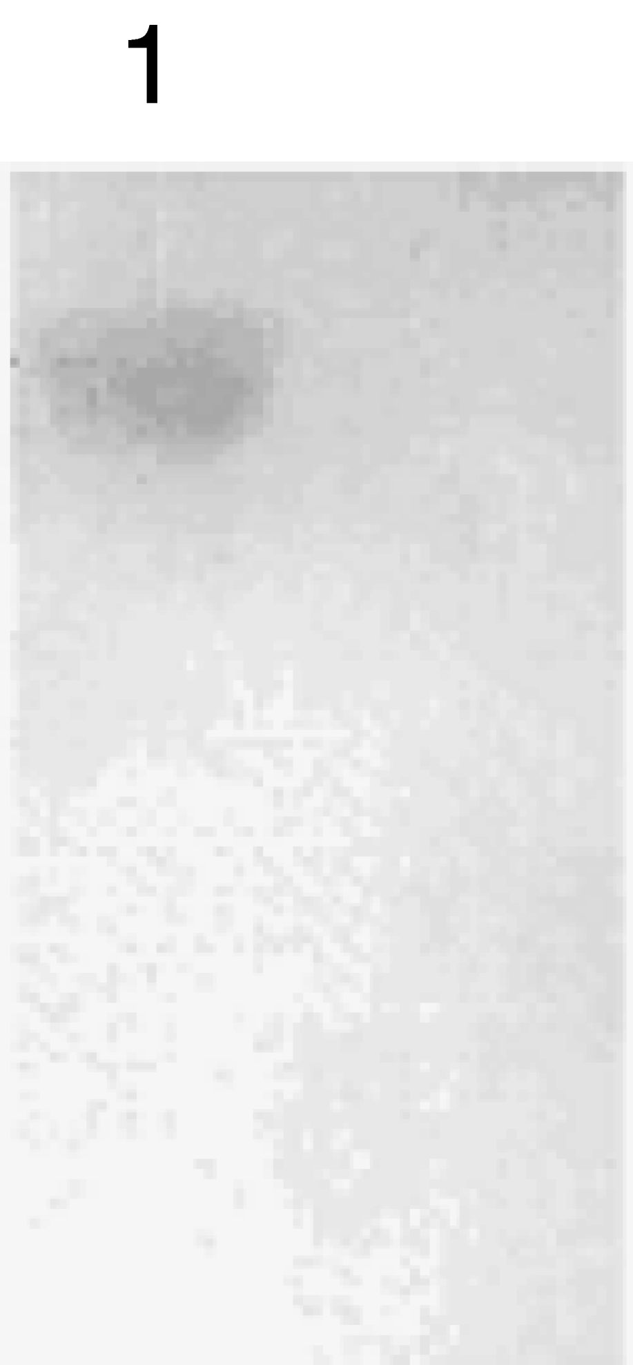
The targeted protein was transferred to a nylon membrane and analysed by Western blot after induction. The results revealed that the cells containing recombinant vector had a specific band (Fig. 4). The N protein was purified by affinity chromatography using nickel–nitrilotriacetic acid (Ni-NTA) agarose following the manufacturer's instructions (Qiagen). Following the purification procedure, the N protein was studied using SDS-PAGE (Fig. 5).
Result of Western blot analysis of N recombinant protein. Lanes: 1, N recombinant protein.
SDS-PAGE analysis of purified recombinant protein. Lanes: M, protein markers; 1, purified recombinant protein.

Indirect ELISA for recombinant N protein
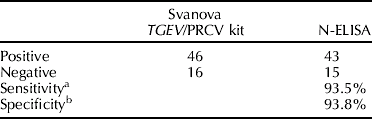
Checkerboard titration revealed that the optimal antigen concentration was 15 μg/ml, the optimal serum dilution was 1:40, the enzyme conjugate dilution was 1:5000 with incubation for 60 min, the blocking solution was 0.5% FBS with the substrate incubated at room temperature for 10 min, when a positive cutoff value (OD ⩾0.35) was applied. According to the established ELISA reaction conditions, known positive sera were detected, the results showing that the OD value of the five sera were all within the standard negative determination range. Sixty-two field serum samples were collected and compared with the TGEV/PRCV antibody diagnosis kit (Svanova). The recombinant N protein ELISA demonstrated a specificity of 93.8% among (n=16) TGEV-free individuals, and a sensitivity of 93.5% among (n=46) clinical samples with TGEV infections (Table 1).
Test results for N-ELISA with comparison using the Svanova TGEV/PRCV kit
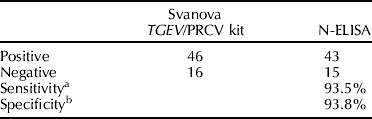
a Sensitivity (%)=number of N-ELISA positive sera/number of TGEV/PRCV kit positive.
b Specificity (%)=number of N-ELISA negative sera/number of TGEV/PRCV kit negative.
Discussion
The use of affinity tags simplifies the purification of the recombinant fusion proteins by using Ni-NTA metal-affinity chromatography methods. In this study, recombinant N protein with a polyhistidine tag at the end of the amino terminus was induced by IPTG, and SDS-PAGE revealed protein bands corresponding to the expected molecular mass of 66 kDa.
TGEV causes fatal diarrhoea in newborn piglets. Although most infected young and adult pigs recover from the disease, they are carriers of the virus, which allows infection for a longer period in affected herds. To detect carrier animals, a simple and reliable diagnostic method for monitoring the status of TGEV infection in animals was required. The nucleoprotein is the most abundantly expressed structural protein and highly conserved among all TGEV isolates, so it is a suitable target for serodiagnosis of the disease. The results of Western blot analysis showed that the target protein possessed immunogenicity, and could be used as an antigen in ELISA to detect serum antibody against the N protein. Recombinant protein-based ELISA appeared to be 93.8% specific and 93.5% sensitive for the diagnosis of TGE using the TGEV/PRCV antibody diagnosis kit, which is extensively used in China to detect TGE antibody. The reason for the discrepancy between the ELISAs may be related to differences in the target antigens.
A non-specific reaction is often observed in ELISA when total cell protein is used as the antigen. It is therefore necessary to replace viral antigens with a recombinant protein in a diagnostic ELISA. In this study, recombinant protein-based ELISA had high sensitivity and specificity. In conclusion, our findings suggested that E. coli-derived recombinant N proteins would be suitable diagnostic antigens for quick and reliable screening of TGEV infections.
Acknowledgements
This work was supported by Programs from Changjiang Scholars and Innovative Research Team in University (no. IRT0555).