Percutaneous device closure has become the preferred procedure for treatment of patients with secundum-type atrial septal defects. Generally, the sheath and the device are advanced through the femoral vein to the inferior caval vein and thereby to the right atrium and the interatrial septum. Rarely, transfemoral approach might not be possible due to congenital or acquired venous anomalies. Alternative methods of device delivery have been described in these patients and achieved successful results. Here, we present modified transfemoral atrial septal defect device closure in a patient with interrupted inferior caval vein.
Case report
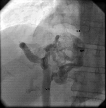
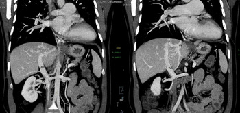
A 50-year-old asymptomatic male had cardiac consult before a non-cardiac surgery and was referred due to incidental finding of the right ventricle enlargement and a large secundum atrial septal defect in transthoracic echocardiography. Past medical history was negative, and he had history of occasional drug use for lumbar discopathy. In physical examination, wide fixed splitting of the second heart sound and a II/VI systolic ejection murmur was audible. There was no ascites or pedal oedema. Twelve lead ECG showed normal sinus rhythm with incomplete right bundle branch block and T wave inversion in V2-V3 precordial leads. Chest X-ray depicted evidence of right ventricular enlargement and pulmonary overflow. Transesophageal echocardiography was performed and reported moderate right ventricle enlargement with mild systolic dysfunction and a large secundum-type atrial septal defect measuring 2.6 cm × 1.40 cm in 3D. There was significant left to right shunt through the atrial septal defect and sufficient all rims for device closure except for absent anterosuperior (aortic) rim. The pulmonary arterial pressure was normal. The patient was scheduled for device closure. During catheterisation, the multipurpose catheter could not be advanced to the right atrium. Inferior caval vein injection showed segmental interruption of inferior caval vein with continuation to the right atrium via multiple collaterals draining to enlarged hepatic veins (Fig 1). CT angiogram was performed for better evaluation and showed segmental agenesis of the intra-hepatic portion of the inferior caval vein. The infrahepatic inferior caval vein was connected to the supra hepatic portion via collaterals and hepatic veins (Fig 2). The patient refused surgical repair we decided to proceed through transjugular or transhepatic access for device closure. As the atrial septal defect was reported to be large and the anterosuperior rim was absent, we thought proper device alignment and deployment via transjugular approach might not be feasible.
Angiogram of inferior caval vein showing partial absence of the inferior caval vein and continuation to RA via collaterals and hepatic veins, RA = Right atrium, HV = Hepatic vein, IVC = inferior caval vein.

CT angiogram depicting absent intra-hepatic segment of the inferior caval vein and continuation to RA by hepatic veins, RV = renal vein, HV = hepatic vein.

Therefore through the femoral access and under fluoroscopy guide, a hydrophilic 0.035 guide wire and a multipurpose catheter were gently advanced to the right upper pulmonary vein and then the wire was exchanged for a super-stiff guide wire. Distensibility of the largest collateral vein was checked with a low-pressure 8 × 30 mm peripheral balloon and revealed full balloon expansion in 4 atm. A 12 F sheath was passed through the atrial septal defect safely. Under transesophageal echocardiography, guidance closure with a 30-mm occluder was attempted but the device failed to align with the interatrial septum and was retrieved. Then, a 33-mm device was used and could be implanted successfully (supplementary material). Post-procedural transesophageal echocardiogram showed proper device position with no residue or compressive effect on adjacent structures. The patient was monitored for 48 hours and then discharged home with aspirin and clopidogrel. At one and six months follow-up, he was doing well and the transthoracic echocardiogram showed the device in the proper position and no complications.
Discussion
Congenital abnormalities of the systemic veins are rare. Inferior caval vein interruption is reported in about 1 in 5000 of the general population and results from failure of the embryonic inferior caval vein segments to fuse normally. The blood from the lower parts of the body drain to the right atrium via azigous system.Reference Hardwick, Belcher, Sabharwal and King1,Reference Singh, Neuzil and Skoka3 In these patients, it might become necessary to use alternative routes for percutaneous interventions. Device closure of large atrial septal defects particularly those requiring devices ≥28 mm is demanding as some of the rims might be absent, deficient or highly redundant.Reference Saedi, Aliramezany, Khajali and Sanati2
The majority of reported patients with interrupted inferior caval vein and atrial septal defects underwent percutaneous intervention via transjugular access. There are also cases in which the delivery sheath was passed from the azigous vein to the superior caval vein and then to the right atrium and the left atrium, respectively. However, almost all these patients had a relatively small atrial septal defect with sufficient rims and the authors agree on the unsuitability of the mentioned methods for sizeable atrial septal defects.Reference Alizade, Karaduman, Balaban, Keskin and Kalkan4
Our patient had a remarkably large atrial septal defect with absent anterosuperior rim that made transjugular or trans-azigous approach and necessary manoeuvres potentially troublesome. In transhepatic approach, the venous access is obtained through hepatic veins in the right upper abdomen under fluoroscopy or ultrasonography guide. The transhepatic approach was initially utilised for procedures like cholangiography or access to portal veins for sclerotherapy of bleeding oesophageal varices. It was later used for catheterisation in children with venous anomalies or obstruction.Reference McLeod, Houston, Richens and Wilson5 In adults, intervention through transhepatic access is rarely performed and there are reports of utilisation of this access for patent foramen ovale device closure and electrophysiology procedures of complex arrhythmia ablation.Reference Singh, Neuzil and Skoka3,Reference Hussain, Strumpf, GhandForoush, Jamal and Diethrich6 There are pertaining risks including intra-abdominal haemorrhage, but the overall reported rate of the complications is low. In a rare anatomic variation, as in our patient, remnants of suprarenal segment of inferior caval vein drain into the right atrium via hepatic veins. In these cases, a modification of the transhepatic method could be used through femoral access.Reference Singh, Neuzil and Skoka3 The acceptable size of the veins connecting inferior caval vein to the right atrium made the percutaneous intervention possible.
Supplementary material
To view supplementary material for this article, please visit https://doi.org/10.1017/S1047951121000755
Acknowledgements
The authors would like to thank the RHC imaging department staff for their kind assistance.
Financial support
This research received no specific grant from any funding agency, commercial or not-for-profit sectors.
Conflicts of interest
None.
Ethical standards
Institutional ethics committee approved of the manuscript.