Obesity is considered an epidemic by the WHO, with more than 1·9 billion adults being overweight and 650 million being classified as obese in 2016(1). Changes in body composition, characterised by adipocyte hypertrophy and a consequent chronic state of meta-inflammation associated with low levels of physical activity, may lead to a significant inability to engage in physical activity(2–Reference Sarma, Sockalingam and Dash5).
The chronic state of meta-inflammation associated with obesity is characterised by a significant increase in pro-inflammatory cytokines, including TNF-α and IL-6, and a reduction in anti-inflammatory cytokines, including adiponectin(Reference Schmidt, Weschenfelder and Sander6,Reference Booth, Magnuson and Fout7) . This process is also characterised by an increase in M1 macrophages and a decrease in M2 macrophages, along with a reduction in oxygen concentration in adipose tissue(Reference Trayhurn8–Reference Chylikova, Dvorackova and Tauber10).
Alongside these physiological alterations, dietary habits significantly influence systemic inflammation. The growing consumption of ultra-processed foods, high in palatability and convenience but low in nutritional quality, has been associated with elevated levels of IL-6 and C-reactive protein, reinforcing a chronic pro-inflammatory state(Reference Lopez-Garcia, Schulze and Fung11–Reference Pagliai, Dinu and Madarena14).
Given the increasing prevalence of obesity, especially in developing countries and with a higher incidence among women, it is imperative to implement effective strategies to combat this condition(Reference Schmidt, Weschenfelder and Sander6). Considering its multifactorial nature, a detailed understanding of the clinical conditions associated with obesity, including lipid profiles, inflammatory status and dietary behavioural, is essential for developing more effective treatment approaches.
Although diet plays a fundamental role in weight loss and is the primary strategy for reducing fat mass, physical exercise is essential for the maintenance and increase of lean body mass(Reference Olateju, Opaleye-Enakhimion and Udeogu15). This, in turn, promotes improvements in health-related aspects such as inflammation and metabolic regulation(Reference Christoph and Karsten16). Moreover, regular physical activity is necessary to ensure long-term consistency in body composition outcomes(Reference Christoph and Karsten16).
Given these limitations, particularly among individuals with reduced physical fitness or joint complications, alternative and joint-friendly exercise modalities have been increasingly studied. One such strategy is deep-water running (DWR), a modality that simulates running motions in a deep pool, using a flotation vest to prevent the feet from touching the bottom, thereby eliminating joint impact(Reference Chu and Rhodes17,Reference Reilly, Dowzer and Cable18) . Consequently, DWR has emerged as a viable option for individuals with low physical fitness, offering the benefits of physical activity without the risk of joint injuries(Reference Nagle, Sanders and Franklin19–Reference Kwok, Ng and Man21).
In parallel, high-intensity exercise, particularly when performed as high-intensity interval training, presents advantages compared with moderate-intensity continuous training(Reference Andreato, Esteves and Coimbra22–Reference Wewege, Berg and Ward24). Among the benefits of high-intensity exercise performed as high-intensity interval training are improvements in body composition, including reductions in fat mass and increases in lean mass(Reference Andreato, Esteves and Coimbra22–Reference Guo, Li and Cai25). Furthermore, studies have demonstrated significant improvements in cardiorespiratory fitness(Reference Guo, Li and Cai25,Reference Lunt, Draper and Marshall26) . Finally, high-intensity interval training is generally considered to be more time-efficient and enjoyable than moderate-intensity continuous training(Reference Wewege, Berg and Ward24,Reference Guo, Li and Cai25) .
To further optimise the outcomes, combining physical exercise in normoxia (NOR) with hypoxia during rest periods has emerged as an effective non-pharmacological strategy for managing obesity. This approach enhances both metabolic and cardiovascular adaptations through multiple physiological mechanisms, including the increased activation of pathways related to mitochondrial biogenesis and lipid metabolism(Reference Burtscher, Citherlet and Camacho-Cardenosa27), elevated glycolytic enzyme activity and improved insulin sensitivity(Reference Urdampilleta, González-Muniesa and Portillo28), which together promote greater fatty acid utilisation and better glycaemic control(Reference Urdampilleta, González-Muniesa and Portillo28,Reference Ghaith, Chacaroun and Borowik29) .
Additionally, hypoxic exposure reduces peripheral oxygen saturation (SpO2), thereby increasing cardiorespiratory and metabolic stress at a given mechanical loads(Reference Foresti, Carvalho and Ribeiro30,Reference Costa, Camacho-Cardenosa and Brazo-Sayavera31) . This characteristic may be particularly relevant for individuals with obesity, for whom weight-bearing high-intensity exercise may be limited by joint discomfort and low physical fitness(Reference Ghaith, Chacaroun and Borowik29).
In the present study, high-intensity interval exercise was selected to provide a substantial cardiorespiratory and metabolic stimulus within a relatively short training duration, while the aquatic environment reduced the mechanical and joint loading associated with high-intensity land-based exercise. Hypoxia was applied only during the passive recovery periods to increase physiological stimulus without increasing the mechanical demand during the exercise bouts.
In this regard, the main aim of this study was to investigate the effects of high-intensity exercise combined with intermittent hypoxia on cardiorespiratory parameters, body composition and lipid and inflammatory markers in women with grade I obesity. In addition, we assessed dietary intake and overall nutritional quality to explore potential associations between dietary patterns and the observed physiological responses.
Methods
The experimental model of this research was conducted in accordance with the Declaration of Helsinki and registered with the US National Institutes of Health’s international clinical trial registry (https://clinicaltrials.gov/) under the identifier NCT05380830. The research was approved by the Ethics Committee of the School of Physical Education and Sports of Ribeirão Preto, University of São Paulo (CAAE: 39719620.0.0000.5659). All participants signed an informed consent form. This research followed the CONSORT-2025 guideline.
Experimental design
The study was conducted as a parallel-group clinical study using a block randomisation approach. To ensure a single-blind protocol, participants remained unaware of whether normoxic or hypoxic air was administered during the rest periods. All outcomes were analysed as changes from baseline and compared between groups. The 12-week experimental timeline comprised 8 weeks of supervised high-intensity interval training using tethered deep-water running (T-DWR), preceded by 2 weeks of DWR familiarisation and evaluations, and followed by final evaluations. Assessment protocols included body composition analysis, performance testing, 3-d food records and venous blood sampling to determine lipid profiles and inflammatory biomarkers. Baseline assessments were completed before participants were assigned to either normoxic or hypoxic groups. At the end of the study, the participants were asked to identify the type of air they believed they had breathed to confirm that the groups remained single-blind throughout the training.
Participants
Participants were women with grade I obesity aged between 25 and 49 years. Initially, potential participants completed an interest form containing questions regarding their general health status. The inclusion criteria were (1) age between 25 and 49 years, (2) grade I obesity defined as a BMI between 30 and 34·9 kg/m2, (3) no participation in structured exercise or dietary programmes during the previous 12 months, (4) premenopausal status and (5) no use of thyroid medications. The exclusion criteria were (1) arterial hypertension, (2) metabolic syndrome or other obesity-related co-morbidities, (3) alcohol abuse or smoking and (4) exposure to altitudes above 1500 m during the 3 months preceding enrolment.
A total of thirty-one women were recruited and underwent familiarisation and baseline assessments. After completing the baseline assessments, each participant received a unique coded identification number. Participant recruitment and generation of the random allocation sequence were conducted by different members of the research team. The principal investigator was not involved in participant recruitment, sequence generation or group allocation. The researcher responsible for recruitment and enrolment had no access to the allocation sequence.
Participants were allocated in a 1:1 ratio using block randomisation with a fixed block size of 4. For each block, one of the possible balanced allocation sequences was randomly selected using the RAND() function in Microsoft Excel®, thereby generating the complete allocation sequence a priori. The master allocation list linking participants’ coded identification numbers to their assigned experimental conditions was stored on a restricted-access computer and was accessible only to the research team member responsible for randomisation. Allocation was revealed only after each participant had been enrolled and had completed all baseline assessments. For operational reasons, during the intervention, the principal investigator was aware of whether hypoxic or normoxic air was being delivered during each training session and therefore was not blinded to the condition being administered.
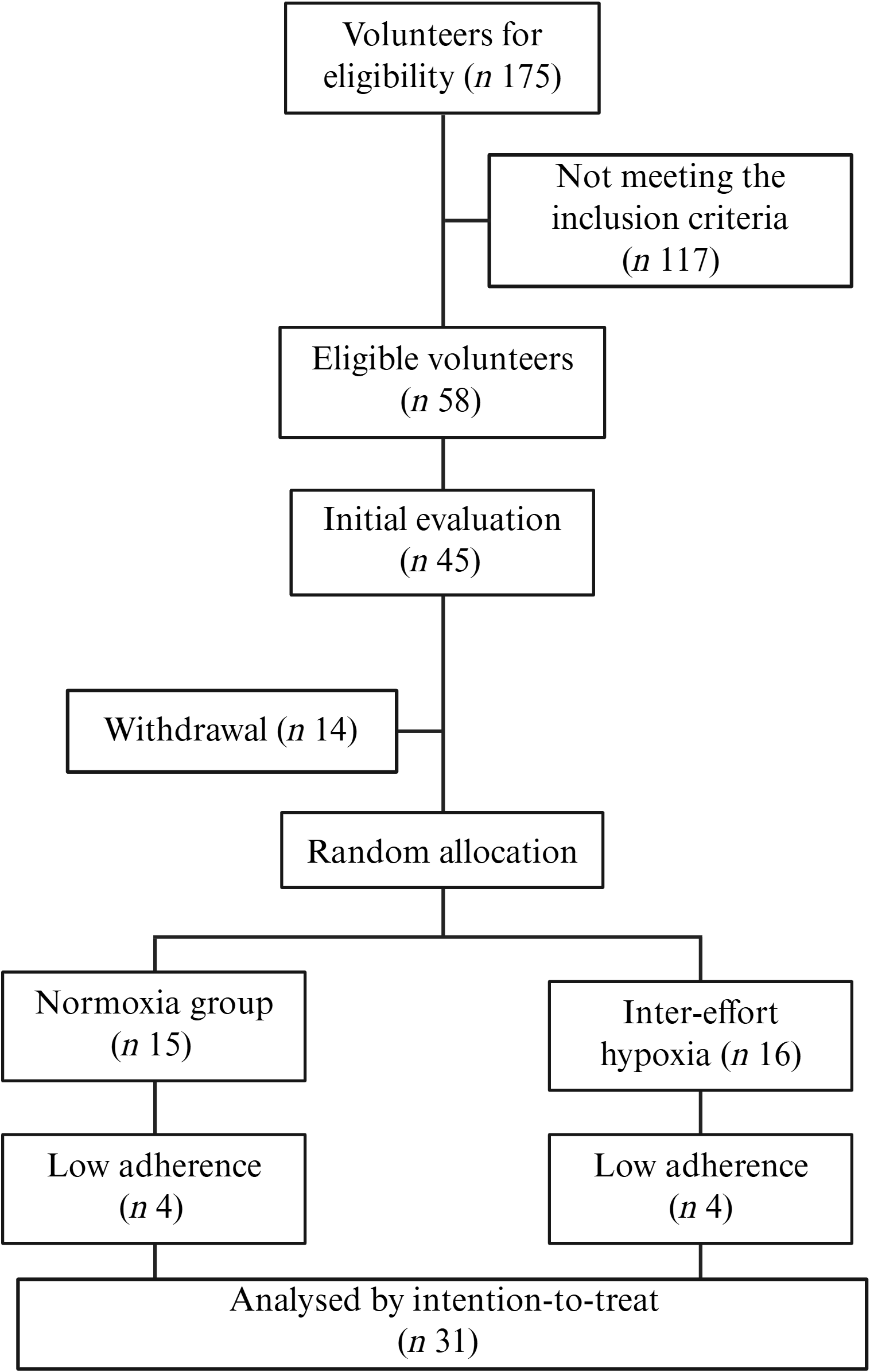
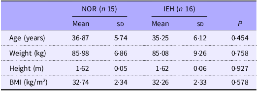
Participants were allocated to either the NOR (n 15) or the inter-effort hypoxia (IEH) group (n 16) (Figure 1). Baseline characteristics according to group allocation are presented in Table 1.
Flow chart.

Figure 1. Long description
Flowchart.
Characterisation of participants before tethered deep-water running training of normoxia (NOR) and inter-effort hypoxia (IEH) groups (Mean values with their standard deviation)

Table 1. Long description
Table 1 – Characterization of participants before tethered deep-water running training of normoxia (NOR) and inter-effort hypoxia (IEH) groups. Mean values (Standard deviation).
Student’s t test was used for the analysis.
Familiarisation and tethered deep-water running training
The familiarisation, training sessions and assessments were conducted at the School of Physical Education and Sport of Ribeirão Preto (EEFERP/USP) and were fully supervised by a qualified professional. The familiarisation phase spanned 2 weeks, followed by an 8-week T-DWR training protocol. This consisted of twenty-four sessions conducted three times per week, with each session involving 1 h of T-DWR. Training sessions were held in groups of up to ten people, during morning and evening periods, based on participants’ availability and interest.
During the first visit to the pool environment, participants were instructed on the proper use of a buoyancy vest (Actual), secured at the waist to prevent contact with the bottom of the pool and to keep their heads above the water surface. Subsequently, the DWR technique was demonstrated, and the category ratio 0–10 (CR-10) Borg scale, which ranges from 0 (rest) to 10 (maximum), was introduced. The familiarisation process occurred three times over non-consecutive days, in sessions lasting from 30 to 50 min. The DWR was performed freely and, at different intensities, determined by the participant according to their adaptation to the running. To ensure scale proficiency, perceived exertion was assessed using the CR-10 Borg scale every 10 min.
In the second week, a single 45-minute session was held to adapt T-DWR, during which participants wore the flotation vest already attached to an elastic cord fixed near the edge of the pool; the tension of this cord determined the intensity of the external load. The intensities and durations were self-selected. During the last 10 min of the session, an adapted graded exercise test was simulated using shorter stimuli, encouraging participants to reach the maximum possible extension of the elastic cord. The graded exercise test was conducted 24 to 48 h after the T-DWR familiarisation, and the training started the following week.
Following the familiarisation and baseline evaluations, the T-DWR training was initiated. Prior to each training session, heart rate (HR), SpO2 and rating of perceived exertion (RPE) were recorded. The warm-up consisted of 5 min of T-DWR at RPE 2–3, followed by 5 min of passive rest in hypoxia or NOR. The main training block was performed at an intensity of 100–110 % of the HR at the 4mM lactate threshold (HRL4mM), corresponding to RPE of 9–10, consisting of four efforts of 5 min in T-DWR followed by 5 min of passive rest (adapted from Lunt et al. (Reference Lunt, Draper and Marshall26)) (Figure 2).
Protocol of the training programme in T-DWR. The first rectangle shows the 5 min warm-up, with subsequent 5 min effort at 100 % HR at L4mM, intercalated with 5 min at passive rest, in normoxia or inter-effort hypoxia. At the end of each effort, the individual RPE, HR and SpO2 initial, minimum and final were evaluated. Image created with BioRender. T-DWR, tethered deep-water running; HR, heart rate; L4mM, [La-] 4mM; RPE, rating of perceived exertion; SpO2, oxygen saturation.

Figure 2. Long description
Protocol of training program in T-DWR. The first rectangle shows the 5 minutes warm-up, with subsequent 5 minutes effort at 100% HR at L4mM, intercalated with 5 minutes at passive rest, in normoxia or inter-effort hypoxia. At the end of each effort the individual RPE, HR and SpO2 initial, minimum and final were evaluated. Image created with BioRender.
During the rest period, participants in the IEH group wore a mask connected to an air storage system for hypoxia exposure, whereas the NOR group’s mask was connected to a placebo system(Reference Norberto, Torini and Firmino33). The entire system was covered with a dark cloth to maintain the blinding of the participants.
Training intensity was monitored using HR and RPE at rest, after each effort and at the end of the session(Reference Costalat, Lemaitre and Ramos32). Peripheral SpO2 during rest was monitored using a pulse oximeter (G-Tech Portable Oled) at three points: beginning of rest (initial SpO2), the lowest point of saturation during rest (SpO2min), and the end of rest (final SpO2)(Reference Costa, Camacho-Cardenosa and Brazo-Sayavera31,Reference Trapé, Camacho-Cardenosa and Camacho-Cardenosa34) . The training impulse measurement (TRIMP) was calculated in arbitrary units (a.u.), individually, for all sessions, using the average HR, TRIMPHR = intensity (bpm)*effort (minutes) and the average RPE, TRIMPRPE = intensity (rpe)*effort (minutes)(Reference Foster, Glorhaug and Franklin35).
Instruments to generate the hypoxic environment and hypoxia dose
An air storage manufactured by our laboratory was used to provide hypoxia while in the pool – the hypoxia generator. This structure connects to a hypoxia generator to channel airflow into the apparatus airbags(Reference Norberto, Torini and Firmino33,Reference Carvalho, Ferronato and Coelho36) . Participants wore a mask connected to the airbags during all training sessions to ensure blinding, so they would not know which condition the session was in (NOR or IEH). The airflow through the valve system during hypoxia exposure was controlled for each exercise session, as appropriate. Valves supplying ambient air were closed, while valves for the hypoxic airbags were opened during hypoxia training sessions; the opposite was done for NOR training sessions.
Considering the principle of individuality, the same fraction of inspired oxygen can elicit different responses in SpO2. The reduction in oxygen concentration under hypoxic conditions, as well as the increase in oxygen consumption during exercise, are individual. Thus, the calculation of hypoxia dose (HD) was individual and done for each session, as the product of exposure time in hours (t) multiplied by the average reduction in SpO2 during the training session, using the formula HD = (98 – SpO2mean) *t, adapted from Millet et al. (Reference Millet, Brocherie and Girard37).
Graded exercise test
Before and after the training, the graded exercise test was used to determine maximum VO2 (VO2max), exercise intensity and HR. The tests were carried out in an indoor pool, with a mean temperature of 28 °C. The graded exercise test consisted of progressive efforts until voluntary exhaustion, assumed to be the inability to continue exercising at the intensity determined by the protocol.
The initial intensity was 4·9N, with increments of 4·9N every 2 min and a 30-s interval between increments, with the intensity controlled using a load cell with a digital display fixed in the swimming starting block and pre-calibrated cones as reference samples along the edge(Reference Carvalho, Ferronato and Coelho36) (Figure 3). HR, RPE using the CR-10 Borg scale(Reference Borg38) and blood samples were collected from the earlobe for lactate concentration at rest and after each increment. VO2max was determined immediately after exhaustion using the retro-extrapolation technique (VO2000 Inbramed gas analyser), with the peak oxygen intake as the highest value(Reference Montpetit, Léger and Lavoie39–Reference Luches-Pereira, Kalva-Filho and Barbieri43).
Study protocol of evaluation in T-DWR, showing the load cell with digital display connected with the buoyancy vest by an elastic rope. The pre-calibrated cones of each stage were placed on the board of the swimming pool. After each stage, the RPE, HR and earlobe blood for [La–] were collected, and immediately after exhaustion, the VO2max was determined with the gas analyser. Image created with BioRender. T-DWR, tethered deep-water running; RPE, rating of perceived exertion; HR, heart rate; [La–], blood lactate concentration; VO2max, maximum VO2.

Figure 3. Long description
Study protocol of evaluation in T-DWR, showing the load cell with digital display connected with the buoyancy vest by an elastic rope. On the board of the swimming pool the pre-calibrated cones of each stage. After each stage the RPE, HR and earlobe blood for [La-] were collected, and immediately after exhaustion the VO2max was determined with the gas analyzer. Image created with BioRender.
Subsequently, the blood sample (25 µl) was collected from the earlobe before the first stage and after the end of each stage until the volunteer’s exhaustion for the determination of blood lactate concentration ([La–]) (YSI 2300 STAT analyser (Yellow Springs)). It was assumed to be the anaerobic threshold when [La–] achieved 4 mM (L4 mM), and then the HR (HRL4mM), the intensity (IL4mM) and the percentage of VO2peak (%L4mMVO2peak) at the L4mM were calculated.
Body composition
Total and regional body composition was determined by DXA (Lunar GE Medical Systems Prodigy Advance, Encore software, version 13.6). The examination followed standardised procedures(Reference Venturini, Silva and Abdalla44). Measurements included total weight in kilograms, lean mass and fat mass in percent (%) and visceral fat mass in grams (g). Participants were instructed to remove metallic objects (earrings, rings, necklaces, etc.) and were positioned supine, centred on the scanner table, with lower limbs fastened together by Velcro straps. Participants’ hands were kept open, with palms resting on the table and arms extended along the body (within the table’s scanning lines), as per the manufacturer’s instructions.
Blood sample
Blood samples were collected by a trained nurse in accordance with proper hygiene and antisepsis protocols. Participants were instructed to fast for 8 to 10 h. A total of 15 mL of blood was drawn from the right basilic median vein using a vacuum system. The samples were immediately placed in specific tubes for each analysis and then centrifuged and stored at −80 °C in a biofreezer for later analysis.
Lipid profile
Total cholesterol, TAG, HDL-cholesterol, LDL-cholesterol and glucose were measured using Labtest Diagnostica® kits: Total Cholesterol Liquiform, HDL Cholesterol, Triglycerides Liquiform and Glucose Liquiform, with an enzymatic system and an absorbance spectrophotometer. The LDL-cholesterol fraction was calculated using Friedewald’s formula (LDL-c = (TC – HDL-c) – (TG/5))(Reference Friedewald, Levy and Fredrickson45).
Inflammatory profile
Adiponectin was measured in serum using Millipore’s MILLIPLEX® Human Adipokine Panel 1, HADK1MAG-61K kit. IL-6, leptin and TNF-α were measured using Millipore’s MILLIPLEX® Human Adipokine Panel 2, HADK2MAG-61K (Merck Millipore, EMD Millipore Corporation). The analyses were performed at the School of Pharmaceutical Sciences of Ribeirão Preto – USP using Luminex (Magpix System).
Dietary intake analysis
All participants were instructed to complete 3-d food records to assess their dietary intake. The participants completed food records at two distinct time points (before and after the interventions), filling out three non-consecutive days of records for each time point, with one of them completed on a weekend day(Reference Willet46). They were instructed to fill out the food records with as much detail as possible, recording everything consumed using household measures. The average of the three records for each participant was calculated for variables such as energy intake, protein, lipids, carbohydrates, Na, sugar and fibre intake, and the average intake for each group was then computed(Reference Willet46). The analysis of energy content and macronutrients was performed using the online software DietBox®.
Diet quality analysis
Diet quality was assessed using the NOVA classification system. According to NOVA, food was classified into three groups: Group 1 – unprocessed and minimally processed foods; Group 2 – processed foods; and Group 3 – ultra-processed foods(Reference Monteiro, Cannon and Lawrence47). For each category, the total energy content consumed was calculated, and subsequently, the percentage (%) contribution of each food group to the overall diet was determined(Reference Louzada, Baraldi and Steele48).
All participants were instructed to maintain their usual food intake throughout the intervention period.
Statistical analysis
The a priori sample size calculation was performed using ANOVA_power software, based on the fat-free mass values reported by Wiesner et al.(Reference Wiesner, Haufe and Engeli50) : hypoxia group, n 24, before 69·0 (1·9)% and after 71·0 (2·0)%; and NOR group, n 21, before 68·0 (1·4)% and after 68·0 (1·6)%. The calculation indicated that a minimum of ten participants per group would provide 86·9 % statistical power to detect an effect size of 0·25, considering α = 0·05. To account for an expected withdrawal rate of 30 %, the planned sample size was increased to fifteen participants per group.
Based on the study design, a priori analyses were performed to assess differences in the outcome variables based on randomisation group. Baseline characteristics and training session variables were compared using independent Student’s t tests.
The data are presented as means and standard deviation. Normality was assessed using the Shapiro–Wilk test, and homogeneity of variances was evaluated using Levene’s test.
Linear mixed-effects models were used to examine the effects of group, time and the group × time interaction as fixed effects, with participants as a random effect to account for within-subject correlations across repeated measurements. Four participants from each group withdrew before the follow-up assessments. The primary analyses followed the intention-to-treat principle, with all randomised participants analysed according to their originally assigned groups. Mixed-effects models were fitted using all available observations, including data from participants with incomplete follow-up, without imputing missing values, under the assumption that data were missing at random. When significant main or interaction effects were identified, post hoc comparisons were performed using Tukey’s multiple-comparisons test.
Mean differences and corresponding 95 % CI are reported. Statistical analyses were performed using GraphPad Prism version 8.0.1 (GraphPad Software), with statistical significance set at α < 0·05.
Results
After the baseline assessments, thirty-one women were randomly allocated to the NOR and IEH groups, with fifteen participants assigned to NOR and sixteen to IEH. No significant between-group differences were observed at baseline for age, body mass, height or BMI (Table 1). Participant flow and adherence to the intervention are presented in Figure 1.
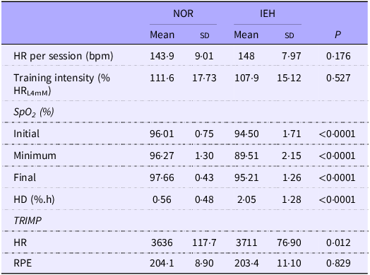
Table 2 summarises the training session characteristics. Both groups achieved the target training intensity, reaching 111·6 % of the prescribed intensity in the NOR group and 107·9 % in the IEH group. SpO₂ (%) and HD (%.h) differed significantly between groups (p < 0·0001), and HR-derived TRIMP was also significantly higher in the IEH group (p < 0·05).
Characterisation of the 8-week training monitoring of tethered deep-water running of normoxia (NOR) and inter-effort hypoxia (IEH) groups (Mean values with their standard deviation)

Table 2. Long description
Table 2 – Characterization of the eight-week training monitoring of tethered deep-water running of normoxia (NOR) and inter-effort hypoxia (IEH) groups. Mean values (Standard deviation).
HR, heart rate; %HRL4mM, percent of the heart rate at La- [4mM]; SpO2, peripheral oxygen saturation; HD, hypoxia dose; TRIMP, training impulse; RPE, rating of perceived exertion.
Student’s t test was used for the analysis.
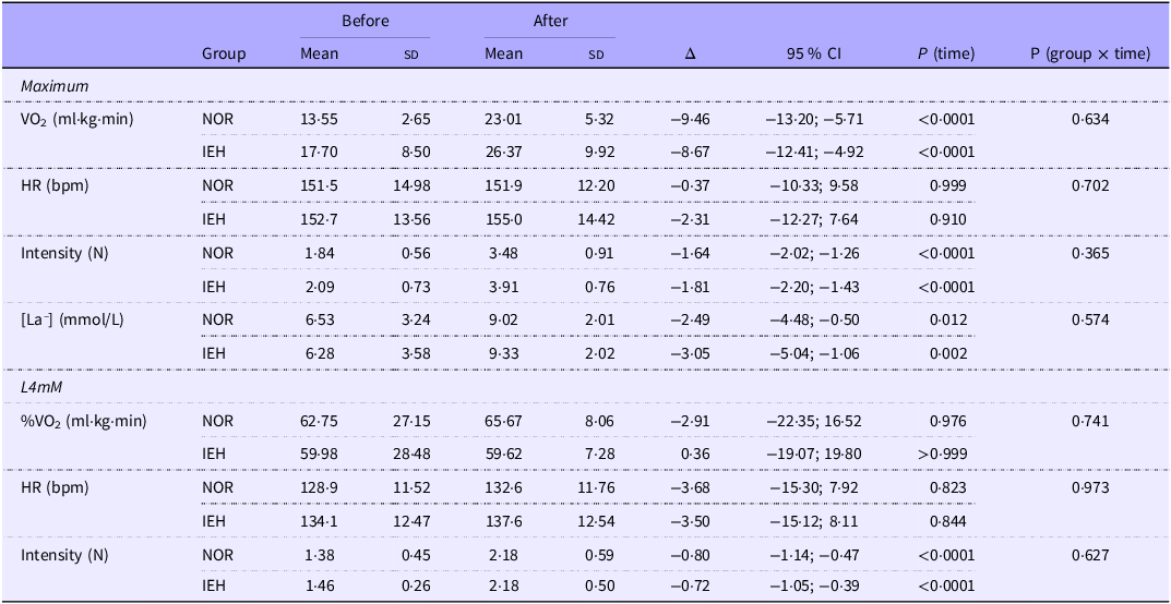
Following the T-DWR intervention, no significant group × time interaction effects were observed for cardiopulmonary, physiological, or performance outcomes (Table 3).
Cardiopulmonary, physiological and performance responses during the graded exercise test before and after the training intervention in normoxia and inter-effort hypoxia (Mean values with their standard deviation; Δ and CI-95 % lower; upper bound)

Table 3. Long description
Table 3 – Cardiopulmonary, physiological and performance responses during the graded exercise test before and after the training intervention in normoxia and inter-effort hypoxia. Mean values (Standard deviation). δ (CI-95% lower; upper bound).
Δ (IC 95 %), mean difference + 95 % CI; VO2, oxygen uptake; HR, heart rate; bpm, beats per minute; N, newton; [La–], blood lactate concentration; L4mM, [La-] 4mM; %VO2, percentage of VO2peak at L4mM.
A mixed-effect model with repeated-measures ANOVA was used for the analysis.
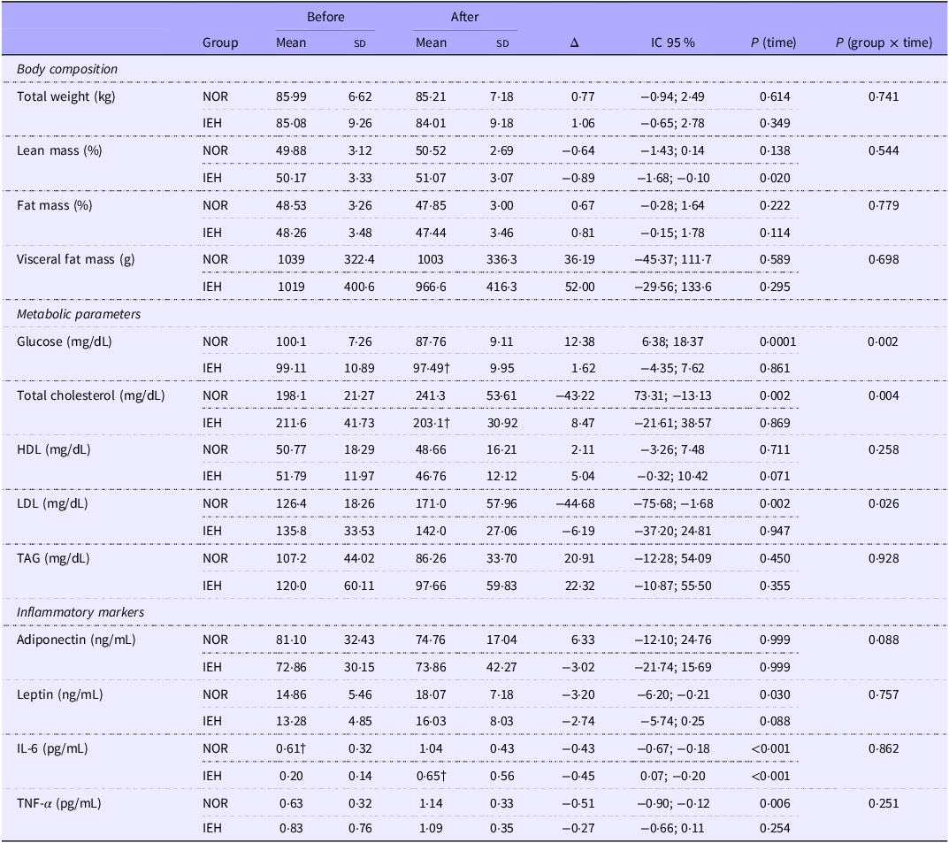
Health-related outcomes, including body composition, metabolic parameters and inflammatory markers, are presented in Table 4. No significant group × time interaction effects were observed for any body composition variable.
Body composition, metabolic parameters and inflammatory markers before and after 8 weeks of tethered deep-water running training. Normoxia and inter-effort hypoxia groups (Mean values with their standard deviation; Δ and CI-95 % lower; upper bound)

Table 4. Long description
Table 4 – Body composition, metabolic parameters and inflammatory markers before and after eight weeks of tethered deep-water running training. Normoxia and inter-effort hypoxia groups. Mean (Standard deviation). δ (CI-95% lower; upper bound).
Δ (IC 95 %), mean difference + 95 % CI.
A mixed-effect model with repeated-measures ANOVA was used for the analysis.
†Significant between-group difference at the same time point (P < 0.05).
Among the metabolic parameters, significant group × time interactions were found for glucose (F(1,15) = 13·37, p = 0·002, η 2 p = 0·47), total cholesterol (F(1,15) = 10·91, p = 0·004, η 2 p = 0·42) and LDL-cholesterol (F(1,15) = 6·09, p = 0·026, η 2 p = 0·29). Post hoc analyses showed that glucose decreased in the NOR group (p = 0·0001), whereas total cholesterol and LDL-cholesterol increased from baseline to post-intervention (both p = 0·002).
No significant group × time interaction effects were detected for any inflammatory marker (p > 0·05).
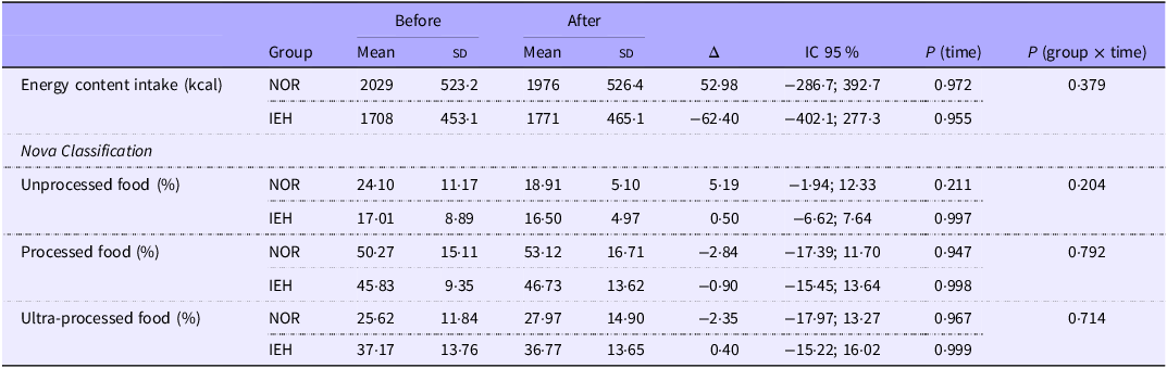
Energetic intake remained stable throughout the intervention in both groups, with no significant main effect of time (p = 0·965) or group × time interaction (p = 0·379) (Table 5). According to the NOVA classification, no significant changes were observed in the consumption of unprocessed, processed or ultra-processed foods in either group. Overall, both groups maintained a dietary pattern characterised by high consumption of processed and ultra-processed foods and low consumption of unprocessed foods throughout the study.
Discussion
The main findings of the present study were as follows: (i) SpO₂ was lower and the hypoxic dose was higher during passive recovery in the IEH group; (ii) heart-rate-derived internal training load was higher in the IEH group; and (iii) significant group × time interactions were observed for glucose, total cholesterol and LDL-cholesterol, primarily reflecting a reduction in glucose and increases in total cholesterol and LDL-cholesterol in the NOR group.
Obesity is associated with poor cardiorespiratory capacity coupled with high body fat percentages(Reference Laukkanen and Kujala4). The American College of Sports Medicine (ACSM) suggests that VO2max below 30 ml/kg/min is classified as poor cardiorespiratory fitness(2), consistent with the profile of the participants of this study. The ACSM also recommends water-based exercises, such as DWR, for individuals with obesity(Reference Donnelly and Blair51), resulting in less risk of joint injury and consequently withdrawing from physical exercise.
Although DWR is related to lower VO2max and HRmax values(Reference Phillips, Legge and Jones52), Azevedo et al.(Reference Azevedo, Lambert and Zogaib53) demonstrated that DWR performed at or above anaerobic threshold intensity has shown promising results in maintaining or improving aerobic capacity, comparable to land-based running. Although the present study did not demonstrate a statistically significant additional effect of hypoxia on cardiorespiratory capacity, familiarisation remains important to ensure adequate responses to the maximal effort test and to enable participants to perform the exercise at the prescribed intensity, as observed in the present study(Reference Frangolias and Rhodes54).
Exposure to hypoxia during rest proved effective in triggering physiological responses, such as a reduction in SpO2 and consequently greater HD. As proposed by Papoti et al.(Reference Papoti, Manchado-Gobatto and Gobatto55), exposure to hypoxia during rest was sufficient to trigger a greater internal training load with a perception of effort similar to remaining in NOR, as observed by the TRIMP values calculated with HR and RPE. Several cardiorespiratory and performance-related outcomes improved over time in both groups. Despite eliciting an additional physiological response, hypoxia did not produce greater improvements in cardiorespiratory capacity or physical performance. These findings suggest that the improvements observed in the present study were predominantly associated with T-DWR training, whereas the addition of hypoxia did not provide a detectable benefit beyond that achieved with exercise alone.
Studies exploring body composition changes in association with clinical conditions, exercise and hypoxia commonly report alterations in energy balance and metabolic markers(Reference Wiesner, Haufe and Engeli50,Reference Kayser and Verges56,Reference Kayser and Verges57) . However, they often fail to observe significant changes in the proportions of lean and fat mass, as did the present study. Because this is a population with obesity, a multifactorial condition involving tissue hypoxia and systemic inflammation, the lack of dietary control may result in confounding results or the absence of significant results(Reference Burtscher, Citherlet and Camacho-Cardenosa27,Reference Kayser and Verges56,Reference Park, Kim and Park58–Reference Pham, Parikh and Heinrich60) . While physical activity plays a crucial role in altering energy balance and promoting changes in body composition, energetic intake is equally important for achieving these goals and improving health markers, emphasising the need to control dietary consumption to attain more significant results(Reference Olateju, Opaleye-Enakhimion and Udeogu15).
Uncontrolled dietary intake poses challenges in modifying the lipid profile, which, even with high-intensity physical activity, remains directly influenced by diet(Reference Kelley, Kelley and Roberts61,Reference Doewes, Gharibian and Zaman62) . A meta-analysis comparing diet alone, exercise alone and their combination demonstrates that both isolated diets and the combination of diet and exercise improve parameters such as total cholesterol, TAG and LDL(Reference Doewes, Gharibian and Zaman62). In contrast, exercise alone predominantly reduces TAG, which are more sensitive to physical activity(Reference Singh, Shishehbor and Ansell63). Furthermore, changes in HDL levels require continuous exposure to physical activity, with volume being more impactful than intensity(Reference Kodama, Tanaka and Saito64).
In the present study, the group not exposed to recovery hypoxia reduced glucose levels but increased total cholesterol and LDL-cholesterol. Ultra-processed food consumption has been associated with adverse cardiometabolic profiles, including higher total cholesterol levels(Reference Martinez-Perez, San-Cristobal and Guallar-Castillon66). These foods may also contribute to saturated fat intake exceeding the recommended limit of 10 % of total energy intake(65). However, in the present study, the relative contribution of the NOVA food categories to total energy intake did not change significantly over time, and no significant group × time interactions were observed. Therefore, although the high consumption of processed and ultra-processed foods may have influenced the overall metabolic context, the dietary data obtained in the present study does not support attributing the increases in total cholesterol and LDL-cholesterol in the NOR group specifically to ultra-processed food consumption. Possible dietary underreporting(Reference McKenzie, Coyle and Santos67) and the absence of dietary control should also be considered as sources of residual confounding. Despite the increase in cholesterol levels, a significant reduction in blood glucose was observed only in the NOR group. This reduction may be partly explained by the implementation of T-DWR.
Physical exercise is known to enhance whole-body insulin sensitivity, particularly in skeletal muscle, by increasing glucose uptake and reducing blood glucose levels(Reference Beals, Kayser and Smith68). Physical exercise has been shown to improve lipid profiles, insulin sensitivity, glucose tolerance, glycated Hb and cardiometabolic health markers in individuals with obesity or overweight(Reference Singh, Shishehbor and Ansell63,Reference Kodama, Tanaka and Saito64,Reference Beals, Kayser and Smith68–Reference Ross, Hudson and Stotz70) .
The findings of the present study suggest that physical exercise performed under NOR was associated with a reduction in glucose and increases in total cholesterol and LDL-cholesterol, whereas these responses were attenuated under intermittent hypoxia. Although the molecular mechanisms underlying these effects were not directly assessed, several physiological pathways may plausibly contribute to the observed responses. Therefore, these mechanisms should be regarded as potential physiological explanations rather than definitive pathways, given that the molecular responses involved were not directly evaluated in the present study.
Hypoxia may enhance glucose uptake by skeletal muscle and promote glucose transporter type 4 (GLUT4) translocation, while altering the relative contribution of glycolytic and oxidative pathways according to oxygen availability, thereby influencing the systemic glycaemic response during and after exercise(Reference Burtscher, Citherlet and Camacho-Cardenosa27–Reference Ghaith, Chacaroun and Borowik29,Reference Wiesner, Haufe and Engeli50,Reference Kayser and Verges56–Reference Park, Kim and Park58) . In parallel, reduced oxygen availability stabilises HIF-1α and activates hypoxia-responsive transcriptional signalling, which can alter substrate utilisation and may influence lipid metabolism in peripheral tissues and the liver(Reference Urdampilleta, González-Muniesa and Portillo28,Reference Ghaith, Chacaroun and Borowik29,Reference Kayser and Verges56–Reference Park, Kim and Park58) .
Thus, the different changes in glucose, total cholesterol and LDL-cholesterol observed after the training with intermittent hypoxia may reflect differences in the metabolic response to exercise under hypoxia, potentially involving adaptations in glucose transport, substrate selection and lipid metabolism(Reference Wiesner, Haufe and Engeli50,Reference Kayser and Verges56,Reference Park, Kim and Park58,Reference Morin, Goulet and Mauger73) .
The effects of hypoxia on lipid metabolism are complex and may vary according to HD, as well as the metabolic status of the individual. Although sustained or severe hypoxia has been associated with impaired lipid homeostasis and greater lipid accumulation in hepatic and adipose tissues(Reference Lewis, Carpentier and Adeli71–Reference Morin, Goulet and Mauger73), intermittent hypoxia combined with exercise may elicit distinct metabolic responses. In this context, the absence of increases in total and LDL-cholesterol in the IEH group may reflect the interaction between the intermittent hypoxic stimulus and the metabolic effects of high-intensity physical exercise rather than a direct cholesterol-lowering effect of hypoxia alone.
Beyond metabolic parameter changes, the pro-inflammatory state in obesity is primarily driven by tissue hypoxia, as oxygen pressure in the adipose tissue of individuals with obesity is 20 % lower than in their eutrophic counterparts(Reference Karczewski, Śledzińska and Baturo74). Poor vascularisation is the leading cause of tissue hypoxia, driving increased production of reactive oxygen species and oxidative stress. This oxidative stress can lead to the oxidation of molecules such as DNA, proteins and lipids, as well as the phosphorylation and degradation of inhibitors of pro-inflammatory proteins like NF-κB, which signals cytokines such as IL-6 and TNF-α (Reference Trayhurn8,Reference Karczewski, Śledzińska and Baturo74–Reference McGarry, Biniecka and Veale78) .
Elevated levels of these cytokines are characteristic of low-grade systemic inflammation, a condition commonly associated with obesity(Reference Trayhurn8,Reference McGarry, Biniecka and Veale78) . Although IL-6 is commonly classified as a pro-inflammatory cytokine, its transient increase following exercise may also represent an adaptive response involving anti-inflammatory signalling and beneficial metabolic regulation, particularly in response to skeletal muscle contraction(Reference Pedersen and Febbraio79,Reference Cerqueira, Marinho and Neiva80) . Post hoc analyses indicated increases in leptin and TNF-α in the NOR group and in IL-6 in both groups. However, no significant group × time interactions were observed for any inflammatory marker, indicating that changes over time did not differ significantly between NOR and IEH. Therefore, these within-group changes should not be interpreted as condition-specific effects. High-intensity exercise is recognised to induce acute increases in inflammatory mediators associated with the physiological response to exercise and muscle remodelling(Reference Cerqueira, Marinho and Neiva80,Reference Farinha, Ferreira and Serrano81) . Moreover, specific nutritional characteristics of ultra-processed foods, including high energy density, high glycaemic load and elevated saturated fat content, may contribute to the activation of inflammatory pathways(Reference Barbaresko, Koch and Schulze82,Reference Bahrampour, Shiraseb and Noori83) .
It is well established in scientific literature that the benefits of physical exercise are enhanced when combined with dietary programmes aimed at weight loss(Reference Beals, Kayser and Smith68). Our findings align with current evidence regarding the effects of physical exercise on metabolic and inflammatory parameters. Several factors can negatively affect health markers, such as metabolic and inflammatory parameters, extending beyond tissue hypoxia or the high-intensity exercise performed in this study. Notably, dietary patterns are also recognised as a significant influence in this context(Reference Asensi, Napoletano and Sofi84). These patterns are often characterised by high consumption of foods rich in added sugars, Na, SFA and ultra-processed foods, which are known for their hyperpalatable properties(Reference Asensi, Napoletano and Sofi84,Reference Contreras-Rodriguez, Reales-Moreno and Fernández-Barrès85) .
In their study, Askari et al. (Reference Askari, Heshmati and Shahinfar86) highlighted that the consumption of ultra-processed foods is associated with weight gain, a phenomenon linked to the high intake of energy-dense foods, the low consumption of whole and natural foods and prolonged periods of sedentary behaviour. These findings align with the present study, which, although not measuring sedentary behaviour duration, observed high consumption of processed and ultra-processed foods in both groups and further identified inadequacies in macro- and micronutrient intake, as suggested by WHO recommendations(87).
This study stands out for its use of T-DWR as a high-intensity intermittent training, suitable for individuals with obesity, while exploring exposure to hypoxia during rest periods. The findings reinforce the need for safe and consistent physical activity and highlight the importance of nutritional guidance regarding both quality and quantity, as these factors directly influence health markers.
The findings suggest that future interventions should integrate individualised physical training with targeted dietary strategies to enhance clinical outcomes. Thus, this study provides an important foundation for future research exploring combined lifestyle interventions in women with obesity. The authors emphasise that, although no health issues associated with hypoxia exposure, such as headache, nausea, loss of appetite or insomnia, were reported, it is important to maintain systematic monitoring and documentation of potential side effects.
Limitations
The relatively small sample size and the withdrawal of eight participants reduced the number of post-intervention observations, which may have limited the precision of the estimates and the ability to detect group × time interactions. Nevertheless, all randomised participants were retained in the primary analyses according to their originally assigned groups, using mixed-effects models based on all available data and without imputation of missing values. Another limitation is the relatively large number of outcomes assessed, which may have increased the risk of type I error due to multiple statistical comparisons. Although the outcomes were selected a priori based on the study objectives and relevant physiological domains, no formal adjustment for multiple testing across outcomes was performed. Therefore, the possibility of chance findings cannot be completely excluded, and the results should be interpreted with appropriate caution.
Conclusion
This study demonstrated that T-DWR is a safe and feasible exercise modality for women with obesity. Exposure to hypoxia during recovery reduced blood SpO2 and increased internal load, as reflected by a higher internal load calculated from HR, without altering RPE compared with the NOR group or compromising the ability to perform high-intensity exercise. However, this hypoxic stimulus was insufficient to promote additional improvements in cardiorespiratory, physiological or performance-related outcomes. Likewise, no significant group × time interactions were observed for body composition or inflammatory markers, indicating that IEH did not provide additional effects on these outcomes compared with NOR. In contrast, significant group × time interactions were observed for glucose, total cholesterol and LDL-cholesterol. These interactions primarily reflected a reduction in glucose and increases in total and LDL-cholesterol in the NOR group, whereas these changes were not observed in the IEH group. However, these metabolic findings should be interpreted cautiously and considered exploratory, particularly given the relatively small sample size and the absence of dietary control.
Diary energy content intake and NOVA classification before and after 8 weeks of tethered deep-water running training. Normoxia (NOR; n 11) and inter-effort hypoxia (IEH; n 12) groups (Mean values with their standard deviation; Δ and CI-95 % lower; upper bound)

Table 5. Long description
Table 5 – Diary calories intake and NOVA classification before and after eight weeks of tethered deep-water running training. Normoxia (NOR; n=11) and inter-effort hypoxia (IEH; n=12) groups. Mean (Standard deviation). δ (CI-95% lower; upper bound).
Δ (IC 95 %), mean difference + 95 % CI.
A mixed-effect model with repeated-measures ANOVA was used for the analysis.
Data availability statement
The data that support the findings of this study are available from the corresponding author upon request. The data are not publicly available due to privacy and ethical restrictions regarding participant confidentiality.
Acknowledgements
The authors appreciate the School of Physical Education and Sport of Ribeirão Preto (EEFERP) and the University of São Paulo for the support. The authors also thank the Laboratory of Exercise Physiology and Metabolism (LAFEM) and the Study Group on Physiological Sciences and Exercise (GECIFEX) members for their support.
Author contributions
M.C.L.V. Conceptualisation, Investigation; Writing – Original Draft and Tables; B.M.S. Writing, Review and Tables; G.F.A. Writing; Review and Editing; G.U.O. Writing; Review and Editing; S.G.T. Review and Editing; M.P. Conceptualisation, Editing and Supervision; E.C.F. Conceptualisation, Editing and Supervision.
Financial support
This research was funded by the São Paulo Research Foundation (FAPESP), grant numbers 2020/14126-0, 2022/00221-6, 2022/06638-6; National Council for Scientific and Technological Development (CNPq) process number 303766/2022-0; and Coordenação de Aperfeiçoamento de Pessoal de Nivel Superior – Brasil (CAPES) finance code 001.
Competing interests
The authors declare no conflict of interest.