Introduction
Artemisinin compounds have attracted an appreciable amount of interest recently as possible alternatives to triclabendazole (TCBZ) for the treatment of infections with the liver fluke Fasciola hepatica. This is an important area of work, given the issue of TCBZ resistance that has emerged in many areas of the world (see Fairweather, Reference Fairweather2011b for pre-2011 reports; also, Olaechea et al., Reference Olaechea, Lovera, Larroza, Raffo and Cabrera2011; Gordon et al., Reference Gordon, Zadoks, Skuce and Sargison2012; Ortiz et al., Reference Ortiz, Scarcella, Cerna, Rosales, Cabrera, Guzmán, Lamenza and Solana2013; Brockwell et al., Reference Brockwell, Elliott, Anderson, Stanton, Spithill and Sangster2014; Hanna et al., Reference Hanna, McMahon, Ellison, Edgar, Kajugu, Gordon, Irwin, Barley, Malone, Brennan and Fairweather2015). The artemisinins were originally developed as antimalarial compounds, but they have activity against other diseases as well, including cancer, inflammatory conditions and viral infections (Haynes, Reference Haynes2006; Utzinger et al., Reference Utzinger, Xiao, Tanner and Keiser2007; Cui & Su, Reference Cui and Su2009; Charturvedi et al., 2010; Ding et al., Reference Ding, Beck and Raso2011; Lai et al., Reference Lai, Singh and Sasaki2013; Ho et al., Reference Ho, Peh, Chan and Wong2014). Among non-malarial parasites, artemisinins are effective against a number of helminth parasites, notably the human blood fluke Schistosoma spp. (Keiser & Utzinger, Reference Keiser and Utzinger2012). In relation to F. hepatica, there have been several studies to establish efficacy both in vitro and in vivo (Keiser et al., Reference Keiser, Xiao, Tanner and Utzinger2006a, Reference Keiser, Utzinger, Tanner, Dong and Vennerstromb, Reference Keiser, Utzinger, Vennerstrom, Dong, Brennan and Fairweather2007, Reference Keiser, Rinaldi, Veneziano, Mezzino, Tanner, Utzinger and Cringoli2008, Reference Keiser, Veneziano, Rinaldi, Mezzino, Duthaler and Cringoli2010a, Reference Keiser, Kirchhofer, Haschke, Huwyler, Dong, Vennerstrom, Vanhoff, Kaminsky and Malikidesb; Zhao et al., Reference Zhao, Vargas, Dong, Zhou, Wang, Sriraghavan, Keiser and Vennerstrom2010; Kirchhofer et al., Reference Kirchhofer, Vargas, Braissant, Dong, Wang, Vennerstromm and Keiser2011, Reference Kirchhofer, Vargas, Hawler and Keiser2012; Wang et al., Reference Wang, Zhao, Vargas, Dong, Sriraghavan, Keiser and Vennerstrom2011; Duthaler et al., Reference Duthaler, Rinaldi, Cringoli and Keiser2012; Meister et al., Reference Meister, Duthaler, Huwyler, Rinaldi, Bosco, Cringoli and Keiser2013). In contrast, there is little information on the cellular actions of the compounds.
The aim of the present study was to examine changes to the spermatogenic cells in the testis of F. hepatica following in vivo treatment with two artemisinin derivatives, artemether and artesunate. The fluke produces large numbers of sperm to underpin its huge output of eggs (Happich & Boray, Reference Happich and Boray1969). Sperm development proceeds via spermatogenesis and spermiogenesis and the process involves a series of three mitotic and two meiotic divisions, a rapid rate of cell turnover and the cells display a high level of metabolic activity. As a consequence of these factors, the testis is likely to be highly susceptible to drug treatment and represents a valuable model tissue for studying drug actions. As such, it has been the focus of a number of studies with different fasciolicides (Dawes, Reference Dawes1966a, Reference Dawesb, Reference Dawes1967, Reference Dawes1968; Stammers, Reference Stammers1975a, Reference Stammersb, Reference Stammers1976; Stitt & Fairweather, Reference Stitt and Fairweather1992; Hanna et al., Reference Hanna, Cromie, Taylor and Couper2006, Reference Hanna, Edgar, McConnell, Toner, McConville, Brennan, Devine, Flanagan, Halferty, Meaney, Shaw, Moffett, McCoy and Fairweather2010, Reference Hanna, Scarcella, Solana, McConnell and Fairweather2012, Reference Hanna, Forster, Brennan and Fairweather2013; Toner et al., Reference Toner, Brennan, Wells, McGeown and Fairweather2008, Reference Toner, Brennan, Hanna and Fairweather2011; McConville et al., Reference McConville, Hanna, Brennan, McCoy, Edgar, McConnell, Castillo, Hernández-Campos and Fairweather2010; Scarcella et al., Reference Scarcella, Fiel, Guzman, Alzola, Felipe, Hanna, Fairweather, McConnell and Solana2011; Savage et al., Reference Savage, Meaney, Brennan, Hoey, Trudgett and Fairweather2014; Hanna, Reference Hanna2015).
The study is part of a wider investigation into the morphological and physiological changes to the liver fluke resulting from artemisinin treatment, in order to gain a more complete understanding of artemisinin action. Previous papers have dealt with changes to the tegumental system, somatic musculature, gut and different components of the female reproductive system (Halferty et al., Reference Halferty, O'Neill, Brennan, Keiser and Fairweather2009; O'Neill et al., Reference O'Neill, Johnston, Halferty, Brennan, Keiser and Fairweather2009, Reference O'Neill, Johnston, Halferty, Brennan and Fairweather2015a, b). Two well-defined TCBZ-resistant fluke isolates were used in the study; they share a common mechanism of resistance (Meaney et al., Reference Meaney, Savage, Brennan, Hoey, Trudgett and Fairweather2013; Savage et al., Reference Savage, Meaney, Brennan, Hoey, Trudgett and Fairweather2013a, Reference Savage, Meaney, Brennan, Hoey, Trudgett and Fairweatherb, Reference Savage, Meaney, Brennan, Hoey, Trudgett and Fairweather2014).
Materials and methods
Fluke material
Two laboratory-maintained TCBZ-resistant isolates of F. hepatica were used for these experiments: the Sligo and Oberon isolates. The reader is referred to the review on fluke provenance by Fairweather Reference Fairweather(2011a) for details of in vivo efficacy trials and in vitro experiments carried out with the two isolates, the results of which demonstrate their resistance status (see also more recent papers by Devine et al., Reference Devine, Brennan, Lanusse, Alvarez, Trudgett, Hoey and Fairweather2011, Reference Devine, Brennan, Lanusse, Alvarez, Trudgett, Hoey and Fairweather2012; Fairweather et al., Reference Fairweather, McShane, Shaw, Ellison, O'Hagan, York, Trudgett and Brennan2012; Meaney et al., Reference Meaney, Savage, Brennan, Hoey, Trudgett and Fairweather2013; Savage et al., Reference Savage, Meaney, Brennan, Hoey, Trudgett and Fairweather2013a, Reference Savage, Meaney, Brennan, Hoey, Trudgett and Fairweatherb, Reference Savage, Meaney, Brennan, Hoey, Trudgett and Fairweather2014). Adult male Sprague–Dawley rats were each infected orally with 20 metacercarial cysts of either isolate of F. hepatica.
In vivo drug treatment
Artemether
At 5 months post infection (pi), 15 rats infected with the Sligo isolate were weighed individually and dosed orally with artemether at 200 mg/kg body weight. The stock solution used comprised 1.0 g of artemether dissolved in 7% (v/v) Tween 80 and 3% (v/v) ethanol. The dose used has been shown to effect a 100% adult worm burden reduction in the TCBZ-susceptible Cullompton isolate (Keiser et al., Reference Keiser, Xiao, Tanner and Utzinger2006a). Following dosing, flukes were recovered from the bile ducts at necropsy 24, 48 and 72 h post treatment (pt). Eighty-three flukes were recovered in total, each of which was washed briefly in NCTC 135 culture medium at 37°C. Zero-hour specimens were recovered from three untreated rats and prepared as controls: the flukes were fixed immediately following their recovery from infected rats. Thirty artemether-treated specimens were prepared for histology and 23 for transmission electron microscopy (TEM), as described below, together with two and four control flukes for histology and TEM, respectively. Artemether was obtained from Shaanxi Sciphar Biotechnology Co. Ltd, Xi'an, China.
Artesunate
At 5 months pi with the Oberon isolate, the rats were weighed individually and 16 were dosed orally with artesunate at 200 mg/kg body weight. The stock solution used comprised 1.0 g of artesunate dissolved in 7% (v/v) Tween 80 and 3% (v/v) ethanol. The dose used has been shown to effect a 71% adult worm burden reduction in the TCBZ-susceptible Cullompton isolate (Keiser et al., Reference Keiser, Xiao, Tanner and Utzinger2006a). Following dosing, flukes were recovered from the bile ducts at necropsy 24, 48, 72 and 96 h pt. Thirty-two flukes were recovered in total, each of which was washed briefly in NCTC 135 culture medium at 37°C. Zero-hour untreated specimens were prepared as controls: they were fixed immediately following their recovery from infected rats. Twelve artesunate-treated and four control specimens were prepared for transmission electron microscopy (TEM), as described below. Artesunate was obtained from Shaanxi Sciphar Biotechnology Co. Ltd.
Tissue preparation for histology and TEM
For histology, the flukes were each lightly flat-fixed beneath a glass plate for 24 h with 10% (v/v) neutral-buffered formalin. Each fluke was then divided by way of a single transverse cut into head and tail sections at the level of the ootype. The fluke pieces were dehydrated through an ascending series of ethanol, cleared and embedded in wax. Sections ~5 μm in thickness were stained with haematoxylin and eosin using standard histological protocols, examined and the reproductive tissues photographed using a Leica DM LB2 microscope (Leica Microsystems Wetzlar GmbH, Wetzlar, Germany) with a Nikon Coolpix 5000 camera system (Nikon Corporation, Tokyo, Japan). For a more detailed protocol, see the descriptions by Hanna et al. (Reference Hanna, Edgar, Moffett, McConnell, Fairweather, Brennan, Trudgett, Hoey, Cromie, Taylor and Daniel2008, Reference Hanna, Edgar, McConnell, Toner, McConville, Brennan, Devine, Flanagan, Halferty, Meaney, Shaw, Moffett, McCoy and Fairweather2010).
For TEM, flukes were lightly flat-fixed for 1 h at room temperature in 4% (w/v) glutaraldehyde in 0.1 m sodium cacodylate buffer (pH 7.4). Specimens were then dissected into oral cone, midbody and tail regions, which were further split into transverse slices approximately 3 mm in thickness. These slices were free-fixed in fresh fixative for 3 h, before being washed several times in sodium cacodylate buffer. The samples were stored in buffer overnight. Following washing, fluke samples were post-fixed for 1 h in 1% osmium tetroxide, washed in buffer and dehydrated through an ascending series of ethanol. Fluke material was then embedded using Agar 100 resin. Ultrathin sections (~60–75 nm in thickness) were cut on a Reichert Ultracut E ultramicrotome (C. Reichert Optische Werke AG, Vienna, Austria) and mounted on uncoated 200-mesh copper grids. The sections were double-stained with alcoholic uranyl acetate (9 min) and aqueous lead citrate (6 min) and viewed in a FEI CM100 transmission electron microscope (FEI UK Ltd, Cambridge, UK) operated at 100 keV.
Results
Visual observations
Details of visual observations on flukes recovered following treatment with artemether are given in O'Neill et al. (Reference O'Neill, Johnston, Halferty, Brennan, Keiser and Fairweather2009). Briefly, flukes were highly active at 24 and 48 h pt, resembling control flukes in terms of their motility but, by 72 h pt, the flukes had become very sluggish and jerky in their movement, although none were totally inactive.
Shortly after oral treatment with 200 mg/kg artesunate, two rats died. Flukes recovered 24 h pt were either dead or very sluggish. At 48 h pt, the flukes recovered from one rat were all dead, but flukes recovered from other rats displayed a high level of activity. At 72 h pt, the majority of flukes retrieved were dead, although a couple retained a reasonable level of activity. At 96 h pt, one rat did not contain any flukes, but the bile duct was swollen and filled with fluid and debris, suggesting that the initial infection had been successful. Flukes extracted from other rats were all pale and very sluggish in their movement. For further details, see O'Neill et al. Reference O'Neill, Johnston, Halferty, Brennan and Fairweather(2015a).
Histology
For economy of space, micrographs of artemether-treated flukes will not be included here, but the changes observed will be described briefly. For the images, see supplementary fig. S1. The testis tubules of control flukes were normal, containing all developmental stages of spermatogenesis and spermiogenesis. At 24 h pt with artemether, testis tubules appeared to be mostly normal, although some minor disruption was evident in a number of sections. This took the form of a low number of abnormally large, rounded bodies with eosinophilic cytoplasm and very dense nuclei. They have been described as eosinophilic bodies and are believed to be abortive material resulting from aberrant secondary spermatocyte cells (Hanna et al., Reference Hanna, Edgar, Moffett, McConnell, Fairweather, Brennan, Trudgett, Hoey, Cromie, Taylor and Daniel2008).
Testis tubules from specimens recovered 48 h pt displayed a fairly normal morphology, although later developmental stages – particularly the 32-cell spermatid stages – seemed to have declined in number. The eosinophilic bodies observed 24 h pt were present in much higher numbers, with the nuclei appearing karyorrhectic. Also present in a number of tubules were abnormal multinucleate spermatogonia, which contained dense nuclei and a highly eosinophilic cytoplasm. These figures may be the result of aberrant tertiary spermatogonia which have failed to undergo mitosis.
Flukes recovered 72 h pt displayed distinct changes to the morphology of the testis. Tubules appeared increasingly empty of normal developmental cell stages. Primary and secondary spermatogonia were present in very low numbers, as were tertiary spermatogonia. A number of spermatocyte stages were present, but very few spermatids were observed. The numbers of abnormal eosinophilic bodies observed had increased markedly and this was also true for abnormal multinucleate spermatogonia. Large vacuoles were observed throughout the majority of tubules.
Transmission electron microscopy
Control flukes
All developmental stages of spermatogenesis and spermiogenesis were present in the tubules of the two fluke isolates, and the organization of the tubules was as described by Hanna et al. (Reference Hanna, Edgar, Moffett, McConnell, Fairweather, Brennan, Trudgett, Hoey, Cromie, Taylor and Daniel2008, Fig. 1a–i; Reference Hanna, Edgar, McConnell, Toner, McConville, Brennan, Devine, Flanagan, Halferty, Meaney, Shaw, Moffett, McCoy and Fairweather2010, Fig. 1g; Reference Hanna, Gordon, Moffett, Edgar, Oliver, McConnell, Shaw, Brennan and Fairweather2011, Fig. 1b). The ultrastructure of the spermatogenic cells in the testis tubules of control specimens appeared to be normal and matched the descriptions by Stitt & Fairweather (Reference Stitt and Fairweather1990, Figs 1–6) and by McConville et al. (Reference McConville, Hanna, Brennan, McCoy, Edgar, McConnell, Castillo, Hernández-Campos and Fairweather2010, Figs 5–7).
Artemether
Twenty-four hours pt with artemether
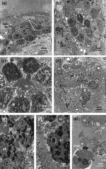
Testis tubules examined at this time-period showed evidence of distinct changes. The spermatogonia, denoted by a high ratio of nucleus to cytoplasm and located close to the tubule wall, retained a relatively normal morphology, although the mitochondria in the cells were enlarged and electron-lucent (fig. 1a). The mitochondria in the processes of the sustentacular cells surrounding the spermatogonia had a similar morphology (fig. 1a). The majority of primary and secondary spermatocytes were normal (fig. 1b–d) although, in a number of specimens, abnormal developmental stages were observed in close proximity to them. These abnormal cells, which appeared to be aberrant secondary spermatocyte stages (fig. 1b), contained large, heteromorphic nuclei with dense, fragmentary patches of heterochromatin and appeared to be undergoing early-stage karyorrhexis; most likely, they represent the eosinophilic bodies observed at the light microscope level. At high magnification, the mitochondria in the cytophore region of the spermatocyte rosette and in the cytoplasm of the cells themselves were distended, electron-lucent and contained swollen cristae (fig. 1c and d). Spermatid cells, denoted by the arrangement of the chromatin in lamellar-like sheets, were few in number. In certain cases, the spermatid cells were clearly apoptotic, with nuclear material appearing fragmented and electron-dense, representing late pyknosis/early karyorrhexis (fig. 1e and f). Mature spermatozoa were observed toward the centre of a number of tubules (fig. 1f and g), although they were far fewer in number than those observed in the controls and appeared morphologically abnormal. In some specimens, bi-nucleate spermatogonia, which had failed to undergo cytokinesis, were observed (fig. 1g). Large vacuoles and evidence of cell debris were widespread throughout the tubules. This debris is likely to represent the eosin-stained protein-filled vacuoles observed at the light microscope level, 72 h pt.
Transmission electron micrographs of the testis of triclabendazole-resistant Fasciola hepatica (Sligo isolate), 24 h post treatment in vivo with artemether at 200 mg/kg. (a) Primary and secondary spermatogonia (1°/2°SG) are located at the periphery of the tubule. Mitochondria (m) in these cells and in the surrounding sustentacular cells (SuC) appear to be disrupted and electron-lucent. N, nucleus. (b) Rosette of primary spermatocytes (1°SC) joined together at the central cytophore (cp). The cytophore contains electron-lucent mitochondria (m). Adjacent to the rosette are cells containing nuclei with condensed masses of chromatin (arrows). N, nucleus. (c) Primary spermatocyte rosette. The mitochondria (m) in the cytophore (cp) and in the cytoplasm of the cells are distended and contain swollen cristae. N, nucleus. (d) A primary spermatocyte (1°SC) is present, as characterized by the nuclear synaptonemal chromosomal complex (arrow). Adjacent, are primary and secondary spermatogonia (1°/2°SG). The mitochondria (m) in both cell types appear to be swollen and electron-lucent. N, nucleus. (e) Abnormal early-stage spermatid cells (ST), which appear to be breaking down. Arrow, nucleus. (f) Peripheral region of an abnormal spermatid cell (ST). Adjacent to the cell, abnormal mature spermatozoa (SP) with single axonemes are visible. m, mitochondrion. (g) Tubule overview showing a bi-nucleate secondary spermatogonium (2°SG) which has failed to undergo cytokinesis. Profiles of mature spermatozoa (SP) are present, although cellular debris and large vacuoles (arrows) occupy much of the lumen of the tubule. N, nucleus.

Forty-eight hours pt with artemether
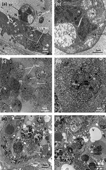
All developmental stages were observed in the tubules, from spermatogonia to mature spermatozoa, although they displayed a number of changes (fig. 2a–d). While the spermatogonial (fig. 2a) and spermatocyte (fig. 2c and d) cells retained a largely normal morphology, they contained swollen, electron-lucent mitochondria and the incidence of cells undergoing breakdown (having aborted development) had increased. The spermatocyte cells were loosely grouped together, rather than being organized in typical rosette structures (fig. 2c). Nuclear changes indicative of karyorrhexis were evident in the spermatocyte cells (fig. 2c). Cells belonging to the spermatid stage (fig. 2a and b) were few in number and contained swollen, abnormal mitochondria with dilated cristae and an electron-lucent appearance. In many specimens, these cells contained abnormally shaped nuclei, demonstrating an error in the elongation process. Moreover, the spermatid cells occurred as separate cells and not as part of a syncytial cluster. Mature spermatozoa were present, although the numbers were low, and they appeared to be abnormal, with the profiles containing only a single axoneme.
Transmission electron micrographs of the testis of triclabendazole-resistant Fasciola hepatica (Sligo isolate), 48 h (a–d) and 72 h (e and f) post treatment in vivo with artemether at 200 mg/kg. (a) Testis tubule overview showing primary/secondary spermatogonia (1°/2°SG) at the periphery of the tubule. An early-stage spermatid cell (ST) is more centralized. Mature spermatozoa (SP) are also present. The mitochondria (m) in the cells are swollen and electron-lucent. N, nucleus. (b) Early-stage spermatid cell, showing the nucleus (N) extending into characteristic lamellar-like sheets. The mitochondria (m) are substantially disrupted, with swollen cristae and an electron-lucent appearance. An abnormal single axoneme of a mature spermatozoon (SP) is present in the lumen of the tubule. (c) A primary spermatocyte rosette (1°SC). Cells of the rosette appear to be loosely grouped. A synaptonemal complex (arrow) is present in the nucleus (N) of one of the cells. The mitochondria (m) in the cells are swollen and electron-lucent. The nuclei are karyorrhectic. (d) Primary spermatocyte cell. A synaptonemal complex (arrow) is present within the nucleus (N). (e) Tubule overview showing disrupted secondary spermatogonia (2°SG) with nuclear material (N) beginning to condense and fragment. A tri-nucleate spermatogonium (arrow) has failed to undergo cytokinesis. Swollen, electron-lucent mitochondria (m) and autophagic vacuoles (AV) are present in sustentacular cell processes (SuC) along the edge of the tubule lumen. (f) A testis tubule containing cells which are undergoing breakdown. Mitochondria (m) appear to be swollen and empty, with no visible cristae. Autophagic vacuoles (AV) and cytoplasmic debris (arrow) are abundant. Some apoptotic cells contain nuclei (N) which are undergoing karyorrhexis. Large, seemingly empty vacuoles (V) are also present.

Seventy-two hours pt with artemether
Substantial changes were observed in the spermatogenic cells of flukes recovered at this time-point. The tubules examined were virtually empty of normal developmental cell stages and, instead, much of the lumen was occupied by cell debris and abnormal cells (fig. 2e and f). Both spermatogenic and sustentacular cells contained numbers of autophagic vacuoles. Mitochondria, present in all cells of the tubules, appeared swollen and lacked visible cristae. For the most part, only spermatogonial cell stages were recognizable and often they were multinucleate (fig. 2e), having failed to complete mitosis successfully. Many of the cells examined appeared to be apoptotic, with cell nuclei undergoing karyorrhexis (fig. 2f). Large, seemingly empty vacuoles were present throughout many of the tubules.
Artesunate
Twenty-four hours pt with artesunate
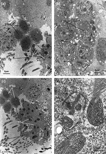
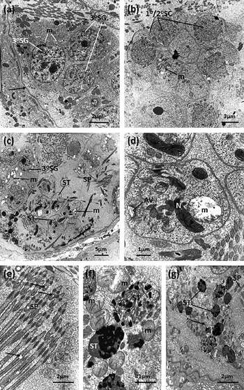
Testis tubules examined at this time-period showed little evidence of disruption. All spermatogenic and spermiogenic cell stages were observed, from spermatogonia (fig. 3a and b) to mature spermatozoa (fig. 3a and c). The spermatogonia, residing close to the walls of the tubules, displayed a normal morphology. Primary and secondary spermatocytes, which were situated more centrally within the tubules, also appeared to be fairly normal. In some cases, abnormal, electron-lucent mitochondria were observed in the cytoplasm of spermatocyte cells and in the cytophores of the spermatocyte rosettes (fig. 3b). Spermatid cells appeared to be normal and the developing spermatozoa were observed in various stages of elongation (fig. 3c). Spermatid nuclei, undergoing the typical processes of scrolling and elongation to form spermatozoa, were observed at high magnification. Central bodies were present in the zone of differentiation, together with neighbouring basal bodies, the latter displaying a characteristic 9+0 structure (fig. 3d). Spermatozoa were present, but some profiles were abnormal, lacking axonemes, mitochondria and a nucleus (fig. 3a and c).
Transmission electron micrographs of the testis of triclabendazole-resistant Fasciola hepatica (Oberon isolate), 24 h post treatment in vivo with artesunate at 200 mg/kg. (a) Tertiary spermatogonia (3°SG) are located close to the tubule wall. Spermatids (ST) and mature spermatozoa (SP) are present in the lumen of the tubule. The cells appear normal, although some of the spermatozoa are abnormal, lacking axonemes, mitochondria and a nucleus (arrows). (b) Rosette of primary spermatocytes (1°SC) joined together at the central cytophore (cp). The cytophore contains electron-lucent mitochondria (m). The rosette lies close to the tubule wall (arrow). N, nucleus; SG, spermatogonium. (c) Central region of a testis tubule. Present are normal, early-stage spermatid cells (ST), as well as mature spermatozoa (SP). Some of the spermatozoon profiles are abnormal, in that they lack axonemes, mitochondria and a nucleus (arrows). 3°SG, tertiary spermatogonia. (d) Profile of a normal, developing spermatozoon. In the periphery, the elongating nuclei of spermatid cells (arrows) are present. bb, basal body; cb, central body; N, nucleus.

Forty-eight hours pt with artesunate
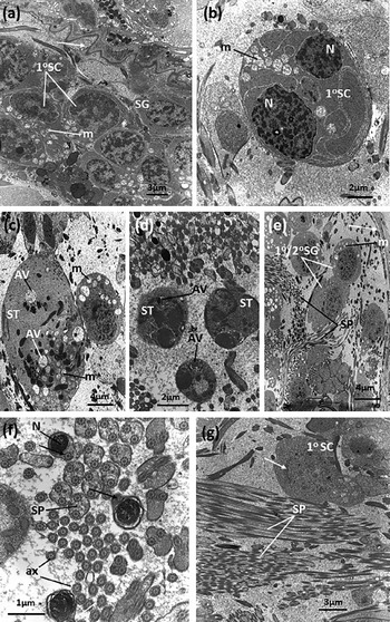
A number of changes were observed at this time-point. Spermatogonia, although mostly normal, often possessed nuclei with fragmented chromatin (fig. 4a). The primary spermatocytes contained high numbers of morphologically abnormal mitochondria, which were swollen and electron-lucent (fig. 4a and b). A number of abnormal bi-nucleate spermatocytes were also observed in some specimens (fig. 4b). They may be cells that have failed to undergo cytokinesis after cell division. Fragmentation of nuclear material, indicative of karyorrhexis, was seen in the spermatocyte cells (fig. 4a and b). Some aberrant, separated spermatid cells were present: they contained high numbers of autophagic vacuoles and electron-lucent mitochondria (fig. 4c and d). The normal advanced stages of the nuclear elongation process were not observed. Despite these changes, many spermatozoa were present in the tubules (figs 4e–g). However, some contained only one axoneme, while other profiles lacked axonemes, mitochondria and a nucleus (fig. 4f). Free axonemes were present in the tubule lumen (fig. 4f).
Transmission electron micrographs of the testis of triclabendazole-resistant Fasciola hepatica (Oberon isolate), 48 h post treatment in vivo with artesunate at 200 mg/kg. (a) A group of primary spermatocytes (1°SC). In their cytoplasm are abnormal swollen and electron-lucent mitochondria (m). SG, spermatogonium. (b) An abnormal primary spermatocyte (1°SC), which has failed to undergo cytokinesis and possesses two nuclei (N). The nuclear material is fragmented. The mitochondria (m) within the cell are abnormally swollen and electron-lucent. (c) Aberrant spermatid cells (ST), which are separate from each other. Autophagic vacuoles (AV) and electron-lucent mitochondria (m) are present in the cytoplasm. (d) Abnormal early-stage spermatid cells (ST). The cells are separate and contain autophagic vacuoles (AV). (e) Primary/secondary spermatogonia (1°/2°SG) lie close to the tubule wall (arrow). They contain electron-lucent and swollen mitochondria (m). Numerous spermatozoa (SP) are present in the tubule lumen. (f) Profiles of spermatozoa (SP) within the lumen of a testis tubule. Some of the profiles appear to contain one axoneme (arrows), while others lack axonemes, mitochondria and a nucleus. Numbers of free axonemes (ax) are present in the tubule lumen. N, nucleus. (g) The lumen of a tubule. Large numbers of spermatozoa (SP) are present. A synaptonemal complex (arrow) is present in a primary spermatocyte cell (1°SC).

Seventy-two hours pt with artesunate
Flukes recovered at this time-point displayed considerable changes to the morphology of the spermatogenic cells in the testis tubule. The spermatogonia appeared to be fairly normal, although they contained electron-lucent mitochondria and the nuclear chromatin was fragmented (fig. 5a). Rosettes of primary and secondary spermatocytes were few in number. Those that were present appeared to be morphologically abnormal, in that they contained mitochondria which were swollen and electron-lucent (fig. 5b). Spermatid cells were also present in abnormally low numbers and possessed greatly enlarged and electron-lucent mitochondria (fig. 5c and d). Some spermatid cells appeared to be separate from each other, rather than having a common syncytial cytoplasm (fig. 5c), although evidence of the sperm formation process, such as the scrolling of chromatin material in the nuclei, was observed (fig. 5c and d). Free spermatozoa were present in the tubules in normal numbers, although many of the profiles appeared to be morphologically abnormal, with the mitochondria being swollen and electron-lucent (fig. 5e). In certain cases, tubules contained spermatogenic cells resembling spermatids, but which were clearly apoptotic, having nuclear material which was fragmented and electron-dense, indicative of karyorrhexis (fig. 5f and g). Quantities of cellular debris were present in the lumen of the tubule (fig. 5g).
Transmission electron micrographs of the testis of triclabendazole-resistant Fasciola hepatica (Oberon isolate), 72 h post treatment in vivo with artesunate at 200 mg/kg. (a) Normal tertiary spermatogonia (3°SG), which reside close to the wall of the tubule (arrow). Mitochondria (m) in the cells are electron-lucent. (b) A rosette of spermatocyte cells (1°/2°SC). Some of the cells contain mitochondria (m) which appear to be swollen and electron-lucent. (c) Tubule overview showing tertiary spermatogonia (3°SG), spermatids (ST) and mature spermatozoa (SP). The mitochondria (m) in the cytoplasm of the spermatogonia and spermatids appear to be swollen and electron-lucent. The spermatid cells are separate from one another. (d) Spermatid cell, which appears to be separate from other spermatid cells. The scrolling nuclear material within the nucleus (N) indicates the process of sperm formation. The mitochondrion (m) is enlarged and does not contain visible cristae. AV, autophagic vacuole. (e) Profiles of mature spermatozoa (SP), some of which appear to be morphologically abnormal, in that they contain a mitochondrion (arrows) which is electron-lucent. (f) Abnormal early-stage spermatid cells (ST). The nuclear material is undergoing condensation and fragmentation. The mitochondria (m) within these cells are abnormal. (g) Tubule overview showing a number of aberrant spermatid cells (ST) undergoing apoptosis. Cellular debris (arrow) occupies much of the lumen of the tubule (arrow). SP, spermatozoa.

Ninety-six hours pt with artesunate
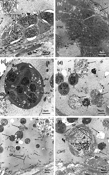
By 96 h pt, the number of spermatogenic cells of all types present in the tubules had decreased markedly, leaving the tubules empty-looking, with a lack of cells (fig. 6a–f). Identification of specific cell types was very difficult. Spermatogonial cells, which retained the most normal morphology of all cell types, were observed to contain electron-lucent vacuoles within their cytoplasm (fig. 6a). No normal spermatocyte or spermatid cells were present in the tubules. In their place, spermatocytes containing abnormal mitochondria were observed (fig. 6b), along with multinucleate cells, which had failed to separate successfully after division (fig. 6c). Many of the spermatogenic cells in the centre of the tubule were separated from each other, lying free in the lumen, and appeared to be in the advanced stages of apoptosis, with the nuclei undergoing karyorrhexis and karyolysis (fig. 6c–f). High numbers of autophagic vacuoles were observed in the cytoplasm of the cells (fig. 6c) and large quantities of cell debris were present in the lumen of the tubules (fig. 6d and e). No evidence of sperm formation was observed. Free spermatozoa were present (fig. 6f), but were often morphologically abnormal, containing only a single axoneme.
Transmission electron micrographs of the testis of triclabendazole-resistant Fasciola hepatica (Oberon isolate), 96 h post treatment in vivo with artesunate at 200 mg/kg. (a) Micrograph showing primary/secondary spermatogonia (1°/2°SG) along the inside of the wall of the tubule. Large, electron-lucent vacuoles (arrows) are present in the cytoplasm of these cells. (b) Rosette containing abnormal primary spermatocytes (1°SC). The cells, which reside close to the tubule wall (arrow), contain mitochondria (m) which are swollen and electron-lucent. (c) Abnormal multinucleate cell which has failed to undergo successful cytoplasmic division. The cell contains high numbers of autophagic vacuoles (AV). N, nucleus. (d) Testis tubule. Very low numbers of spermatogenic cells are present. The tubule, instead, contains apoptotic cells and cell debris (arrows). N, nucleus. (e) A sparsely populated testis tubule. With the exception of free spermatozoa (SP), those spermatogenic cells which are present are breaking down (arrows). ST, spermatid cell. (f) Free spermatozoa (SP) are present in the testis tubule. Electron-lucent vacuoles (arrows) are present in the lumen of the tubule. The remnants of spermatids (ST) can be seen; these cells are breaking down. Other cells in the tubule contain nuclei that are undergoing apoptosis.

Discussion
Treatment with artemether and artesunate caused severe disruption to the testis of TCBZ-resistant F. hepatica, leading to the cessation of sperm formation and resulting in empty, vacuolated testis tubules. The fine structural changes undergone by the cells have provided information relevant to the mechanism of action of this group of compounds. Adding the present results to those from previous studies on different tissue systems in the fluke has enabled a possible time-course of drug action to be constructed. These topics will be discussed below.
In the testis, marked changes to cellular morphology were observed as early as 24 h pt with artemether. They included bi-nucleate spermatogonia, disruption of mitochondria in the spermatocytes, a decrease in the number of later developmental stages (spermatids, spermatozoa), abnormalities in the sperm and evidence of apoptosis (pyknosis and karyorrhexis) in the nuclei of the spermatogonia, spermatocytes and spermatids. The cell population had declined further at 48 h pt, with few spermatids and spermatozoa being seen, and the organization of the spermatocyte and spermatid cell clusters had become abnormal. There was evidence of karyorrhexis in the nuclei of spermatogonia and spermatocytes. By 72 h pt, the testis tubules were filled with cell debris and abnormal cells, and large vacuoles were present. Only spermatogonia could be recognized; other cells were multinucleate, with autophagic vacuoles and abnormal mitochondria in their cytoplasm. There was widespread evidence of apoptosis in the cells (karyorrhexis and karyolysis). In a separate paper, it has been shown that no sperm were present in the uterus of treated flukes from 24 h onwards (O'Neill et al., Reference O'Neill, Johnston, Halferty, Hanna, Brennan and Fairweather2015b). The results of the histological and TEM analysis were in agreement with each other.
Following treatment with artesunate, the overall organization of the testis tubules and the morphology of the different cell types appeared to be relatively normal at 24 h pt. The only significant change was the presence of swollen, electron-lucent mitochondria in the spermatocyte cells. Sperm formation was seen to be taking place in the spermatid cells, although some of the sperm profiles lacked axonemes, mitochondria and a nucleus. Greater changes were observed at 48 h pt, in all cell types. The spermatogonia contained abnormal mitochondria and a number of bi-nucleate spermatocytes were seen; nuclear changes in these cells were compatible with karyorrhexis. The spermatid cells had become separated from each other, rather than being in a syncytial mass, and they contained autophagic vacuoles and abnormal mitochondria. Evidence of sperm formation was seen in these cells, but some sperm profiles were abnormal, in that axonemes, mitochondria and a nucleus were missing. Numbers of free axonemes were present in the tubule lumen. Those spermatozoa that were present may represent residual sperm, that is, sperm formed earlier. By 72 h pt, the number of cells had declined, particularly the number of spermatocytes and spermatids, and the tubule contained cell debris. Abnormal mitochondria were a feature of all cell types, from spermatogonia to spermatozoa. The spermatid cells were separate from each other, and their nuclei were undergoing apoptotic (karyorrhectic) changes, but sperm formation was seen to be taking place, although the sperm were abnormal. At 96 h pt, the testis tubules had a very empty appearance and few cells were present; instead, the tubules contained much cell debris. Spermatogonia and spermatocytes were present, but abnormal, the latter often taking the form of multinucleate cells containing numerous autophagic vacuoles. Otherwise, individual cells occupied the tubule lumen and they were (seen to be) undergoing apoptotic breakdown (karyolysis). There was no evidence of sperm formation; some residual sperm were seen, but they were abnormal, possessing a single axoneme.
Comparing the impact of the two drugs on the testis at matching time-periods, it seems that changes occurred more quickly and there was greater disruption following artemether treatment. This is in contrast to their relative action against the tegument and vitelline cells, but similar to their effects on the gut (O'Neill et al., Reference O'Neill, Johnston, Halferty, Brennan, Keiser and Fairweather2009, Reference O'Neill, Johnston, Halferty, Brennan and Fairweather2015a, Reference O'Neill, Johnston, Halferty, Hanna, Brennan and Fairweatherb). Nevertheless, the changes that were seen in the testis were common to both compounds, that is, disruption of the mitochondria, the presence of autophagic vacuoles, a block in the normal developmental sequence of the spermatogenic cells (resulting in a distinct decline in sperm production) and widespread evidence of cell apoptosis. Furthermore, comparable changes were seen in the testis tubules of F. hepatica following treatment in vitro with the artemisinin derivative, OZ78 (Halferty et al., Reference Halferty, O'Neill, Brennan, Keiser and Fairweather2009). The same organelle and cellular changes have been seen in other fluke tissues as well, namely, the tegumental, gut and vitelline cells, and therefore seem to be a consistent feature of artemisinin action (Halferty et al., Reference Halferty, O'Neill, Brennan, Keiser and Fairweather2009; O'Neill et al., Reference O'Neill, Johnston, Halferty, Brennan, Keiser and Fairweather2009, Reference O'Neill, Johnston, Halferty, Brennan and Fairweather2015a, Reference O'Neill, Johnston, Halferty, Hanna, Brennan and Fairweatherb). Moreover, they can be related to what is generally known about the action of artemisins (see below).
There have been a number of studies detailing changes in the testis following flukicidal action. For example, following nitroxynil and rafoxanide treatment, premature elongation of the nucleus occurred in the spermatocytes and retention of spermatids was seen in the sperm morulae (Stammers, Reference Stammers1975a, Reference Stammersb, Reference Stammers1976). With bithionol, spermatogenesis was inhibited at the spermatid stage: individual spermatids were released prematurely from the sperm morulae and failed to complete their development, becoming comma-shaped and only slightly elongate (Dawes, Reference Dawes1966a, Reference Dawesb, Reference Dawes1968). Triclabendazole treatment in vitro and in vivo led to a drop in the number of cells in the testis tubules, disruption of the cytophore of the spermatocyte and spermatid stages, and a block in spermatozoon formation (Stitt & Fairweather, Reference Stitt and Fairweather1992; Toner et al., Reference Toner, Brennan, Hanna and Fairweather2011). These changes are typical of microtubule inhibition and have been seen following treatment of flukes with a microtubule inhibitor (Stitt & Fairweather, Reference Stitt and Fairweather1992). The combined data suggest that drug effects vary from drug to drug and are not necessarily a generalized response to drug action. This conclusion argues against the idea proposed by Dawes (Reference Dawes1968), that ‘… flukes which are threatened by drug action on the host will tend first to sacrifice non-vital organs and to conserve vital organs’. It is likely that the disruption to the testis and reduction in sperm production would have an impact on egg production. Following artemether treatment, it has been shown that the eggs in the uterus became abnormal and no sperm were present, suggesting that the eggs may not have been viable (O'Neill et al., Reference O'Neill, Johnston, Halferty, Hanna, Brennan and Fairweather2015b). Egg viability could be tested by means of the egg hatch assay designed by Fairweather et al. (Reference Fairweather, McShane, Shaw, Ellison, O'Hagan, York, Trudgett and Brennan2012). In turn, the production of non-viable eggs would have a serious effect on the reproductive capacity of the fluke and transmission of the disease.
It is generally accepted that artemisinin activation proceeds via the generation of free radicals that, in turn, induce mitochondrial changes (Wakabayashi & Karbowski, Reference Wakabayashi and Karbowski2001; W. Li et al., Reference Li, Mo, Shen, Sun, Wang, Lu, Gitschier and Zhou2005; Wang et al., Reference Wang, Huang, Li, Fan, Long, Li and Zhou2010). The swelling of mitochondria in the spermatogenic cells was a consistent feature of drug action and has been seen in other fluke tissues (Halferty et al., Reference Halferty, O'Neill, Brennan, Keiser and Fairweather2009; O'Neill et al., Reference O'Neill, Johnston, Halferty, Brennan, Keiser and Fairweather2009, Reference O'Neill, Johnston, Halferty, Brennan and Fairweather2015a, Reference O'Neill, Johnston, Halferty, Hanna, Brennan and Fairweatherb). Swelling and lysis of mitochondria as a result of artemisinin treatment have also been observed in malarial and schistosome parasites (Ellis et al., Reference Ellis, Li, Gu, Peters, Robinson, Tovey and Warhurst1985; Kawai et al., Reference Kawai, Kano and Suzuki1993; Maeno et al., Reference Maeno, Toyoshima, Fujioka, Ito, Mesnick, Benakis, Milhous and Aikawa1993; Xiao et al., Reference Xiao, Binggui, Utzinger, Chollet and Tanner2002a, Reference Xiao, Binggui, Utzinger, Chollet and Tannerb). Mitochondria are believed to be involved in the induction (and execution) of apoptosis and this may be initiated by lysosomal enzyme action (Wakabayashi & Karbowski, Reference Wakabayashi and Karbowski2001; W. Li et al., Reference Li, Mo, Shen, Sun, Wang, Lu, Gitschier and Zhou2005; Efferth et al., Reference Efferth, Glaisi, Merling, Krammer and Li-Weber2007; P.C.H. Li et al., Reference Li, Lam, Roos, Zdzienicka, Kaina and Efferth2008; Hamacher-Brady et al., Reference Hamacher-Brady, Stein, Turschner, Toegel, Mora, Jennewein, Efferth, Eils and Brady2011; Crespo-Ortiz & Wei, Reference Crespo-Ortiz and Wei2012). A build-up of autophagic vacuoles in the spermatogenic cells was seen in the present study and in previous studies on the gut and vitelline cells of F. hepatica (Halferty et al., Reference Halferty, O'Neill, Brennan, Keiser and Fairweather2009; O'Neill et al., Reference O'Neill, Johnston, Halferty, Brennan, Keiser and Fairweather2009, Reference O'Neill, Johnston, Halferty, Brennan and Fairweather2015a, Reference O'Neill, Johnston, Halferty, Hanna, Brennan and Fairweatherb). Mitochondrial disruption either preceded or paralleled the apoptotic changes occurring in the nuclei of the spermatogenic cells, and leading to the (autophagic) breakdown of the cells. The linkage between the processes was particularly evident in the testis and the observations would lend support to this mechanism of action operating in the fluke.
This is the last paper in a series describing the morphological changes in F. hepatica following treatment of the rat host with artemether and artesunate. It provides an opportunity to collate and compare the changes in several different tissues, and to try to assess their relative significance (in terms of drug efficacy) against what is known about the pharmacokinetics of the drug and the speed of elimination of the flukes. Taking artemether first, a reduction in motility was only noticed at 72 h pt, but none of the flukes retrieved were totally inactive and there was no evidence of elimination from the host. In a separate study on artemether, ‘dead’ flukes were recovered at 48 and 72 h pt, although other flukes from the same rats displayed some activity, and by 96 h pt all flukes were ‘dead’; furthermore, ‘dead’ flukes could still be recovered 12 days pt, suggesting that elimination is a slow process (Keiser et al., Reference Keiser, Xiao, Tanner and Utzinger2006a). The first tissue to show signs of disruption in our experiments was the testis, at 24 h pt. At 48 h pt, the tegument, gut and vitellaria exhibited changes, which progressed in severity, but the disruption to the tegumental surface remained slight, even at 72 h pt. The gut was always more severely affected than the tegument, which is indicative of oral entry of artemether into the fluke. The changes to the gut would force the fluke into a state of starvation and this, together with the reduced levels of movement, would exacerbate the more direct effects of artemether on its cellular targets. Disruption of the cells in the testis and vitellaria resulted in abnormal egg formation, but that only became apparent in the uterus at 72 h pt. So, the speed of drug action is relatively slow, given that the peak plasma concentration (t max) of artemether following oral treatment is reached after only 29 min in the rat and the elimination half-life (t 1/2) is ~2 h (Q.-G. Li et al., Reference Li, Peggins, Fleckenstein, Masonic, Heiffer and Brewer1998). A similar paradox between drug pharmacokinetics and drug action has been seen in studies with clorsulon, another flukicide (Meaney et al., Reference Meaney, Fairweather, Brennan, McDowell and Forbes2003, Reference Meaney, Fairweather, Brennan and Forbes2004). In contrast, the morphological changes induced by TCBZ and nitroxynil more closely match their pharmacokinetic profile in the host (McKinstry et al., Reference McKinstry, Fairweather, Brennan and Forbes2003, Reference McKinstry, Brennan, Halferty, Forbes and Fairweather2007; Halferty et al., Reference Halferty, Brennan, Hanna, Edgar, Meaney, McConville, Trudgett, Hoey and Fairweather2008; Toner et al., Reference Toner, Brennan, Hanna and Fairweather2011).
For artesunate, (some of) the trends are less clear-cut. For example, inactive flukes were recovered from rats as early as 24 h pt, although other flukes were reasonably active. This was the case at 48 and 72 h pt as well. At 96 h pt, flukes were very sluggish, although they were still recovered, and it was at this time-point that evidence was obtained to indicate that elimination had taken place. Such variation has been seen in other studies and flukes were still recovered at 12 days pt (Keiser et al., Reference Keiser, Xiao, Tanner and Utzinger2006a; Keiser & Morson, Reference Keiser and Morson2008). This variability in response may be due to the different (and inconsistent) way in which infected animals metabolize the drug, as a result of liver damage caused by fluke infection, and to their individual sensitivity to the toxic effects of artesunate. As with artemether, the t max and t 1/2 of artesunate are short: 20 min and 42.6 min, respectively, in the rat after an oral dose (Keiser et al., Reference Keiser, Gruyer, Perrottet, Zanolari, Mercier and Decosterd2009). The first morphological changes were observed at 24 h pt, in the gut and vitellaria. At 48 h pt, changes were evident in the tegument and testis as well, and changes in all tissues became more severe with time. Surface changes were limited, even after 96 h treatment, although a severely damaged fluke was recovered at 72 h pt, and it is possible that other, similarly affected, flukes may already have been eliminated from the rats before they could be collected. Disruption to the sub-tegumental musculature was only observed at 96 h pt. As with artemether, then, the speed of action of artesunate is slow.
In conclusion, the present study has shown that artemether and artesunate cause severe disruption of the testis of F. hepatica, with artemether being the more disruptive, and more immediately disruptive, of the two compounds. An explanation has been put forward to account for the cellular changes seen, which were common to both drugs. The loss of sperm production is likely to affect egg formation, and the lack of sperm in the uterus post artemether treatment supports this idea. Integrating the results with those from other studies on the two drugs, it has been possible to obtain a more complete picture of drug action, create a potential time-line of drug action and assess which changes are more important than others in relation to drug action. This was less easy to do with artesunate, due to the variation in drug response observed. Morphological studies have been carried out with three different compounds, namely, artemether, artesunate and OZ78 (Halferty et al., Reference Halferty, O'Neill, Brennan, Keiser and Fairweather2009; O'Neill et al., Reference O'Neill, Johnston, Halferty, Brennan, Keiser and Fairweather2009, Reference O'Neill, Johnston, Halferty, Brennan and Fairweather2015a, Reference O'Neill, Johnston, Halferty, Hanna, Brennan and Fairweatherb, present study; O'Neill, Reference O'Neill2014). From the results, a number of general statements can be made about artemisinin action against the fluke: that drug action is relatively slow; that internal changes are more severe than those visible externally; that drug entry is oral rather than trans-tegumental; that reproductive capacity is severely impaired, shutting down egg production; that the mitochondria, granular endoplasmic reticulum and lysosomes are prime cellular targets in several tissues; and that changes to these organelles may trigger the apoptotic breakdown of the cells. Moreover, the compounds were active against TCBZ-resistant F. hepatica, which is significant in relation to any future use for artemisinins in treating fasciolosis.
Supplementary material
To view supplementary material for this article, please visit http://dx.doi.org/10.1017/S0022149X16000079
Financial support
This work was supported by a postgraduate studentship awarded to J. O'N. by the Department of Agriculture and Rural Development, Northern Ireland (DARDNI).
Conflict of interest
None.
Ethical standards
The authors assert that all procedures contributing to this work comply with the ethical standards of the relevant national guides on the care and use of laboratory animals (rats) and has been approved by the Animal Welfare and Ethical Review Body, The Queen's University of Belfast.