Introduction
Recurrent vertigo is one of the most incapacitating symptoms of Ménière's disease, and currently available therapy is poorly effective. In severe cases of Ménière's disease, attacks of vertigo accompanied by nausea and vomiting can sometimes be controlled only by eliminating the vestibular reflexes of the affected ear.Reference Cheng, Cunningham and Rubel1–Reference Rybak and Whitworth3 Many methods have been used to eliminate vestibular function. Destructive surgical procedures, in the form of labyrinthectomy, reduce vertigo but severely impair hearing; furthermore, section of the vestibular nerve risks serious intracranial complications.Reference De La Cruz, Teufert and Berliner4 Thus, new, minimally invasive methods have been developed for vestibular ablation based on local application of ototoxic substances.Reference Stokroos and Kingma5, Reference Beck and Schmidt6 Since vertigo often occurs due to dysfunction of only one inner ear, and since systemic application of ototoxic substances induces damage to both inner ears, topical application of ototoxic agents may have a more selective action by affecting only the inner ear of interest. For this reason, transtympanic administration of ototoxic agents may be a suitable way to achieve unilateral vestibular ablation for the control of unilateral Ménière's disease.
Gentamicin has been reported to be toxic to both the cochlea and the vestibular system,Reference Black, Pesznecker and Stallings7 but appears to damage selectively the vestibular system when applied at low doses, with relative sparing of the cochlea. Thus, it would appear to be the aminoglycoside of choice for vestibular ablation.Reference Stokroos and Kingma5
Few published studiesReference Roland, Rybak and Owens8 have quantified the histological evidence of vestibular toxicity following a single transtympanic application of gentamicin intended to damage the vestibular neuroepithelium of guinea pigs. Thus, the objective of the present study was to quantitate the dose–effect relationship of a single intratympanic infusion of varying doses of gentamicin in guinea pigs, as observed histologically with scanning electron microscopy.
Materials and methods
This study used 25 albino guinea pigs aged four months, of both sexes, weighing 450–550 g. Before the experiment, the animals were evaluated and selected according to their weight, otoscopic appearance and gait.
The selected animals were divided into five groups of five animals each. The animals in the five groups received a single dose of 1, 5, 10 or 25 mg gentamicin sulphate solution, prepared as a sterile saline dilution and applied transtympanically. Gentamicin (40 mg/ml) was buffered with sodium bicarbonate (pH 6.4).
A control group received sterile saline by the same route as experimental groups.
Before drug administration, the animals were anaesthetised with sodium pentobarbital (30 mg/kg body weight) injected intraperitoneally. The right ear was preferentially used for gentamicin application, while the left ear was used as a self-control. The tympanic membrane was visualised with the aid of a speculum and a light microscope, and gentamicin was injected through the tympanic membrane with a 1 ml insulin syringe needle. The needle was inserted into the posteroinferior quadrant of the tympanic membrane and then advanced towards the medial wall of the middle ear. A bolus of 0.1 ml of the buffered antibiotic solution was then injected over a period of 5 to 10 seconds. An application was considered to be successful when no extravasation of fluid through the puncture site was observed. The animal was then positioned with the injected side upwards for about 3 hours, with the snout elevated 45o to minimise escape of the solution through the auditory tube and to insure that the injected fluid remained in contact with the round and oval windows.
Two weeks after injection, the guinea pigs were euthanized after anesthesia with ketamine hydrochloride (65 mg/kg) and xylazine (6.5 mg/kg). The animals were then decapitated and their temporal bones processed for microscopy. After wide opening of the bulla, the vestibular system was microdissected and the lateral ampullar crista and the utricular macula prepared for scanning electron microscopy.
If any evidence of middle- or inner-ear infection was observed at the time of sacrifice, the animal was excluded from the study.
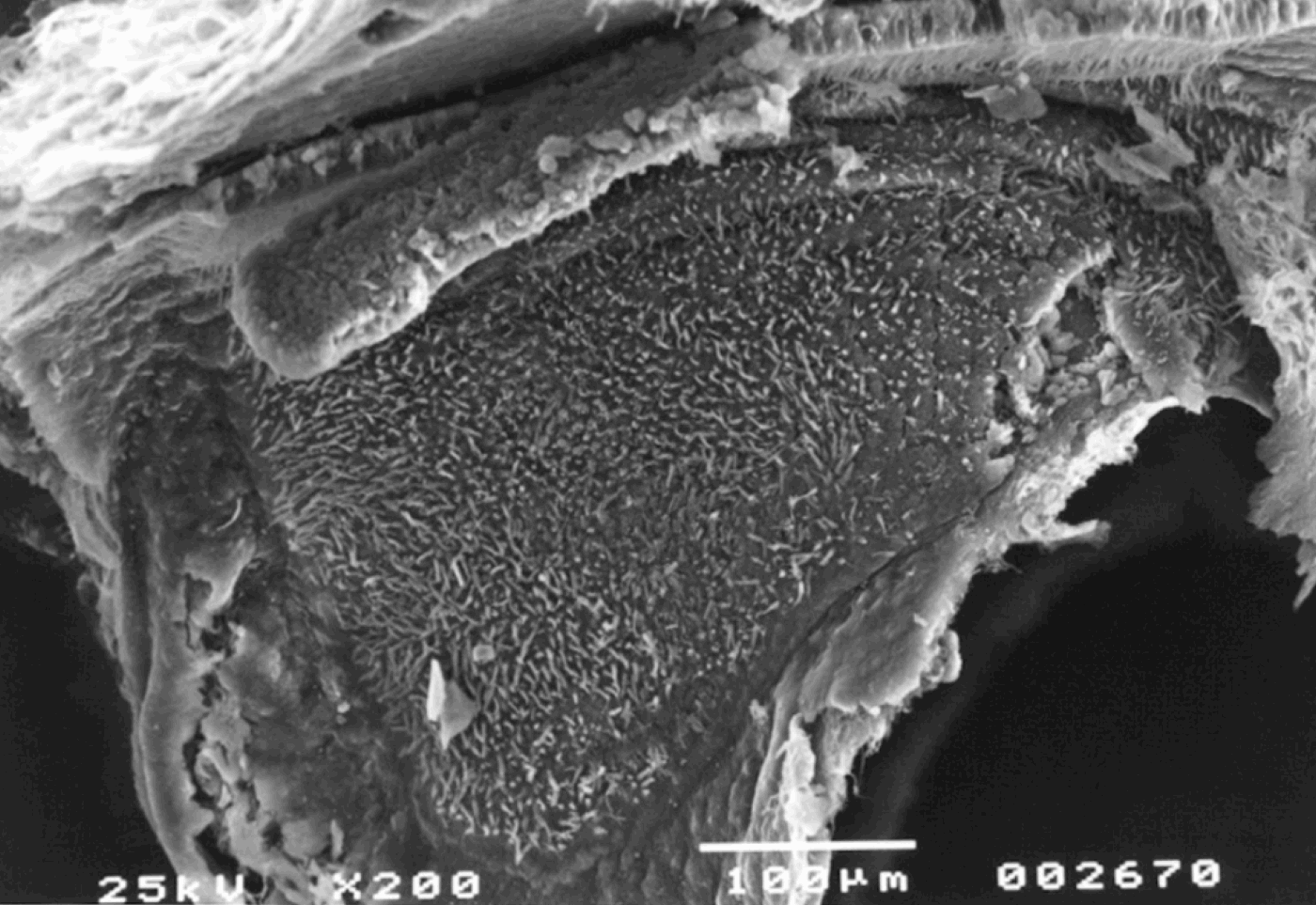
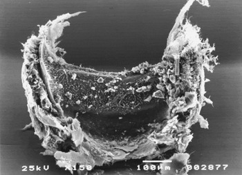
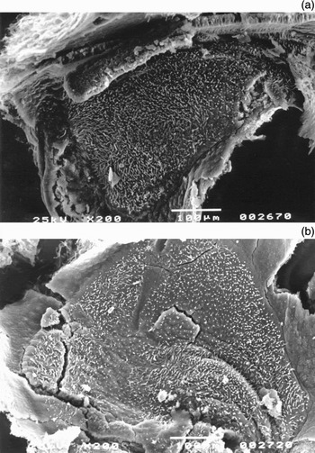
A blinded analysis of the histological material was performed. Structural lesions occurring as a result of gentamicin ototoxicity were determined, and differentiated from any artefacts caused by preparation or post-mortem autolysis. A method for quantitative evaluation of ototoxic damage was needed, in order to compare the animals' vestibular lesions. It was decided that the diagnosis of vestibular neuroepithelial damage would be based upon disappearance of the stereociliary bundles of the hair cells, as determined by scanning electron microscopy,Reference Parnes, Sun and Freeman9 according to the method of Nakayama.Reference Nakayama10 We recorded a tally for each animal which represented the arithmetic mean of the number of hair cells without stereociliary bundles seen in at least four serial samples of the central (striolar region), intermediate and peripheral portions of the utricular macula (Figure 1) and four serial samples of the central, intermediate and peripheral portions of the median part of the lateral ampullar crista (Figure 2), studied at ×7500 magnification.
Scanning electron micrograph of the normal guinea pig utricular macula (×200).
Scanning electron micrograph of the normal guinea pig lateral ampullar crista (×150).

The area from which cell counts were obtained was kept constant for all samples, and the angle of view was kept perpendicular to the cellular surface viewed, in order to avoid variation in the size of the area examined.
In this way, the total number of cell contours viewed and the number of stereociliary bundles present were recorded. The mean number of stereociliary bundles per viewing area was calculated for each guinea pig. The mean number of stereociliary bundles per viewing area was then established for each experimental animal group.
Student's t-test was used for statistical analysis, calculated using the Statistical Analysis System 2001 version 9.1.3 software program.
Results and analysis
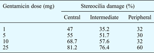
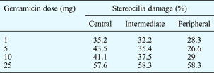
All animals showed damage to neuroepithelial structures on the injected side (p < 0.05), as determined by Student's t-test when compared with control group. Table I shows the mean proportion of damage to the stereocilia of the lateral ampullar crista, for the various antibiotic doses employed.
Stereocilia damage: ampullar crista
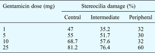
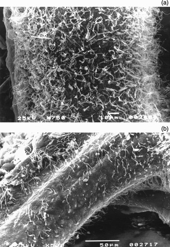
In the central portion of the crista of the lateral ampulla, damage to the stereocilia increased with increasing gentamicin concentration (Figure 3). In the intermediate portion, dose-dependent damage was also observed, but was less intense than in the central portion. In the peripheral portion, there was little variation in the degree of damage with increasing gentamicin doses up to 10 mg. However, this relative resistance to damage disappeared when the highest dose was applied (25 mg): at this dosage, 60 per cent of stereociliary bundles were damaged.
Scanning electron micrographs of the lateral ampullar crista. (a) Normal ampullar crista (×750); (b) damaged ampullar crista, more severe in the central region, with stereocilia still present in the peripheral portion (×500).

Within-group analysis showed that the degree of damage increased in intensity in the more central portions of the ampullar crista. Thus, damage to the hair cells was more severe in the central and intermediate portions of the ampullar crista, compared with the peripheral portion (p < 0.05).
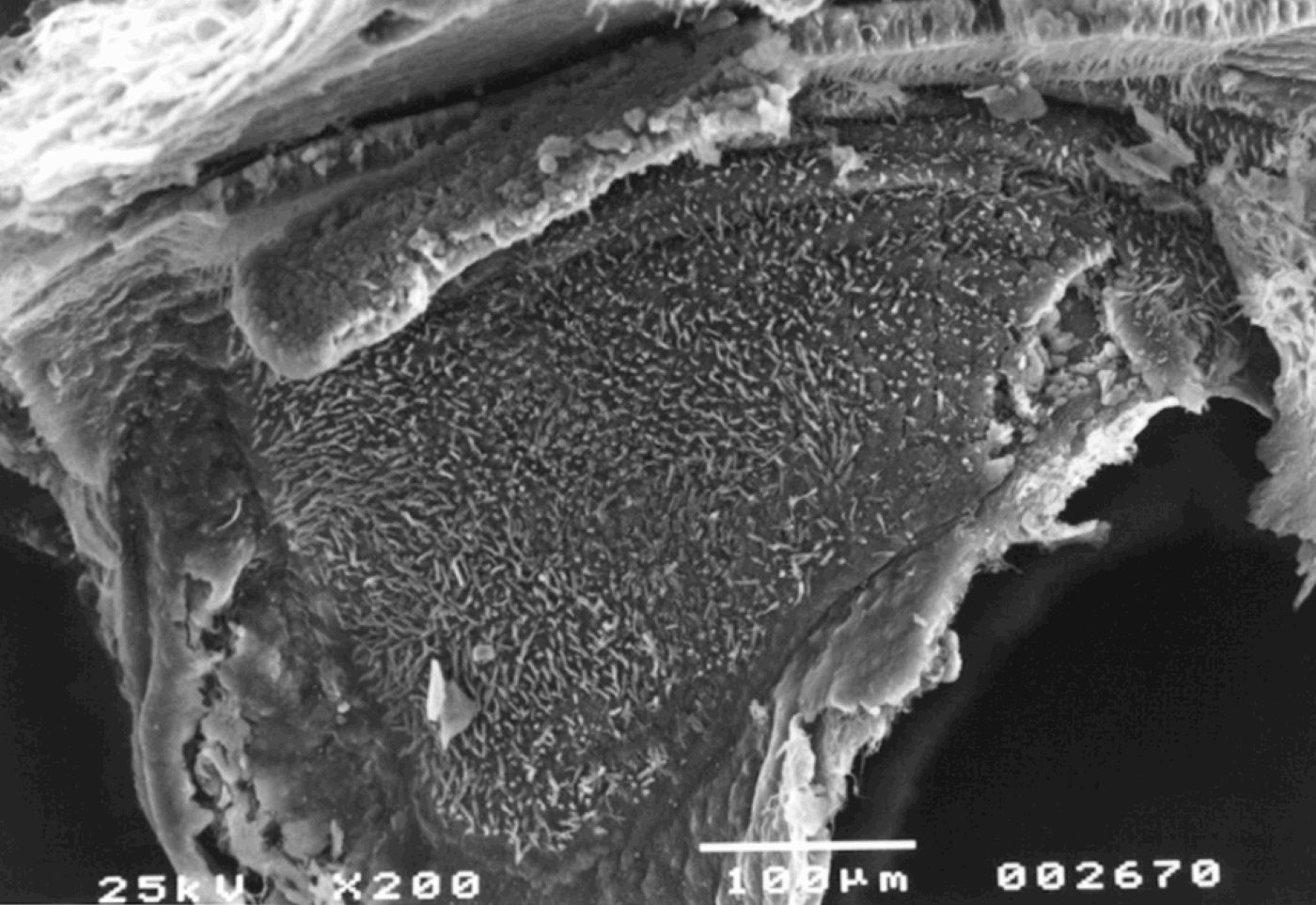
In the utricular macula, the mean levels of neurovestibular epithelial damage were similar to those observed in the ampullar crista (Table II). In the utricular macula, damage to the stereociliary bundles was also dose-dependent, with more intense damage occurring in more central regions, similar to the appearance in the ampullar crista (Figure 4). Within-group analysis showed that more damage was present in the central portion compared with the peripheral portion (p < 0.001), but with a smaller range of variation than that observed in the ampullar crista.
Scanning electron micrographs of the utricular macula. (a) Normal utricular macula (×200); (b) damaged utricular macula, showing rarefaction of the stereocilia especially in the central or striolar region, in an animal receiving 5 mg gentamicin (×200).

Stereocilia damage: utricular macula
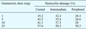
In the central and intermediate portions of both vestibular structures (i.e. the ampullar crista and the utricular macula), there was a dose-dependent increase in the degree of stereociliary damage. The peripheral portions of both structures showed a certain degree of resistance to aminoglycoside toxicity, even when the gentamicin dose increased from 1 to 10 mg. It was only after administration of the highest gentamicin dose (25 mg) that marked damage was observed. Thus, general analysis indicated that the central region of the ampullar crista and the utricular macula was more affected than the peripheral regions (Figures 3b and 4b). We also observed that the ampullar crista was more vulnerable to gentamicin ototoxicity than the utricular macula (p < 0.05). We observed no morphological evidence of damage to the dark cell areas of the ampullar crista.
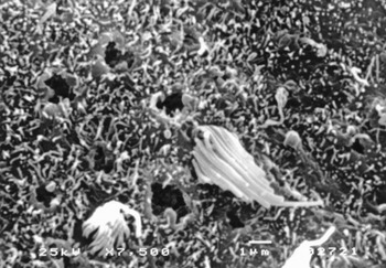
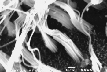
In the group receiving 5 mg gentamicin, the hair cell stereocilia morphology varied from no evidence of damage to little damage to the sensory epithelium (Figures 5 and 6).
Scanning electron micrograph of the utricular macula, showing damage to the macular sensory cells (central portion), with scars where stereociliary bundles were formerly present, in an animal receiving 5 mg gentamicin. The remaining cells show loss of the axis of stereociliary support (×7500).

Scanning electron micrograph showing sensory cells in the lateral portion of the ampullar crista (×7500).

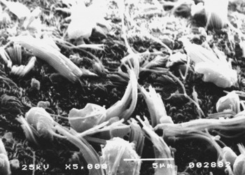
Where stereocilia were still present on the apical surface of the hair cells, they showed worsening degeneration as the gentamicin dosage increased, with cilia fusion and rupture, loss of the cilia supporting axis, and fusion and rarefaction of the cell surface (Figure 7).
Scanning electron micrograph of the utricular macula, showing qualitative signs of ototoxicity, i.e. fusion, oedema and loss of the axis of stereociliary support (×5000).

Discussion
Few published studies have quantified gentamicin vestibular toxicity in guinea pigs. Also, much of the data on the pharmacokinetics and ototoxicity of aminoglycosides has been obtained after systemic administration.
Gentamicin damages the hair cells of both the utricular macula and the ampullar crista. In general, the severity of cellular damage worsens as the antibiotic dose increases. Therefore, the concentration of gentamicin in the perilymph may depend on the dose applied to the middle ear.
We observed that hair cell damage was prevalent in the central region of both the ampullar crista and the utricular macula, and that this damage extended progressively to the periphery with increasing gentamicin doses. Thus, the peripheral regions of these vestibular structures may be less susceptible to such ototoxic effects, in contrast to the central portion of these structures, which showed progressive damage.Reference Lindeman11
These differences in sensitivity may be related to the fact that type I hair cells are more sensitive than type II hair cells, and that type I cells are much more abundant in the central portion of the ampullar crista and the utricular macula compared with the periphery. The observed sensitivity may also correlate with the higher protein metabolic rate of type I cells. Watanuki and Meyer Zum GottesbergeReference Watanuki and Meyer Zum Gottesberge12 have suggested that type I hair cells are phylogenetically more differentiated than type II cells, a fact that would render them more susceptible to any environmental stress, such as the presence of ototoxic drugs. However, the initial resistance of the peripheral portions of the two vestibular structures seems to be lost when higher ototoxic doses are used, as observed in the present study.
Our study found that gentamicin-induced vestibular damage was much more severe in the lateral ampullar crista than in the utricular macula, thus suggesting greater resistance of the otolithic organ to gentamicin ototoxicity.
Direct application of aminoglycosides to the middle ear elicits different responses compared with systemic injection. SchuknechtReference Schuknecht13 suggested that streptomycin has selective vestibulotoxicity after systemic administration, an effect that was not observed when the antibiotic was administered transtympanically. Many other studies of other aminoglycoside antibiotics have led to the conclusion that, if the objective of treatment is to affect primarily the vestibular structures with a minimal effect on the cochlea, gentamicin should be used since it appears to have greater efficacy in producing selective vestibular damage.
Transtympanic application is simple technically and limits treatment to the affected ear (in the case of unilateral involvement). The transtympanic route of administration promotes more uniform effects of the same dose, compared with systemic administration, since it reduces variation in inner-ear drug dosage due to individual differences in systemic compartmentalisation; transtympanic administration also eliminates the synergism of the nephrotoxic effect of gentamicin.Reference Roland, Rybak and Owens8, Reference Wagner, Caye-Thomasen, Laurell, Bagger-Sjoback and Thomsen14 When applied directly to the middle ear, the drug only needs to cross the structures of the medial wall of the middle ear and the membranous labyrinth in order to reach the sensory vestibular cells.Reference Wagner, Caye-Thomasen, Laurell, Bagger-Sjoback and Thomsen14
The membrane of the round window has been described as the main route via which drugs reach the labyrinth.Reference Harada15, Reference Saijo and Kimura16 Other studies have reported that a drug may also be absorbed around the stapes footplate.Reference Selimoglu17 Thus, the drug would be absorbed into the inner ear via both the round and oval windows; this absorption would presumably be more intense through the round window than through the oval window, because of the greater surface area of the former and because of a passive diffusion mechanism.Reference Plontke, Mynatt, Gill, Borgmann and Salt18 The dose that reaches the inner ear may also be proportional to the amount of drug applied to the middle ear.Reference Harada15, Reference Saijo and Kimura16
Some investigatorsReference Rudnick, Ginsberg and Huber19 have reported that stereocilia structural changes are not reliable indicators of ototoxicity, since there is not always a direct relationship with the ototoxic dose employed, and since such changes may sporadically be detected in normal controls. However, HaradaReference Harada15 and Proctor and el-KashefReference Proctor and el-Kashef20 have reported that both ciliary and cellular changes can be used to determine epithelial damage. In our study, we observed structural changes (e.g. fusion, oedema and loss of the stereocilia supporting axis) in all animals treated with gentamicin, and we found that the presence or absence of stereocilia could be used as a reliable quantitative indicator of the extent of ototoxic damage. Thus, this was the histological criterion we used to determine the degree of vestibular ototoxicity.
We selected the utricular macula for study, from the available otolithic structures, because it was easier to dissect than the saccular macula. We also selected for study the lateral ampullar crista as it too was easier to dissect, and also because it is frequently evaluated clinically in humans undergoing otoneurological examination.
• Low dose transtympanic gentamicin appears to damage the vestibular system more than the cochlear system
• Transtympanic gentamicin administration is technically simple, with more uniform effects than systemic administration, possibly due to reduced variation in the amount of drug reaching the inner ear
• This study confirms that the intensity of vestibular neuroepithelial damage due to gentamicin otoxicity is dose-dependent
The present study had some limitations. We evaluated only the histological effects of topical gentamicin application, and did not address the physiological effects. Due to the complexity of preparation and evaluation of the vestibular structures, only five guinea pigs were used in each study group. Scanning electron microscopy was selected as it permitted precise analysis of ototoxic changes, especially the structural lesions created in sensory hair cells.
The small number of animals in each dosage group led to some statistical abnormalities, such as more severe lesions occurring in animals receiving lower doses. Variations in round window membrane permeability and perilymph flow could have accounted for this observation.Reference Plontke, Mynatt, Gill, Borgmann and Salt18
Conclusion
On the basis of these findings, we conclude that significant vestibular neuroepithelial damage occurred in those guinea pig ears receiving transtympanic gentamicin application, although the doses employed did not cause total neuroepithelial destruction. The extent of vestibular neuroepithelial damage was dose-dependent, being more severe at higher doses. Damage was more severe in central versus peripheral vestibular structures, and the mean damage to the utricular macula was significantly less than that observed in the ampullar crista of the lateral semicircular canal.
Acknowledgements
The authors wish to thank the Laboratory of Electron Microscopy, Department of Cellular and Molecular Biology and Pathogenic Bioagents, and the Laboratory of Experimental Surgery, Department of Surgery and Anatomy, Faculty of Medicine of Ribeirão Preto, for technical assistance.