Introduction
In vivo oocyte maturation comprises the period just after the luteinizing hormone (LH) surge, when the oocyte resumes meiosis and undergoes the final modifications that make it capable of being fertilized by a sperm, a mechanism that includes nuclear maturation (linked to changes observed during the meiotic division) and cytoplasmatic maturation (Adona et al., Reference Adona, Pires, Quetglas, Schwarz and Leal2008). Meiosis resumption is observed as the germinal vesicle breakdown (GVBD) stage, followed by the first metaphase plate (MP) formation, indicating that the oocyte reached metaphase I (MI). Then, the oocyte completes its first meiotic division and starts the second, which again will stop the process at divisional metaphase II (MII), until fertilization occurs. The sequence of events that starts with GVBD and ends with the first polar body (PB) extrusion enables the production of suitable oocyte fertilization, and is called oocyte maturation (Dekel, Reference Dekel1995).
Prior to the maturation process, the oocyte must undergo numerous changes during oogenesis and folliculogenesis, which together comprise the so-called growth phase. At the end of this phase the oocyte will be developmentally competent to undergo maturation, both in vivo or in vitro (Hyttel et al., Reference Hyttel, Fair, Callesen and Greve1997).
Oocyte maturation can be mimicked in vitro by the technique of in vitro maturation (IVM) of oocytes—a very promising technology whose use is becoming more prominent (Grøndahl, Reference Grøndahl2008). Although promising, this technique still presents some difficulties surrounding its implementation. The main obstacle is the heterogeneity of oocytes recovered from the gonad, resulting in levels of competency development that are higher in the oocytes matured in vivo, when compared with their counterparts matured in vitro (Wu et al., Reference Wu, Liu, Zhou, Lan, Han, Miao and Tan2007). For this reason, the election of competent oocytes for IVM use is recommended, as there are oocytes that are ready to mature and therefore will bring better results. The search for a more appropriate technique selection of oocytes depends on the fact that the developmental capacity of oocytes is the major determinant in establishing success rates of pregnancy (Jiang et al., Reference Jiang, Xiong, Cao, Xia, Sirard and Tsang2010).
The use of the dye blue cresyl brilliant (BCB) is being proposed as a method to select competent oocytes. BCB is able to interact with the enzyme glucose-6-phosphate dehydrogenase (G6PDH), which presents increased activity before the oocyte growing phase, being characteristic of oocytes that did not undergo such a process. G6PDH activity decreases during the growth phase. Consequently, the oocytes that finished this phase and, therefore, are ready to tolerate the maturation, have low G6PDH activity (Mangia and Epstein, Reference Mangia and Epstein1975; Tsutsumi et al., Reference Tsutsumi, Satoh, Taketani and Kato1992). G6PDH has the ability to metabolize the dye BCB, neutralizing its bluish colour, resulting in colourless oocytes. Thus, the staining of oocyte after exposure to dye will determine the enzymatic activity of G6PDH and the oocyte competence level to undergo IVM (Alm et al., Reference Alm, Torner, Lohrke, Viergutz, Ghoneim and Kanitz2005; Bhojwani et al., Reference Bhojwani, Alm, Torner, Kanitz and Poehland2007). Oocytes that have finished their growth phase and that, accordingly, have decreased G6PDH activity will be stained with BCB (cytoplasm will appear blue, being classified as BCB positive; BCB +), whereas oocytes that are still in a growth phase have high levels of G6PDH activity and therefore will not be stained (cytoplasm will appear colourless because the enzyme metabolizes the dye and the oocyte will be classified as BCB negative; BCB−) (Torner et al., Reference Torner, Ghanem, Ambros, Holker, Tomek, Phatsara, Alm, Sirard, Kanitz, Schellander and Tesfaye2008).
BCB staining has been used as a non-invasive selection method to detect competent oocytes in various species, with such application described for cattle (Alm et al., Reference Alm, Torner, Lohrke, Viergutz, Ghoneim and Kanitz2005), pigs (Wongsrikeao et al., Reference Wongsrikeao, Otoi, Yamasaki, Agung, Taniguchi, Naoi, Shimu and Nagai2006), buffalo (Manjunatha et al., Reference Manjunatha, Gupta, Devaraj, Ravindra and Nandi2007), mice (Wu et al., Reference Wu, Liu, Zhou, Lan, Han, Miao and Tan2007), dogs (Rodrigues et al., Reference Rodrigues, Rodriguez, Silva, Cavalcante, Feltrin and Rodrigues2009), among other animal species. Despite the widespread use of this dye, the literature reports some divergent results, pointing not only to the need to develop new techniques for oocyte selection, but also—and most importantly—indicating the need to improve the techniques that have already been described for that purpose (Opiela et al., Reference Opiela, Lipiński, Słomski and Katska-Ksiazkiewicz2010).
The aim of this study was to evaluate the use of BCB as a selection method of immature rat oocytes to IVM, comparing the rate of meiosis resumption and nuclear maturation in vitro of oocytes selected using this dye. It should be pointed out that—as far as we know—this is the first report on the use of BCB staining in rat oocytes.
Materials and methods
Unless otherwise indicated, all chemicals were obtained from Sigma-Aldrich Chemical Co. Ltd, which will be identified according to their catalog numbers.
Animals
Sexually mature (100 to 150 days old) Wistar female rats (n = 72) from the animal research facility of Universidade Federal de Ciências da Saúde de Porto Alegre (UFCSPA) were used in this work. The experiment followed the rules of international animal experiments and was approved by the Ethics Committee of the Institution (968/09). In order to exclude females in estrus from the experiment, vaginal secretion was collected and examined under light microscopy (IX51, Olympus Co.) at ×10 to ×40 magnification, without the use of condenser lenses, according to the classification described by Marcondes et al. (Reference Marcondes, Bianchi and Tanno2002). Only the females that were not in estrus were included in the subsequent experimental steps.
Experimental design
The study was conducted in two steps. In the first experiment, cumulus–oocyte complexes (COCs) exposed to BCB with a constant concentration of 26 μM for different exposure times (0, 30, 60 or 90 min) were used; thus, this experiment consisted of seven groups: control (without COCs exposure to BCB; 0 min); COCs exposed to BCB for 30 min with stained oocyte cytoplasm (positive; BCB+) or colourless (negative; BCB−); BCB 60 min BCB+ and BCB−; and BCB 90 min BCB+ and BCB−. The second experiment involved staining of COCs with different concentrations of BCB (0, 13, 20 or 26 μM) with an exposure time of 60 min; thus, the second experiment also consisted of seven experimental groups: control (0 μM BCB; COCs not exposed to BCB), 13 μM BCB+ and BCB−, 20 μM BCB+ and BCB−, and 26 μM BCB+ and BCB−.
Oocyte recovery and selection
The ovaries were collected immediately after euthanasia (performed by means of cervical dislocation), kept in modified phosphate-buffered saline (mPBS; Whittingham, Reference Whittingham1971), and transferred to plastic dishes containing mPBS supplemented with 1% (v/v) fetal calf serum (FCS; Nutricell, Campinas, Brazil). The collection of COCs was performed by the method of slicing the ovarian cortex, with the aid of scalpel blades. The COCs morphologically appropriate (uniform ooplasm and compact cumulus cells) were transferred into a manipulation medium M2 (Quinn et al., Reference Quinn, Barros and Whittingham1982). After the recovery and morphological selection, the COCs were transferred to a maturation medium (M16 medium; Whittingham, Reference Whittingham1971) and immediately submitted to IVM (control group; without the BCB exposure), or were exposed to BCB staining in different protocols for subsequent selection and IVM.
Brilliant cresyl blue staining
For staining, COCs were incubated in M16 medium (without phenol red) containing BCB (Sigma, B-5388), with different exposure times and dye concentrations for each experiment, as mentioned above, at 37°C in a 100% relative humidity and 5% CO2 atmosphere. After the exposure time had elapsed, COCs were examined under a stereo zoom microscope (EMZ-13TR, Meiji Techno Co. Ltd) at ×50 magnification and were divided into two groups, according to oocyte cytoplasm coloration: oocytes with coloured blue cytoplasm (BCB+ group), and oocytes with colourless cytoplasm (BCB− group). The COCs of different cytoplasm staining groups were washed several times in M16 medium in order to remove any remaining dye and then were submitted to IVM as soon as possible.
In vitro maturation (IVM)
The COCs from different groups were cultured separately in groups (five to 20 COCs/group) for 24–25 h in 100-μl droplets of IVM medium, covered with mineral oil (M-8410) at 37°C in a 100% relative humidity and 5% CO2 atmosphere in air. The IVM medium was M16 supplemented with 10% (v/v) FCS, 0.1 IU/ml LH (Chorulon®, Intervet) and 5μg/ml FSHb (Follitropin®, Vetrepharm).
Assessment of the progression of meiosis
After IVM, oocytes were denuded of the cumulus-oophorus cells by incubation for five min in M2 medium with 0.1% hyaluronidase (H3506), transferred to a droplet containing M2 supplemented with 5.0 mg/ml Hoechst 33342 (B2261), and incubated in a dark room for three min. After incubation the disposal of the oocytes’ genetic material was assessed with an inverted fluorescence microscope (IX-51-I, Olympus Co.), under ×400 magnification. The presence of GVBD, MP or PB was regarded as resumption of meiosis. On the other hand, only oocytes with an extruded PB were considered as presenting nuclear maturation.
Diagnostics tests
The rates of sensitivity (SE), specificity (SP), positive predictive value (PPV), negative predictive value (NPV) and accuracy (A) for each experiment were calculated using 2 × 2 tables, taking into account the parameter measured (resumption of meiosis rate or in vitro maturation (IVM) rate) and the variables of each experiment (exposure time and BCB concentration).
Statistical analysis
The influence of different exposure times of COCs to BCB (experiment 1) and different concentrations of BCB (Experiment 2) were evaluated using the chi-squared test, supplemented by adjusted residuals calculation. When necessary, the Fischer's exact test was employed. Statistically significant differences were considered as p ≤ 0.05.
Results
Influence of exposure time to the BCB in the selection of COCs
The percentage of oocytes classified as positive was similar (p = 0.1714) between COCs exposed to BCB for 30 (n = 66/95; 69.47%), 60 (n = 61/102; 59.88%) or 90 min (n = 79/137; 57.66%).
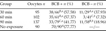
The control group (oocytes not exposed to BCB) showed higher rates of meiosis resumption when compared with BCB+ COCs exposed for 30, 60 and 90 min (p = 0.000). The BCB+ oocytes exposed for 30 and 60 min presented higher rates of meiosis resumption than those exposed for 90 min (p = 0.000). When this parameter was assessed in the oocytes classified as negative, the rates of meiosis resumption observed in the 30-min group were statistically higher when compared with the other BCB− groups, while the rates observed in the 90-min BCB− group were higher than in the group BCB− exposed for 60 min (p = 0.0062). Only the COCs exposed for 30 min did not show increased levels of meiosis resumption in the BCB+ group, when compared with their BCB− counterparts (p = 0.07). Table 1 summarizes the results of the resumption of meiosis rate among the different experimental groups.
Evaluation of meiosis resumption (presence of GVBD, MP or PB) in rat oocytes exposed to 26 μM BCB for 0, 30, 60 or 90 min before IVM

A ,B,C Different letters differ statistically in the column (p ≤ 0.05).
a ,b Different superscripts indicate statistical difference in the line (p ≤ 0.05).
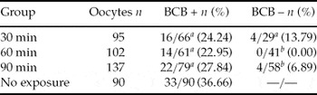
To assess the rates of nuclear maturation, considering only the presence of PB, no differences were found between the BCB+ oocytes of three experimental groups and of the control group (p = 0.2173). Similarly, no differences were observed between the oocytes classified as BCB− (p = 0.0611). Considering the treated groups, only the oocytes exposed for 30 min to BCB did not show higher results of nuclear maturation in the BCB+ group, when compared with their BCB− counterparts (p = 0.2500). Table 2 shows the results concerning the nuclear maturation rates found for these groups.
Evaluation of the rate of nuclear maturation (PB presence only) in rat oocytes exposed to 26 μM BCB for 0, 30, 60 or 90 min before IVM

a ,b Different superscripts indicate statistical difference in the line (p ≤ 0.05).
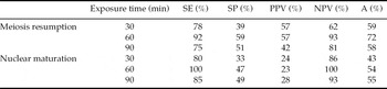
Taking into account the meiosis resumption, the most satisfactory results of diagnostic tests were found in the group exposed to BCB for 60 min, when compared with the other experimental groups (Table 3).
Sensitivity (SE), specificity (SP), positive predictive value (PPV), negative predictive value (NPV) and accuracy (A), according to meiosis resumption and nuclear maturation in rat oocytes exposed to 26 μM BCB for 30, 60 or 90 min before IVM

Similar to what was observed in the diagnostic tests to assess the rate of meiosis resumption, the results concerning the rates of nuclear maturation were more suitable in the group exposed to BCB for 60 min, as shown in Table 3.
Influence of the BCB concentration in the selection of COCs
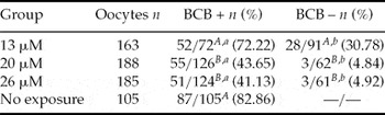
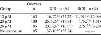
The COCs exposed to 20 μM (n = 126/188; 67.02%) and 26 μM (n = 124/185; 67.03%) BCB presented higher rates of oocytes classified as positive (p = 0.000) when compared with the COCs exposed to 13 μM (n = 72/163; 44.17%). The BCB+ oocytes exposed to 13 μM, similarly to the oocytes not exposed to BCB (control group), showed higher rates of meiosis resumption when compared with other groups classified as positive (p = 0.000). In assessing this parameter between the COCs classified as BCB−, higher results were observed in the COCs exposed to 13 μM BCB as compared with the other BCB concentration groups (p = 0.000). All BCB+ COCs showed rates of meiosis resumption higher than their BCB− counterparts (p = 0.000). Table 4 summarizes the results concerning the meiosis resumption rate in these groups.
Evaluation of meiosis resumption (presence of GVBD, MP or PB) in rat oocytes exposed to 13, 20 or 26 μM BCB for 60 min before IVM

A ,B,C Different letters differ statistically in the column (p ≤ 0.05).
a ,b Different superscripts indicate statistical difference in the line (p ≤ 0.05).
To assess the rates of nuclear maturation, considering only the presence of PB, the control group showed better results than all other experimental groups stained with BCB (p = 0.014), which did not differ from one another. The rates of nuclear maturation among the oocytes classified as BCB− were higher in the COCs exposed to 13 μM BCB and lower in those exposed to 20 μM BCB (p = 0.017). The oocytes classified as BCB+ had better outcomes of nuclear maturation rate when compared with their BCB− counterparts in the 20 μM BCB (p = 0.001) and 26 μM BCB (p = 0.004) groups. On the other hand, the COCs exposed to 13 μM BCB did not show difference between the oocytes classified as BCB+ or BCB− (p = 0.084). Table 5 summarizes the results of the rates of nuclear maturation.
Evaluation of the rate of nuclear maturation (PB presence only) in rat oocytes exposed to 13, 20 or 26 μM BCB for 60 min before IVM

A ,B,C Different letters differ statistically in the column (p ≤ 0.05).
a ,b Different superscripts indicate statistical difference in the line (p ≤ 0.05).
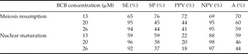
The values for diagnostic tests, independently from the parameter considered (rate of meiosis resumption or of nuclear maturation) were similar among the COCs exposed to 20 μM and 26 μM BCB (high sensitivity and NPV) and divergent from those found in the COCs exposed to 13 μM BCB (high specificity and PPV). The best accuracy results were observed in oocytes exposed to 13 μM BCB, as shown in Table 6.
Sensitivity (SE), specificity (SP), positive predictive value (PPV), negative predictive value (NPV) and accuracy (A), according to meiosis resumption and nuclear maturation in rat oocytes exposed to 13, 20 or 26 μM BCB for 60 min before IVM

No statistically significant differences were observed in the rates of meiosis resumption (p = 0.3720) and nuclear maturation (p = 0.8358), when compared with the control groups of both experiments.
Discussion
Even if there are major differences in the literature regarding the use and the applicability of BCB, although its use in several species has already been described, there is no reference about a gold standard protocol for its implementation. Likewise, to the best of our knowledge, there were no records, before our experiment, about the use of BCB staining in order to select oocytes of Rattus norvegicus, which is widely used in laboratory experiments. Our study was the first to demonstrate the applicability of BCB in the selection of this species’ oocytes intended for IVM. The results show that the staining protocol for rat cannot be the same as that is applied for most species [BCB staining for 90 min with 26 μM concentration, used in buffalo oocytes (Manjunatha et al., Reference Manjunatha, Gupta, Devaraj, Ravindra and Nandi2007), bovine (Alm et al., Reference Alm, Torner, Lohrke, Viergutz, Ghoneim and Kanitz2005; Bhojwani et al., Reference Bhojwani, Alm, Torner, Kanitz and Poehland2007), mice (Wu et al., Reference Wu, Liu, Zhou, Lan, Han, Miao and Tan2007), pigs (Wongsrikeao et al., Reference Wongsrikeao, Otoi, Yamasaki, Agung, Taniguchi, Naoi, Shimu and Nagai2006) and goats (Rodriguez-González et al., Reference Rodriguez-González, Lóopez-Béjar, Velilla and Paramio2002)], as in the present study the best results were obtained after COCs exposure to BCB for 60 min. In accordance with our findings, recent experiments using oocytes staining with BCB studied a 60-min exposure protocol for dog (Rodrigues et al., Reference Rodrigues, Rodriguez, Silva, Cavalcante, Feltrin and Rodrigues2009) and bovine (Mota et al., Reference Mota, Batista, Serapião, Boité, Viana, Torres and de Almeida Camargo2010) oocytes.
The exposure time to the dye has been the object of previous investigation, since this factor may affect the development of the oocytes selected using this technique (Goovaerts et al., Reference Goovaerts, Leroy, Jorssen and Bols2010). Our experiment showed that exposure of COCs to BCB for 60 min (concentration of 26 μM) is sufficient to identify viable oocytes (sensitivity of 100%). The present results also indicate that the oocyte that remains colourless after exposure to BCB is not suitable for IVM (NPV of 100%), according to data presented in Table 3. The exposure of oocytes for 60 min produced the best rates of diagnostic tests, in contrast with the other exposure times (Table 3). Furthermore, exposure of oocytes for that period of time proved to be the most appropriate, as the rate of meiosis resumption in oocytes BCB+ in this group was the closest to that found in the control group. Simultaneously, the rate of meiosis resumption between the oocytes classified as BCB− was lower in this group. The exposure of the oocytes for 30 min is inappropriate, since there was no difference in both rates (meiosis resumption and nuclear maturation) when BCB+ and BCB− oocytes are compared. This result was confirmed by the observation of low specificity (33%) found in this group. Similarly, exposure of oocytes for 90 min is not appropriate, because BCB+ COCs in this group had the lowest meiosis resumption rates, when compared with other oocytes stained.
Our study suggests that the oocytes exposed to 13 and 20 μM BCB produced more satisfactory results than the values observed with 26 μM BCB used for a period of 60 min (standard time defined from Experiment 1). This is due to the fact that the best accuracy was observed in the 13 μM BCB group, besides the fact that meiosis resumption rates in BCB+ oocytes in this group were similar to the control group and higher than those observed in other experimental groups. Nevertheless, we observed lower rates of oocytes classified as BCB+ in this group (when compared with those exposed to 20 or 26 μM BCB) and meiosis resumption rates and nuclear maturation rates in 13 μM BCB oocytes classified as BCB− higher among the other BCB− oocytes. However, the main factor that makes the use of BCB more difficult at this concentration (13 μM) is the observation that the rates of nuclear maturation are similar between these oocytes classified as BCB+ or BCB−.
Oocytes exposed to 20 or 26 μM BCB showed similar meiosis resumption rates, nuclear maturation rates, as well as similar results in diagnosis tests. However, the use of 20 μM BCB afforded advantages: the lowest rate of nuclear maturation among the oocytes classified as negative, and diagnostic tests results slightly higher when compared with the group exposed to 26 μM BCB. Taking into consideration the advantages of BCB used in low concentrations, we suggest that an intermediate concentration (between 13 and 20 μM) could be tested, as the use of 13 μM BCB was not sufficient to afford deselection of oocytes (confirmed by the low sensitivity). Likewise, higher concentrations of the dye presented many false positives (confirmed by the low PPV), perhaps due to the fact that G6PDH is unable to metabolize BCB when used in high concentrations.
The percentage of oocytes classified as positive was similar for different exposure times to BCB, although differences among the groups using different concentrations of the dye were observed (higher rates of positive oocytes in COCs exposed to 20 and 26 μM BCB). The observation of higher percentage of oocytes classified as BCB+, when compared with the percentage of oocytes found BCB−, was also observed in other experiments (Wongsrikeao et al., Reference Wongsrikeao, Otoi, Yamasaki, Agung, Taniguchi, Naoi, Shimu and Nagai2006; Bhojwani et al., Reference Bhojwani, Alm, Torner, Kanitz and Poehland2007; Wu et al., Reference Wu, Liu, Zhou, Lan, Han, Miao and Tan2007; Rodrigues et al., Reference Rodrigues, Rodriguez, Silva, Cavalcante, Feltrin and Rodrigues2009).
In our experiment, oocyte degeneration rates observed in the control group (22.22% in the first experiment and 14.30% in the second) are consistent with the findings published in the literature, which range from approximately 10% (Wongsrikeao et al., Reference Wongsrikeao, Otoi, Yamasaki, Agung, Taniguchi, Naoi, Shimu and Nagai2006), to approximately 20% (Alm et al., Reference Alm, Torner, Lohrke, Viergutz, Ghoneim and Kanitz2005; Rodrigues et al., Reference Rodrigues, Rodriguez, Silva, Cavalcante, Feltrin and Rodrigues2009). Likewise, the number of oocytes retrieved from the gonads of animals (approximately 20 COCs per rat) was similar to results previously reported in the literature (Dekel & Beers, Reference Dekel and Beers1978).
Our experiment did not show higher meiosis resumption rates and nuclear maturation rates of oocytes classified as BCB+, when compared with the control group. Similarly, even other authors (Alm et al., Reference Alm, Torner, Lohrke, Viergutz, Ghoneim and Kanitz2005; Bhojwani et al., Reference Bhojwani, Alm, Torner, Kanitz and Poehland2007; Manjunatha et al., Reference Manjunatha, Gupta, Devaraj, Ravindra and Nandi2007) have observed a significant difference in the rate of formation and hatching of blastocyst between the BCB+ group and the control group (the group stained had better results). The majority of papers shows no significant difference with regard to nuclear maturation rates, when these populations are compared (Alm et al., Reference Alm, Torner, Lohrke, Viergutz, Ghoneim and Kanitz2005; Manjunatha et al., Reference Manjunatha, Gupta, Devaraj, Ravindra and Nandi2007; Wu et al., Reference Wu, Liu, Zhou, Lan, Han, Miao and Tan2007), although these values are higher than those found in the BCB− group—a result confirmed in our experiment. This observation leads us to the fact that oocyte quality may be linked mainly to the oocytes’ ability to give rise to an embryo able to develop at blastocyst stage, i.e. the quality of the oocyte will present long-term repercussions (Goovaerts et al., Reference Goovaerts, Leroy, Jorssen and Bols2010). In agreement with this hypothesis, Bhojwani et al. (Reference Bhojwani, Alm, Torner, Kanitz and Poehland2007) found no statistically significant difference in other parameters measured early in the maturation process (the cleavage rate on day 2), when comparing the BCB group with the control group. Likewise, Alm et al. (Reference Alm, Torner, Lohrke, Viergutz, Ghoneim and Kanitz2005) & Manjunatha et al. (Reference Manjunatha, Gupta, Devaraj, Ravindra and Nandi2007) found no difference between such groups when they compared rates of pronucleus formation and/or embryo cleavage on day 2, despite the significantly difference observed when the rates of blastocyst formation are compared, supporting the hypothesis that oocyte quality will produce important effects especially in the later measurements related to embryonic development. Thus, our results are consistent with those found in the literature.
On the other hand, Wongsrikeao et al. (Reference Wongsrikeao, Otoi, Yamasaki, Agung, Taniguchi, Naoi, Shimu and Nagai2006) observed differences in the meiosis resumption rates and in the number of cleaved embryos between the control group and the BCB+ group, although the blastocyst formation rates were similar between these groups. The difference found not only among the BCB+ and BCB− oocytes, but mainly between the BCB+ group and the control group have been emphasized in recent studies. Although some studies suggest the hypothesis that the selection of oocytes with BCB will bring about benefits in latter assessments, such as the formation and hatching of blastocysts, it is noted that some experiments have found no difference with respect to these parameters between the group of bovine oocytes BCB+ and the control group (Opiela et al., Reference Opiela, Katska-Ksiazkiewicz, Lipiński, Słomski, Bzowska and Ryńska2008; Mota et al., Reference Mota, Batista, Serapião, Boité, Viana, Torres and de Almeida Camargo2010), or between the rates of cleavage (Mota et al., Reference Mota, Batista, Serapião, Boité, Viana, Torres and de Almeida Camargo2010; Opiela et al., Reference Opiela, Lipiński, Słomski and Katska-Ksiazkiewicz2010). Additionally, Opiela et al. (Reference Opiela, Lipiński, Słomski and Katska-Ksiazkiewicz2010) found no statistically significant difference among the three groups, when the authors evaluated the rate of cleavage or blastocyst formation.
The reason why some experiments find the difference between blastocyst formation between the BCB+ group and the control group (and even in other comparisons) may be due to the criteria used in selecting the oocytes, as the morphological evaluation is very subjective. Another possible cause for the difference in these findings is the fact that factors other than G6PDH activity may be involved in the selection of the competence of oocytes (Mota et al., Reference Mota, Batista, Serapião, Boité, Viana, Torres and de Almeida Camargo2010), as demonstrated in mice, an animal system for which oocyte diameter is a very important variable (Wu et al., Reference Wu, Liu, Zhou, Lan, Han, Miao and Tan2007). Moreover, we cannot rule out the idea that the lack of standardization of the staining protocol with BCB—side by side with the subjectivity of the evaluation—may be factors that contribute to the divergence in the results. For this reason, it is necessary to standardize the use of BCB, as our study endeavored to do.
It should be pointed out that, in most cases, the fact that the performance of BCB+ oocytes is not better than performance figures observed in the control group not exposed to the dye (evaluating early parameters such as nuclear maturation, even tough both groups have such a measurement above the BCB− group), has advocated the need not only to develop new techniques for oocyte selection, but also, and more importantly, to improve the techniques that have already been described (Opiela et al., Reference Opiela, Lipiński, Słomski and Katska-Ksiazkiewicz2010).
Our experiment showed results in agreement with those observed in the literature, with respect to higher rates of nuclear maturation and meiosis resumption in the BCB+ and control groups, when compared with the BCB− group. Additionally, we demonstrated that the staining protocols using BCB can drastically alter the results, confirming the idea that the standardization of the use of BCB is essentially important not only for the improvement of the technique, but mainly to obtain better results.