Implications
Because adipose tissue studies have grown over the years due to its huge importance in the context of fat deposition, improving the knowledge of genetic differences between breeds can have a significant impact of beef production. This review summarizes several issues regarding to Wagyu beef and other breeds related to intramuscular fat, since animals of this breed stand out for having higher marbling. The benefits of the use of Wagyu breed for adipogenic studies are also discussed.
Introduction
Fat found in skeletal muscle includes intramyocellular fat droplets (occurring in lower abundance) and adipose tissue between muscle fibers, which is usually referred to as intramuscular fat (IMF). Adipose tissue is derived from the mesenchyme (Fehrer and Lepperdinger, Reference Fehrer and Lepperdinger2005) with a supportive stroma (Romao et al., Reference Romao, Jin, Dodson, Hausman, Moore and Guan2011) and is easily isolated (Hausman and Dodson, Reference Hausman and Dodson2012) for in vitro studies. Adipocytes appear to be dynamic; a renewed search for the origin of adipocyte progenitors has demonstrated high incidences of cellular plasticity, even in adult adipose tissue (Fernyhough et al., Reference Fernyhough, Hausman, Guan, Okine, Moore and Dodson2005 and Reference Fernyhough, Helterline, Vierck, Hausman, Hill and Dodson2008; Hausman et al., Reference Hausman, Dodson, Ajuwon, Azain, Barnes, Guan, Jiang, Poulos, Sainz, Smith, Spurlock, Novakofski, Fernyhough and Bergen2009). As such, adipose tissue may represent a source of stem cells that can have far-reaching effects on several fields (Zuk et al., Reference Zuk, Zhu, Ashjian, De Ugarte, Huang, Mizuno, Alfonso, Fraser, Benhaim and Hedrick2002). Furthermore, adipose tissue has the potential to be a source of cells for tissue engineer purposes, as it appears to contain cells able to act as functional and vascular building blocks for several tissues (Fraser et al., Reference Fraser, Wulur, Alfonso and Hedrick2006). The potential for cellular development of adipocytes is believed to be fixed relatively early in life, with changes thereafter in either the size or number of cells that occur in proportion to the initial cell number and lipogenic proteins (Caserta et al., Reference Caserta, Tchkonia, Civelek, Prentki, Brown, McGarry, Forse, Corkey, Hamilton and Kirkland2001; Pethick et al., Reference Pethick, Barendse, Hocquette, Thompson and Wang2004; Wang et al., Reference Wang, Byrne, Reverter, Harper, Taniguchi, McWilliam, Mannen, Oyama and Lehnert2009). Moreover, dysfunction of the adipose compartment (cells and metabolism) is central to the pathology associated with metabolic diseases such as obesity, type II diabetes (Edelman, 1998), cancer cachexia and lipodystrophies (Cristancho and Lazar, Reference Cristancho and Lazar2011).
Regarding to IMF in production animals, we can highlight the Wagyu beef cattle, which can be considered a model for the studies of adipose tissue deposition. This is a composite breed comprised of four Bos taurus breeds: Japanese Black, Japanese Brown, Japanese Shorthorn and Japanese Polled. Originally bred for strength and endurance, by the 20th century, Wagyu cattle were mostly selected for their desirable marbling characteristics, with strict geographic restraints resulting in a sharp decline in genetic diversity (further amplified by artificial insemination), such that the offspring of five sires accounted for 42% registered Wagyu (Scraggs et al., Reference Scraggs, Zanella, Wojtowicz, Taylor, Gaskins, Reeves, de Avila and Neibergs2014). The importation of Wagyu cattle to the United States in 1973 has allowed for the expansion of the population from the initial individuals in 1994 to an estimated 700 purebred animals in 2009 (Scraggs et al., Reference Scraggs, Zanella, Wojtowicz, Taylor, Gaskins, Reeves, de Avila and Neibergs2014). Wagyu cattle are now highly valued for their tender meat (Yang et al., Reference Yang, Larsen, Powell and Tume1999) and their ability to deposit extremely high amounts of IMF (Fernyhough et al., Reference Fernyhough, Helterline, Vierck, Hausman, Hill and Dodson2008; Shirouchi et al., Reference Shirouchi, Albrecht, Nuernberg, Maak, Olavanh, Nakamura, Sato, Gotoh and Nuernberg2014).
Comparisons between Wagyu cattle and other breeds regarding cell biology and general adipogenesis have been reported in several studies, such as those by Oikawa et al. (Reference Oikawa, Sanehira, Sato, Mizoguchi, Yamamoto and Baba2000), Hausman et al. (Reference Hausman, Dodson, Ajuwon, Azain, Barnes, Guan, Jiang, Poulos, Sainz, Smith, Spurlock, Novakofski, Fernyhough and Bergen2009), Dodson et al. (Reference Dodson, Hausman, Guan, Du, Rasmussen, Poulos, Mir, Bergen, Fernyhough, McFarland, Rhoads, Soret, Reecy, Velleman and Jiang2010 Reference Dodson, Hausman, Guan, Du, Rasmussen, Poulos, Mir, Bergen, Fernyhough, McFarland, Rhoads, Soret, Reecy, Velleman and Jianga) and Duarte et al. (Reference Duarte, Paulino, Das, Wei, Serao, Fu, Harris, Dodson and Du2013). Some studies comparing Wagyu cattle with other breeds (Hausman et al., Reference Hausman, Dodson, Ajuwon, Azain, Barnes, Guan, Jiang, Poulos, Sainz, Smith, Spurlock, Novakofski, Fernyhough and Bergen2009; Dodson et al., Reference Dodson, Hausman, Guan, Du, Rasmussen, Poulos, Mir, Bergen, Fernyhough, McFarland, Rhoads, Soret, Reecy, Velleman and Jiang2010 Reference Dodson, Hausman, Guan, Du, Rasmussen, Poulos, Mir, Bergen, Fernyhough, McFarland, Rhoads, Soret, Reecy, Velleman and Jianga; Duarte et al., Reference Duarte, Paulino, Das, Wei, Serao, Fu, Harris, Dodson and Du2013) have emphasized the uniqueness of this breed and the importance of better understanding adipogenesis, mainly because its increased accumulation of fat within the skeletal muscle, which seems to not follow the same pattern of other adipose tissue depots. Consequently, Wagyu cattle may present a highly marbled beef without an increase of overall fatness, making this breed an unique animal model for the understanding of this phenomena.
Adipogenesis and IMF
In humans, the subcutaneous adipose tissue (SAT) and visceral adipose tissue (VAT) have been identified as the main fat depots. These depots differ in terms of their turnover. Adipose cells derived from VAT, in particular, have higher levels of metabolic activity and have been shown to hypertrophy in obese individuals. In addition to VAT and SAT, intermuscular adipose tissue and IMF are also measured in farm animal species such as swine and cattle (Komolka et al., Reference Komolka, Albrecht, Wimmers, Michal and Maak2014).
IMF deposition is achieved at the cellular level through adipogenesis, the process of proliferation and differentiation of preadipocytes, and lipogenesis, the subsequent assimilation of lipid (Harper and Pethick, Reference Harper and Pethick2004; Hausman et al., Reference Hausman, Dodson, Ajuwon, Azain, Barnes, Guan, Jiang, Poulos, Sainz, Smith, Spurlock, Novakofski, Fernyhough and Bergen2009; Dodson et al., Reference Dodson, Vierck, Hausman, Guan, Fernyhough, Poulos, Mir and Jiang2010b). Previous studies have suggested that the high deposition of IMF in Wagyu is related to the differential expression of specific genes (Hudson et al., Reference Hudson, Reverter, Greenwood, Guo, Cafe and Dalrymple2014). In most cattle breeds, IMF is first visible at about 11 months, with the greatest increases between 15 and 24 months. However, when they are finished on concentrated grain, Wagyu deposit more IMF at an earlier age when compared with other cattle breeds (Wertz et al., Reference Wertz, Berger, Walker, Faulkner, McKeith and Rodriguez-Zas2002; Shirouchi et al., Reference Shirouchi, Albrecht, Nuernberg, Maak, Olavanh, Nakamura, Sato, Gotoh and Nuernberg2014). Dietary conditions, particularly energy density, affect carcass adipose tissue deposition in beef cattle (Yamada et al., Reference Yamada, Kawakami and Nakanishi2009). Thus, cattle that are fed low energy through the finishing phase with forage and pasture type diets seldom show the desired marbling. Moreover, the level of dietary starch that is fed to young cattle may alter gene networks associated with adipocyte differentiation and energy metabolism (Graugnard et al., Reference Graugnard, Berger, Faulkner and Loor2010). Thus, high energy diets (finishing diets usually only during the last 4 to 6 months before harvest) have been used as a production strategy for enhancing intramuscular adipose tissue in beef cattle. Exposure to high-starch diets during the early growth phase of cattle might induce precocious preadipocyte differentiation and lipid filling (Graugnard et al., Reference Graugnard, Berger, Faulkner and Loor2010).
Marbling is an important component of livestock production, as it is a major factor in the overall meat quality (flavor and tenderness), nutrition (protein and fat levels and fatty acid composition) and economic value (Pena et al., Reference Pena, Molina, Aviles, Juarez and Horcada2013; Hudson et al., Reference Hudson, Reverter, Greenwood, Guo, Cafe and Dalrymple2014; Sadkowski et al., Reference Sadkowski, Ciecierska, Majewska, Oprzadek, Dasiewicz, Ollik, Wicik and Motyl2014). An increase in IMF has been proposed to occur either through the myogenic transdifferentiation of myogenic stem cells (MSC) in skeletal muscle through a complex regulatory pathway, which remains unclear, or through the multi-step process of adipogenic determination and differentiation of fibroblast-like preadipocytes into mature adipocytes assimilating lipid (Du and Zhu, Reference Du, Huang, Das, Yang, Duarte, Dodson and Zhu2010; Du et al., Reference Du and Zhu2013).
Myoblasts and adipocytes have been shown to utilize intercellular communication, which directly affects the growth and development of these cells (Kokta et al., Reference Kokta, Dodson, Gertler and Hill2004; Muthuraman, Reference Muthuraman2014). Furthermore, when in close proximity, these cells perform a paracrine function by altering the growth, development or energy storage of each other based on the chemical factors that are released (Kokta et al., Reference Kokta, Dodson, Gertler and Hill2004; Muthuraman, Reference Muthuraman2014). Myoblasts have been shown to regulate the growth, development, differentiation and lipid assimilation of adipocytes through intercellular communication (Muthuraman, Reference Muthuraman2014).
Myostatin, a growth differentiation factor secreted by myoblasts, suppresses proliferation of preadipocytes in muscle tissue through direct down-regulation of adipogenic transcription factors, thus decreasing the overall IMF (Komolka et al., Reference Komolka, Albrecht, Wimmers, Michal and Maak2014). Furthermore, co-culture of myoblasts and adipocytes results in an upregulation of peroxisome proliferator activating receptor γ (PPARγ), CCAAT enhancer binding protein α (C/EBPα) and fatty acid binding protein 4 (FABP4), possibly indicating myoblasts’ promotion of adipogenic specific transcription factors leading to increased IMF or allowing for MSC transdifferentiation as a mechanism for increasing IMF (Muthuraman, Reference Muthuraman2014). In both cases, myogenic and adipogenic contributions to marbling appear to be mediated by the transcription factors, PPARγ, C/EBPα and FABP4. Fibroblasts like preadipocytes, in response to the aforementioned transcription factors, differentiate from progenitor cells and undergo adipogenesis and assimilation of lipid (Du et al., Reference Du, Yin and Zhu2010). Similarly, MSCs respond to high concentrations of the same transcription factors. However, these factors appear to inhibit myogenic differentiation stimulating adipogenesis and also appears to inhibit myogenic differentiation, while ensuring adipogenic differentiation (Teboul et al., Reference Teboul, Gaillard, Staccini, Inadera, Amri and Grimaldi1995; Taylor-Jones et al., Reference Taylor-Jones, McGehee, Rando, Lecka-Czernik, Lipschitz and Peterson2002; Singh et al., Reference Singh, Chae, Hwang, Yoo, Ahn, Lee, Lee, Park and Chung2007; Du et al., Reference Du, Yin and Zhu2010). Considering this, the study of adipogenesis and its effects on the accumulation of IMF can be used not only to understand fat deposition in livestock, but also for a better comprehension of the differentiation process in mesenchymal stem cells.
Stem cells
Skeletal muscle is derived from the mesoderm and is postnatally surrounded by small multipotent myogenic satellite cells (SC) that play an important role in muscle hypertrophy and regeneration (Kook et al., Reference Kook, Choi, Son, Lee, Hwang, Lee, Chang, Choi and Lee2006; Du et al., Reference Du, Yin and Zhu2010; Lee et al., Reference Lee, Lee, Kamli, Pokharel, Bhat, Lee, Choi, Chun, Kang, Lee, Kim, Schnabel, Taylor and Choi2012; Duarte et al., Reference Duarte, Francischetti, Genelhu, Cabello and Pimentel2014). SC are multipotent cells capable of transdifferentiating into intramuscular adipocytes when exposed to local cellular signaling (Taylor-Jones et al., Reference Taylor-Jones, McGehee, Rando, Lecka-Czernik, Lipschitz and Peterson2002; Singh et al., Reference Singh, Chae, Hwang, Yoo, Ahn, Lee, Lee, Park and Chung2007; Du et al., Reference Du, Yin and Zhu2010; Lee et al., Reference Lee, Lee, Kamli, Pokharel, Bhat, Lee, Choi, Chun, Kang, Lee, Kim, Schnabel, Taylor and Choi2012; Ryan et al., Reference Ryan, Daniel, Craggs, Parr and Brameld2013). Moreover, muscle side population (SP) cells are multipotent stem cells that can participate in myogenesis and muscle regeneration upon transplantation (Penton et al., Reference Penton, Thomas-Ahner, Johnson, McAllister and Montanaro2013). Interestingly, SP cells in skeletal muscle tissue have not only the ability to transdifferentiate into cells from myogenic but also in hematopoietic lineage (Reecy et al., Reference Reecy, Miller and Webster2003). In vivo studies using mice as a molecular model, have confirmed the hematopoietic stem cell potential of SP cells, as these present a regenerative capacity for blood, bone and lymph cells (Reecy et al., Reference Reecy, Miller and Webster2003). These studies have implied that SP cells may be an important resident source of transdifferentiation within skeletal muscle, and may be less differentiated than other previously discovered SC populations (Reecy et al., Reference Reecy, Miller and Webster2003).
The regenerative capacity of skeletal muscle decreases with age, thereby resulting in an overall loss of muscle mass over time and an increase in lipid content (Teboul et al., Reference Teboul, Gaillard, Staccini, Inadera, Amri and Grimaldi1995; Kook et al., Reference Kook, Choi, Son, Lee, Hwang, Lee, Chang, Choi and Lee2006; Aguiari et al., Reference Aguiari, Leo, Zavan, Vindigni, Rimessi, Bianchi, Franzin, Cortivo, Rossato, Vettor, Abatangelo, Pozzan, Pinton and Rizzuto2008; Ryan et al., Reference Ryan, Daniel, Craggs, Parr and Brameld2013). This might not be totally accountable by the decrease in stem cell activity; myogenic transdifferentiation into adipocytes also play a role in this phenomena. Since MSCs are multipotent, their transdifferentiation into intramuscular adipocytes is feasible, depending on different cellular signaling exposure, thereby leading to greater amounts of IMF (if the adipocytes invade the perimysium) (Taylor-Jones et al., Reference Taylor-Jones, McGehee, Rando, Lecka-Czernik, Lipschitz and Peterson2002; Singh et al., Reference Singh, Chae, Hwang, Yoo, Ahn, Lee, Lee, Park and Chung2007; Aguiari et al., Reference Aguiari, Leo, Zavan, Vindigni, Rimessi, Bianchi, Franzin, Cortivo, Rossato, Vettor, Abatangelo, Pozzan, Pinton and Rizzuto2008; Du et al., Reference Du, Yin and Zhu2010; Lee et al., Reference Lee, Lee, Kamli, Pokharel, Bhat, Lee, Choi, Chun, Kang, Lee, Kim, Schnabel, Taylor and Choi2012).
Adipogenic markers
A major marker of adipogenesis (or lack thereof) is preadipocyte factor-1 (pref-1), as this membrane protein is expressed on preadipocytes and acts to prevent adipogenesis. It has been reported that pref-1 inhibits adipocyte differentiation via sex determining region Y-box 9, which binds to its binding sites at the C/EBP β and δ promoter regions to suppress their transcription (Sul, Reference Sul2009). In addition, it appears to be depot specific, as high levels of pref-1 expression were observed in smaller adipocytes (Yamada et al., Reference Yamada, Kawakami and Nakanishi2014). Early and late differentiation are marked by the expression of the C/EBP family, which in turn is greater in fat depots of Wagyu when compared with Holstein cattle, as they are responsible for the increased proliferation potential of Wagyu preadipocytes in vitro (Yamada et al., Reference Yamada, Kawakami and Nakanishi2009). Meanwhile, PPARs act as transcription factors to regulate gene expression by acting on lipid metabolism and adipocyte filling. Indeed, both C/EBPα and PPARγ from the IMF of Wagyu are increased when compared with the IMF of Angus cattle (May et al., Reference May, Savell, Lunt, Wilson, Laurenz and Smith1994; Yamada et al., Reference Yamada, Higuchi and Nakanishi2007; Duarte et al., Reference Duarte, Paulino, Das, Wei, Serao, Fu, Harris, Dodson and Du2013).
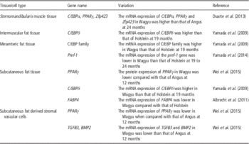
Previous studies have suggested that the high deposition of IMF in Wagyu cattle is related to the differential expression of specific genes such as C/EBPα and PPARγ (late adipogenic markers) as well as an early adipogenic marker zinc finger protein 423 (Duarte et al., Reference Duarte, Paulino, Das, Wei, Serao, Fu, Harris, Dodson and Du2013). Table 1 shows the differences in adipogenic gene expression between Wagyu and other cattle breeds. Moreover, the expression of adipogenesis and the lipid droplet associated genes, perilipin 1 (PLIN1) and adipose differentiation-related protein (ADFP), are upregulated as markers of the overall amount of IMF (Shirouchi et al., Reference Shirouchi, Albrecht, Nuernberg, Maak, Olavanh, Nakamura, Sato, Gotoh and Nuernberg2014). PLIN1 is a major protein that resides on the surface of mature adipocyte lipid droplets and plays an integral role in triacylglycerol storage and breakdown (Shirouchi et al., Reference Shirouchi, Albrecht, Nuernberg, Maak, Olavanh, Nakamura, Sato, Gotoh and Nuernberg2014). The largest lipid droplets in mature adipocytes are exclusively coated with PLIN1. Moreover, PLIN1 promotes skeletal muscle lipid deposition by partitioning excess fatty acids towards triacylglycerol storage (Shirouchi et al., Reference Shirouchi, Albrecht, Nuernberg, Maak, Olavanh, Nakamura, Sato, Gotoh and Nuernberg2014). ADFP functions similarly to PLIN1 but it is ubiquitous, whereas PLIN1 appears to be found only on adipocytes (Shirouchi et al., Reference Shirouchi, Albrecht, Nuernberg, Maak, Olavanh, Nakamura, Sato, Gotoh and Nuernberg2014).
Differences of adipogenic gene expression between Wagyu and other cattle breeds

C/EBPα=CCAAT enhancer binding protein α; PPARγ=peroxisome proliferator activating receptor γ; Zfp423=zinc finger protein 423; Pref-1=preadipocyte factor-1; FABP4=fatty acid binding protein 4; TGFB3 = transforming growth factor 3; BMP2 = bone morphogenic protein 2.
Several genes are well known for their correlation with obesity and other metabolic disorders; these genes include PPARγ (Dodson et al., Reference Dodson, Vierck, Hausman, Guan, Fernyhough, Poulos, Mir and Jiang2010b), leptin (LEP) (Duarte et al., 2007), adiponectin (ADIPOQ), FABP4 (Wang et al., Reference Wang, Bower, Reverter, Tan, De Jager, Wang, McWilliam, Cafe, Greenwood and Lehnert2005), bone morphogenetic protein 4 (BMP4) (Majka et al., Reference Majka, Barak and Klemm2011), fat mass and obesity associated (FTO) (Fischer et al., Reference Fischer, Koch, Emmerling, Vierkotten, Peters, Bruning and Ruther2009), C/EBPβ (Cristancho and Lazar, Reference Cristancho and Lazar2011) and v-akt murine thymoma viral oncogene homolog 2 (AKT2). Three of the genes that are reported above (BMP4, C/EBPβ and AKT2) are found in many reports related to fat storage in humans. Five of them (PPARγ, FABP4, ADIPOQ, FTO and LEP) are mentioned both in connection with human disorders and adipogenesis in B. taurus. In particular, the role of the ADIPOQ gene is well described in Wagyu cattle (Jordan et al., Reference Jordan, Kruger, Willmes, Redemann, Wunderlich, Bronneke, Merkwirth, Kashkar, Olkkonen, Bottger, Braun, Seibler and Bruning2011).
Adipogenesis and lipid metabolism: Wagyu as a model
Although research in humans and farm animals ultimately have different goals – identification of potential drug targets for metabolic diseases v. optimization of meat quality – the same tissue can be the focus of both research efforts (Komolka et al., Reference Komolka, Albrecht, Wimmers, Michal and Maak2014). Bovine adipocytes are a cell model for studying adipogenesis and lipid metabolism for improving animal production and also serving human health (Duarte et al., Reference Duarte, Paulino, Das, Wei, Serao, Fu, Harris, Dodson and Du2013).
Fat deposition in cattle typically follows the order in which perirenal fat is deposited first, followed by intermuscular, subcutaneous and finally by the IMF (Sainz and Hasting, Reference Sainz and Hasting2000; Pethick et al., Reference Pethick, D’Souza, Dunshea and Harper2005 and Reference Pethick, Harper and Oddy2007; Hocquette et al., Reference Hocquette, Gondret, Baéza, Médale, Jurie and Pethick2010). Thus, as IMF deposition is time dependent in most cattle breeds and is dependent on a high energy intake, the use of cattle breeds, instead of Wagyu as models for human dysfunctions is compromised, since it would not be possible to dissociate the effect of energy intake or age from the genetic predisposition for IMF deposition abnormalities. Therefore, the use of animals that have a unique ability to deposit IMF regardless of energy intake or age, such as Wagyu cattle, may be useful in studies of adipogenesis (Lehnert et al., Reference Lehnert, Wang, Tan and Reverter2006).
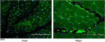
In summary, Wagyu cattle have a great potential to be used as animal models for adipogenesis studies due to their exceptional IMF deposition (Shirouchi et al., Reference Shirouchi, Albrecht, Nuernberg, Maak, Olavanh, Nakamura, Sato, Gotoh and Nuernberg2014), which in turn affects carcass composition. Comparison of Wagyu animals with other popular beef breeds, such as Angus, provides a distinct basis for observation, as Wagyu presented higher feed efficiency, feed-to-gain ratio, and overall greater IMF deposits (Shirouchi et al., Reference Shirouchi, Albrecht, Nuernberg, Maak, Olavanh, Nakamura, Sato, Gotoh and Nuernberg2014). This can be seen in Figure 1, which shows an immunofluorescent staining comparison of the adipogenic marker FABP4 in Angus and Wagyu in the Sternomandibularis muscle, where the number of preadipocytes and adipocytes in Wagyu was greater than that of Angus cattle. Laboratory identification of distinct differences between Wagyu cattle and any comparable beef breed produces valuable data, which can further identify factors that either increase or inhibit adipogenesis and IMF deposition. Research with tissue and cells from Wagyu animals, can be a model to deep understanding of adipogenesis and lipid metabolism, which might be paralleled by similar studies in tissues and cells from other animal types.
Immunofluorescence staining showed a greater number of fatty acid binding protein (FABP4) positive cells between muscle fibers and muscle bundles in Wagyu when compared with Angus skeletal muscle (Sternomandibularis muscle): FABP4 stained green and nuclei counterstained with 4',6-diamidino-2-phenylindole (DAPI). (Adapted from figure 5 in reference Duarte et al., Reference Duarte, Paulino, Das, Wei, Serao, Fu, Harris, Dodson and Du2013, with permission. Figure provided by M. S. Duarte.)

Acknowledgments
The authors acknowledge the financial support of CNPq, CAPES and FAPEMIG.