I. INTRODUCTION
Contamination of water by organic dyes has become an increasing concern to the environment. Reference Liu and Zhang1 Many methods have been used to solve this problem, including physical absorption, oxidation–reduction reaction in chemistry, biological process, and so on. In recent times, the application of heterogeneous solid acid catalysts have attracted extensive interest owing to their easy separation, excellent reusability, environmental benignity, and operational simplicity. Reference Kozhevnikov2 Compared with transition metal building blocks, polyoxometalates (POMs) took on much superiority for their characteristics of oxygen rich surface, high charge density, and controllable size. Reference Zhang, Wu, Wang, Liu and Qian3 POMs can function as photocatalysts through the photoexcitation of the oxygen-to-metal charge transfer bands to separate the electron–hole pair used for reductive and oxidative reactions with surrounding molecules. Reference Yamase4 Due to a number of featured advantages, POMs attracted considerable attentions as photocatalysts for degrading organic dyes. Reference Ismail, Ng, Ahmadi, Razak and Lockman5
The photooxidative behavior of POMs has been studied extensively. However there was a great disadvantage in these systems. High solubility of POMs made the reuse of this kind of photocatalysts difficult. Reference Feng, Zhang, Liu, Lu, Zhao and Yao6–Reference Nikbakht, Yadollahi and Farsani10 To overcome the shortcoming, many exploring experiments were carried out. POMs with many supports such as silica, activated carbons, TiO2, mesoporous molecular sieve, and polymeric membranes Reference Yue, Zhou, Xu, Wu, Zhang, Zou and Jin11–Reference Bonchio, Carraro, Scorrano, Fontananova and Drioli15 have been investigated. Some supports could provide large specific surface area and thus sufficient contact between POMs and pollutants, increasing the catalytic activity of POMs. However, from the standpoint of the reuse of catalysts in practical wastewater treatment, preparation of POM-containing films is the most effective method because no separation process is needed for the films. It was found that the properties as well as the applicability of POM clusters could be fine-tuned by combining them with organic moieties. Reference Reddy, Satyanarayana, Dubey, Ghosh and Pradeep16 There have been reports on POM-containing composite films prepared by self-assembly method and sol–gel technique, Reference Gao, Pan and Cao17 but few reports were made on photocatalytic application of POM-containing films prepared by electrospinning.
As we all know, electrospinning is an effective method to prepare functional materials with tunable architectures such as core–shell nanofibers, molecular sieve fibers, and polymer supported nanocomposite, Reference Zhao, Lv, Pan, Zhang, Zhang and Zhang18 which exhibit excellent performances and promising application prospects. Reference Jing, Mi, Salick, Cordie, McNulty, Peng and Turng19 Zhou Reference Sui, Li, Guo, Cheng, Gao, Zhou, Gong and Du20 prepared Ag/PVA/SiW12 tri-component nanohybrid through electrospinning and photoreduction, and the photocatalytic activity of the electrospun membrane was enhanced significantly. However, PVA nanofibers should be treated before use in water, or it would dissolve in a short time. Electrospun nanofibrous membrane has many remarkable characteristics, such as large specific surface area, high porosity, high permeability, Reference Saeed, Haider, Oh and Park21,Reference Abdal-hay, Mousa, Khan, Vanegas and Lim22 and nonwoven mesh forms, contributing to the high level exposure of the photocatalysts and easy separation. Reference Guo, Shao, Mu, Zhang, Zhang, Zhang, Chen and Liu23 In view of this, electrospun nanofibrous membranes would be promising supports for the immobilization of soluble POMs.
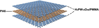
In this work, a sandwich nanofibrous membrane was prepared in which the upper and bottom layers behaved as the supporter of POM. As shown in Fig. 1, both the upper layer and bottom layer are H3PW12O40/polymethylmethacrylate (PMMA) composite nanofibrous membranes with the same thickness, and the intermediate layer is polyamide 6 (PA6) nanofibrous membrane. PMMA is a commonly used polymer with high mechanical strength and good spinnability. As a water-tolerant polymer, PMMA could make the composite membrane stable in water and thus reusable. However, due to the hydrophobicity of PMMA, H3PW12O40/PMMA composite membrane is nonwettable, which hinders the contact between H3PW12O40 and pollutants. Therefore, PA6 was selected as the intermediate layer for increasing the hydrophilicity of the whole membrane. More importantly, the synergistic effect of PA6 and H3PW12O40 would enhance the photocatalytic degradation rate significantly. To the best of our knowledge, this is the first report on the preparation of H3PW12O40/PMMA/PA6 sandwich nanofibrous membrane and its photocatalytic properties and mechanism.
Scheme of H3PW12O40/PMMA/PA6 sandwich nanofibrous membrane.

II. EXPERIMENTAL
A. Materials and instruments
PMMA (M w = 60,000) was obtained from Sumitomo Chemical (Ehime, Japan), and polycaprolactam (PA6) (M w = 30,000) was obtained from Hunan Yueyang Baling Petrochemical Co., Ltd. (Hunan, China). Analytical reagents H3PW12O40, ethanol (C2H5OH), formic and acid (HCOOH) were supplied by Sinopharm Chemical Reagent Co. Ltd. (Shanghai, China). Dimethyl formamide (DMF) and methyl orange (MO) were purchased from Aladdin Industrial Corporation (Shanghai, China). All the solvents were used without further purification, and all the aqueous solutions were prepared with deionized water.
Microstructure of the nanofibrous membranes was analyzed using a Hitachi S-4800 field emission-scanning electron microscope (FE-SEM; Hitachi Corporation, Tokyo, Japan) and with an energy dispersive X-ray spectrometry (EDX; Noran7 EDX spectrometer, Thermo Fisher Scientific Inc., Madison, Wisconsin). FT-IR spectra was recorded using a VERTEX 70 spectrometer (Bruker Corporation, Ettlingen, Germany), which was obtained at a resolution of 4 cm−1. XPS measurement was carried out with an ESCALAB 250Xi spectrometer (Thermo Fisher Scientific Inc., East Grinstead, United Kingdom), with the nonmonochromatised Al Kα X-radiation (hν = 1486.6 eV) and a power of 150 W (10 mA × 15 kV). Concentrations of the MO solutions were measured by a UV-8000S UV–vis spectrophotometer (Shanghai Metash Instruments Co., Ltd., Shanghai, China) over the wave length range of 200–800 nm.
B. Preparation of the sandwich nanofibrous membrane
The sandwich nanofibrous membrane was fabricated by electrospinning from two solutions. One was prepared by adding 1.0 g PMMA in 6 mL DMF and dissolving H3PW12O40 (0.2, 0.6, and 1.0 g, respectively) in 4 mL C2H5OH, respectively. Stirring was continued for additional 40 min after mixing above homogeneous solutions. It was used for fabricating the upper and bottom layers. The other was prepared by dissolving 2.0 g PA6 in 10 mL HCOOH and stirring until PA6 completely dissolved at ambient temperature. It was used for fabricating the intermediate layer.
The as-prepared electrospinning solutions were added to a 5 mL glass syringe with a needle tip (0.5 mm diameter), respectively. For the upper and bottom layers, the flow rate of the solution was 0.6 mL/h, which was controlled by a micro syringe pump. A high voltage power supply was used to apply an electrical potential of 15 kV. The collection distance between the needle tip and aluminum foil was 17 cm. With regard to the intermediate layer, the flow rate was 0.4 mL/h. The used voltage was 20 kV and the collection distance was also 17 cm. The three-layer nanofibrous membranes were compressed for two min at room temperature before being peeled off from the aluminum foil. According to the volume (mL) of electrospinning solutions that the upper, intermediate, and bottom layers consumed, the three-layer nanofibrous membranes are referred to as 0.4/2.0/0.4, 0.6/2.0/0.6, and 0.8/2.0/0.8, respectively.
C. Photocatalytic activity test
The photocatalytic activity of the sandwich nanofibrous membranes was evaluated for decomposition of harmful MO aqueous. A 300 W high pressure mercury lamp with a double walled quartz glass tube (for water cooling) was suspended vertically (the distance between quartz glass reactor and lamp was 10 cm). Photodegradation of MO was carried out at atmospheric pressure and room temperature. In a typical experiment, 50 mL MO aqueous solution (10 mg/L, pH was adjusted to 1.0 using HClO4) was placed in the reactor, and the sandwich nanofibrous membrane was immersed in the solution. Prior to the irradiation, the reactor was shaken (120 min−1) in dark for 30 min to establish adsorption–desorption equilibrium between the organic molecules and the catalyst surface. Decreases in the concentrations of MO were analyzed by UV–vis spectrophotometer at λ = 510 nm. At given intervals of illumination, the samples (3.0 mL) of the reaction solution were taken out and analyzed.
III. RESULTS AND DISCUSSION
A. Characterization of the sandwich nanofibrous membrane
1. Morphology analysis
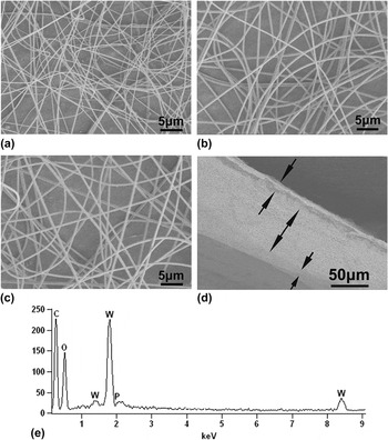
Figures 2(a)–2(c) show the typical SEM images of H3PW12O40/PMMA composite nanofibers (upper layer) with different mass ratio of H3PW12O40 to PMMA. The average diameters of these randomly oriented nanofibers were 310 ± 112 nm, 550 ± 88 nm, and 560 ± 43 nm for the composite fibers in which H3PW12O40:PMMA were 0.2:1.0, 0.6:1.0, and 1.0:1.0, respectively. It clearly showed that the fiber diameters increased with the increase of H3PW12O40 content, yet the diameter distribution became narrow gradually. Given the outstanding morphology and the highest proportion of H3PW12O40, the composite membrane in which H3PW12O40:PMMA was 1.0:1.0 was used in the following study. Figure 2(d) shows the cross section of the sandwich nanofibrous membrane. Three layers are easily recognizable and the intermediate layer is much thicker than the upper and the bottom layers. As shown in EDS spectrum [Figs. 2(e)], C, O, P, and W elements were detected in the upper and bottom layers of the sandwich membrane, supporting that H3PW12O40 has been successfully loaded into the membrane.
SEM images of H3PW12O40/PMMA composite nanofibers with different mass ratio of H3PW12O40 to PMMA (a) 0.2:1.0, (b) 0.6:1.0, (c) 1.0:1.0. Morphology (d) and EDS spectrum (e) of the sandwich nanofibrous membrane.

2. FT-IR spectra analysis
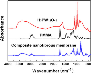
Figure 3 shows the FT-IR spectra of H3PW12O40, PMMA, and H3PW12O40/PMMA composite nanofibrous membrane. Curve (a) was the typical FT-IR spectra of H3PW12O40. Four characteristic absorption peaks at 1083, 984, 902, and 823 cm−1 are attributed to the νas(P–Oa), νas(W=Od), νas(W–Ob–W), and νas(W–Oc–W), respectively, Reference Sui, Li, Guo, Cheng, Gao, Zhou, Gong and Du20,Reference Zhang, Zhao, Gao, Xi and Xu24 attesting to the Keggin structure of H3PW12O40. The PMMA spectra, as shown in curve (b), exhibits the characteristic peaks at 1727 cm−1 (C=O stretching vibration of ester group) and 2946 cm−1 (C–H stretching vibration of alkyl group). Compared with curve (a) and (b), the FT-IR spectrum of H3PW12O40/PMMA composite membrane [curve (c)] has nearly all the key features of H3PW12O40 and PMMA though there are minor shifts of some peaks. Most notably, four discernible peaks between 800 cm−1 and 1100 cm−1 which agree with Keggin unit well are observed in the curve (c), supporting that the primary Keggin structure is not destroyed in the H3PW12O40/PMMA composite membrane. Reference Feng and Shang25
FT-IR spectra of H3PW12O40 (a) PMMA (b) and H3PW12O40/PMMA composite nanofibrous membrane (upper layer) (c).

3. XPS analysis
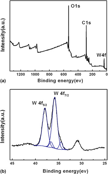
XPS measurement was carried out to identify the surface elements and the valence state of W of the sandwich membrane. The survey spectrum of the upper layer [Fig. 4(a)] shows the presence of O, C, and W as expected. From Fig. 4(b), the shape of the W 4f spectrum suggested two different chemical states of W. The two fitted peaks with binding energies for the W 4f 7/2 and W 4f 5/2 core levels of 35.8 and 37.9 eV were assigned to W (VI), which was ascribed to H3PW12O40 in the membrane. The two peaks account for approximately 91.3% of the total spectral area. The remaining area corresponded to the doublet at 34.5 and 36.6 eV, which may represent the formation of an oxide of the type WO x in which W has an oxidation state lower than VI. According to the previous literature, these values more likely represented the existence of perturbed tungstate environments corresponding to tungsten atoms in terminal W=O bonds that directly coordinate to the support. Reference Berry, Derrick, Marco and Mortimer26
XPS survey spectra (a) and narrow scan W 4f spectra (b) of the H3PW12O40/PMMA composite nanofibrous membrane (upper layer). The binding energy of C–C is 284.8 eV.

B. Photocatalytic property
1. Photocatalytic activity
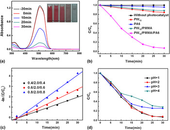
The photocatalytic activities of H3PW12O40/PMMA/PA6 sandwich nanofibrous membranes were tested by degrading aqueous MO under UV irradiation and the results are shown in Fig. 5. From Fig. 5(a), the sandwich membrane made the maximum absorption peaks (around 510 nm) of MO decline rapidly with the increase of the irradiation time. Meanwhile, the color of the aqueous MO solution faded drastically, supporting that MO was nearly degraded completely with the sandwich nanofibrous membrane after 30 min irradiation.
(a) UV–vis spectra of MO versus photoreaction time catalyzed by the sandwich membrane (0.6/2.0/0.6); (b) photodegradation of MO over different catalysts against irradiation time (sandwich membrane, 0.6/2.0/0.6); (c) kinetic linear fitting curves of the sandwich membranes with different H3PW12O40/PMMA layer thickness; (d) effect of pH on photocatalytic activity of the sandwich membrane (0.6/2.0/0.6).

As a comparison, the degradation efficiency of MO with H3PW12O40, PA6 nanofibrous membrane, H3PW12O40/PMMA composite nanofibrous membrane and H3PW12O40/PMMA/PA6 sandwich nanofibrous membranes were evaluated [Fig. 5(b)]. The amount of PW12 was equal in all the samples. No obvious degradation of MO was observed in the absence of photocatalyst under UV irradiation. Only 4.1% of MO was degraded with PA6 nanofibrous membrane, indicating that the removal process was absolute physical adsorption. With equal amounts of H3PW12O40, the degradation efficiency of MO after 30 min UV light irradiation were calculated as about 15.6, 11.6, and 92.7% for H3PW12O40, H3PW12O40/PMMA composite membrane and H3PW12O40/PMMA/PA6 sandwich membrane, respectively. It was clear that the sandwich membrane exhibited superior photocatalytic activities. The possible main reason was that a completely different photocatalytic mechanism took place owing to the synergistic effect of PA6 and H3PW12O40, resulting in the enhancement of the degradation rate (see below). Moreover, the sandwich structure of the laminated membrane doubled the contact area between H3PW12O40 and MO.
Figure 5(c) shows the kinetics linear simulation curves of MO photocatalytic degradation over sandwich membranes with different H3PW12O40/PMMA layer thickness. The results demonstrated that these photocatalytic reactions followed a Langmuir–Hinshelwood apparent first-order kinetics model due to the low initial concentration (10 mg/L) of the MO. Reference Turchi and Ollis27 The model can be expressed by the following equations:
 $$- \ln \left( {{C \over {{C_0}}}} \right) = {k_{{\rm{app}}}}t\quad ,$$
where C 0 is the initial concentration of the reactant (mg/L), C is the concentration of the reactant at time t (mg/L), t is the UV-light illumination time, and k app is the apparent first-order rate constant (min−1). Reference Lee, Park, Lee, Ju and Hong28 The determined k app are 0.068, 0.095, and 0.140 for the 0.4/2.0/0.4, 0.6/2.0/0.6, and 0.8/2.0/0.8 sandwich nanofibrous membranes, respectively. The degradation rate of MO increased with the increase of H3PW12O40/PMMA layer thickness examined, indicating that the photocatalytic activity of the sandwich membranes show catalytic layer thickness dependent behavior and is not yet saturated under the investigated H3PW12O40/PMMA layer thickness range.
Figure 5(d) shows the effect of pH value on the degradation efficiency of MO. It can be observed that increase in pH value led a decrease in the degradation efficiency. 92.7, 91.6, 74.2, and 71.6% degradation of MO were obtained for the solutions whose pH were 1.0, 2.0, 3.0, and 4.0, respectively. [PW12O40]3− is stable when the pH of solution is lower than 2.0. However, at higher pH value, it is partly converted to [PW11O39], Reference Gkika, Kormali, Antonaraki, Dimoticali, Papaconstantinou and Hiskia7−Reference Hori, Takano, Koike, Takeuchi and Einaga8 whose photocatalytic activity is remarkable lower than that of the saturated one. In addition, the photocatalytic degradation of MO by the sandwich membrane was driven by the reductive pathway owing to the presence of PA6. The reductive cleavage of the –N=N– bond in the case of MO dye, requires four electrons and four H+ to be completely reduced (Scheme 1), suggesting that the degradation of MO is highly favored in acidic solutions. Reference Troupis, Triantis, Gkika, Hiskia and Papaconstantinou29
Mechanism of photocatalytic reductive degradation of MO.

2. Stability and reusability
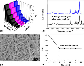
Reusable capacity is one of the key performances of catalysts for long-use in practical application. In view of this, two factors were needed to be considered: one was how easily the catalyst could be separated from the reaction system; the other was the stability of the catalyst to maintain its high activity over time. Reference Zhang, Shao, Li, Zhang, Zhang, Sun and Liu30 For laminated nanofibrous membrane, after photodegradation was complete, it could be directly removed from the aqueous MO solution without any additional separation operation. So the sandwich nanofibrous membrane could be separated from the reaction system easily. As shown in Fig. 6(a), no appreciable decrease in catalytic activity was observed after the sandwich membrane was repeatedly used for three times. It may be attributed to the stability of H3PW12O40 in the membrane. Figure 6(b) shows the FT-IR spectra of the H3PW12O40/PMMA layer after a three-cycle photocatalytic experiment. It is obvious that the four discernible peaks between 800 cm−1 and 1100 cm−1 still exist, supporting that the Keggin structure of H3PW12O40 remains intact in the membrane after catalysis. The morphology of H3PW12O40/PMMA layer after three cycles of reactions was shown in Fig. 6(c). It is clear that the structure of the composite nanofibers was almost unchanged. To further confirm the stability of the catalyst, the sandwich membrane was removed from the reaction system after 10 min irradiation, and the MO solution continued to be exposed to UV light. From Fig. 6(d), in the absence of the sandwich membrane, no obvious degradation of MO was observed, suggesting that there was no catalytically active species leaching into the system.
(a) Photocatalytic activity of the sandwich nanofibrous membrane (0.6/2.0/0.6) for MO degradation with three times of cycling uses. FT-IR spectra (b) and SEM image (c) of the H3PW12O40/PMMA layer after a three-cycle experiment. (d) Photodegradation of MO with and without removing the membrane catalyst.

3. Possible photocatalytic mechanism
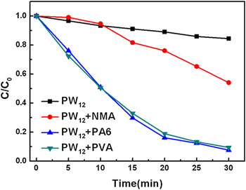
Photocatalytic degradation of azo dyes with POM can be driven by either oxidative or reductive pathway. The photooxidative process involved excitation of POM by UV–vis light, which led to the charge transfer from an O2− ion to W6+ ion at W–O–W bonds and the formation of a strongly oxidizing excited state of POM (POM*) or POM (h+ + e−). This process is in analogy to TiO2, Reference Velegraki, Poulios, Charalabaki, Kalogerakis, Samaras and Mantzavinos31 which took place via the reaction through OH radicals or direct reaction of the POM* with the substrate. Reference Guo and Hu32,Reference Hiskia, Mylonas and Papaconstantinou33 However, some literature Reference Wang, Lu and Feng34–Reference Arslan-Alaton and Ferry36 have reported that the photocatalytic degradation of azo dyes was driven by the reductive pathway due to the presence of alcohol (methanol, ethanol, and isopropanol) as sacrificial electron donor. The degradation rate was much faster in the photoreductive process. Our previous work has found that polyvinyl alcohol (PVA), like other alcohols, could also enhance the photocatalytic activity of H4SiW12O40 and its performance was more outstanding than isopropanol. Reference Li, Zhang, Li, Liu, Wang and An37 Along this direction, the effect of PA6 on the photocatalytic activity of H3PW12O40 was carried out. As seen in Fig. 7, 15.6, 45.9, 92.5, and 90.7% degradation of MO was observed after 30 min irradiation for H3PW12O40 (PW12), PW12 + N-methylacetamide (NMA), PW12 + PA6, and PW12 + PVA redox systems, respectively. The results showed that PA6 could also enhance the degradation rate of MO and performed better than NMA. It was reported that the α-C atoms of PA6 can be oxidized, Reference Liu and Wang38,Reference Hill and Bouchard39 suggesting that PA6, like alcohols, could donate electron to POM and make MO undergo photoreductive decomposition.
Photodegradation of MO by different H3PW12O40 redox systems (H3PW12O40, 50 mg).

According to the literature Reference Troupis, Triantis, Gkika, Hiskia and Papaconstantinou29 and the experimental results, the possible mechanistic scheme for the degradation of MO on the as-prepared sandwich membrane under UV irradiation is proposed:
 $${{\rm{H}}_3}{\rm{P}}{{\rm{W}}_{12}}{{\rm{O}}_{40}} + h\nu \leftrightarrow {{\rm{H}}_3}{\rm{P}}{{\rm{W}}_{12}}{{\rm{O}}_{40}}*\quad ,$$
 $${{\rm{H}}_3}{\rm{P}}{{\rm{W}}_{12}}{{\rm{O}}_{40}}* + \,{\rm{PA6}} \to {{\rm{H}}_4}{\rm{P}}{{\rm{W}}_{12}}{{\rm{O}}_{40}} + {\rm{oxidation\ products}}\quad {\rm{,}}$$
 $${{\rm{H}}_4}{\rm{P}}{{\rm{W}}_{12}}{{\rm{O}}_{40}} + {\rm{MO}} \to {{\rm{H}}_3}{\rm{P}}{{\rm{W}}_{12}}{{\rm{O}}_{40}} + {\rm{reduction\ products}}\quad {\rm{,}}$$
 $${{\rm{H}}_3}{\rm{P}}{{\rm{W}}_{12}}{{\rm{O}}_{40}}\!* + \,{{\rm{H}}_{\rm{2}}}{\rm{O}} \to {{\rm{H}}_4}{\rm{P}}{{\rm{W}}_{12}}{{\rm{O}}_{40}} + {}^ \cdot {\rm{OH}} + {{\rm{H}}^{\rm{ + }}}\quad ,$$
 $${}^ \cdot {\rm{OH}} + {\rm{MO}} \to {\rm{O}}{{\rm{H}}^ - } + {\rm{oxidation\ products}}\quad {\rm{,}}$$
 $${{\rm{H}}_3}{\rm{P}}{{\rm{W}}_{12}}{{\rm{O}}_{40}}* + \,{\rm{MO}} \to {{\rm{H}}_4}{\rm{P}}{{\rm{W}}_{12}}{{\rm{O}}_{40}} + {\rm{oxidation\ products}}\quad {\rm{,}}$$
 $${{\rm{H}}_4}{\rm{P}}{{\rm{W}}_{12}}{{\rm{O}}_{40}} + {{\rm{O}}_{\rm{2}}} \to {{\rm{H}}_3}{\rm{P}}{{\rm{W}}_{12}}{{\rm{O}}_{40}} + {\rm{O}}_2^ - \quad .$$
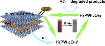
PW12 at the layer boundary of PA6 and PMMA makes MO undergo photoreductive decomposition [(1)–(3)], while PW12 not at the layer boundary photodegrades MO through an oxidative pathway [(1), (4)–(7)]. The photooxidative process is in analogy to TiO2. The photoreductive pathway plays a major role in the degradation rate of MO. Figure 8 shows the photoreductive process of MO. Reactions (1) and (2) describe the photocatalytic oxidation of PA6 and the formation of H4PW12O40 which has strong reducibility, while reaction (3) describes a fast reoxidation of H4PW12O40 in the presence of MO. In a word, the whole process is that H3PW12O40 absorbs light and mediates the electron transfer from PA6, the sacrificial donor, to MO. The electron donating from PA6 to H3PW12O40 may be the rate-limiting step of the photocatalytic reaction, so H4PW12O40 production may play an important role in the degradation rate of MO.
The photoreductive mechanism on H3PW12O40/PMMA/PA6 sandwich nanofibrous membrane.

IV. CONCLUSIONS
In this paper, H3PW12O40/PMMA/PA6 sandwich nanofibrous membranes were prepared through electrospinning. IR, EDX, and XPS characterization confirmed that H3PW12O40 with intact Keggin structure existed in the upper and bottom layers of the sandwich membrane. The sandwich membrane exhibited greatly enhanced photocatalytic activity in the decomposition of MO compared with H3PW12O40 only and H3PW12O40/PMMA film. It may be attributed to the photoreduction mechanism owing to the synergistic effect of PA6 and H3PW12O40, and the double contact area between H3PW12O40 and MO due to the sandwich structure of the laminated membrane. More importantly, the sandwich membranes were stable in water, so that they could be easily separated and reused without appreciable losses in photocatalytic activity. In view of this, H3PW12O40/PMMA/PA6 sandwich nanofibrous membranes exhibit the potential for practical applications to eliminate organic pollutants from wastewater.
Supplementary Material
To view supplementary material for this article, please visit http://dx.doi.org/10.1557/jmr.2016.313.
ACKNOWLEDGMENTS
This work was supported by the National Natural Science Foundation of China (Grant No. 51472074), Natural Science Foundation of Hebei Province (E2016209202), the Scientific Research Foundation for Colleges and Universities of Hebei Province under grant for Project (QN2014051), the Hundred Talents Program of Hebei Province of China (E2012100005), the Graduate Student Innovation Fund of North China University of Science and Technology (2016S08) and the Engagement Fund of North China University of Science and Technology (GP201517), to which the authors would like to express grateful appreciation. We thank Jiayang Li and Zhong Li, Fushun Mining Group of China, for designing the scheme of membranes.