Introduction
Primary distal renal tubular acidosis is a renal tubular disorder of either autosomal dominant or autosomal recessive inheritance.Reference Alper1 The disease is characterised by hyperchloraemic metabolic acidosis and inappropriately alkaline urine. Other findings may include hypokalaemia and mild hypercalcaemia. An inability to acidify the urine is accompanied by low urinary citrate, hypercalciuria and nephrocalcinosis (which may lead to an inability to concentrate urine), nephrolithiasis, and, infrequently, renal failure. An important consequence of distal renal tubular acidosis in children is failure to thrive and delayed growth.Reference Alper1
Two types of autosomal recessive distal renal tubular acidosis are recognised, and a genome-wide linkage screen has recently identified the involvement of a different gene in each. In the first type, distal renal tubular acidosis is accompanied by bilateral, often progressive, sensorineural hearing loss (SNHL) and is caused by mutations in the ATP6V1B1 gene.Reference Karet, Finberg, Nelson, Nayir, Mocan and Sanjad2–Reference Ruf, Rensing, Topaloglu, Guay-Woodford, Klein and Vollmer4 In the second type, patients usually have normal hearing in childhood, although SNHL occasionally develops in early adulthood. It is caused by mutations in the ATP6V0A4 gene.Reference Stover, Borthwick, Bavalia, Eady, Fritz and Rungroj3–Reference Smith, Skaug, Choate, Nayir, Bakkaloglu and Ozen5 The two genes encode two different subunits of the H+ adenosine triphosphatase (ATPase) proton pump, which is found in the alpha-intercalated cells of the kidney,Reference Karet, Finberg, Nelson, Nayir, Mocan and Sanjad2 inner earReference Karet, Finberg, Nelson, Nayir, Mocan and Sanjad2–Reference Dou, Finberg, Cardell, Lifton and Choo6 and male reproductive tract.Reference Herak-Kramberger, Breton, Brown, Kraus and Sabolic7 However, studies have excluded linkage to ATP6V1B1 mutations in some families with distal renal tubular acidosis, and shown linkage to ATP6V0A4 mutations in other families with normal hearing, suggesting that additional genes may be involved in distal renal tubular acidosis.Reference Stover, Borthwick, Bavalia, Eady, Fritz and Rungroj3
The degree of hearing loss in distal renal tubular acidosis varies from severe to profound. Some patients demonstrate a progression of hearing loss despite systemic alkali treatment.Reference Karet, Finberg, Nelson, Nayir, Mocan and Sanjad2 Two recent studies described an enlarged vestibular aqueduct in four patients with distal renal tubular acidosis and progressive, nonsymmetrical hearing loss.Reference Berrettini, Forli, Franceschini, Ravecca, Massimetti and Neri8, Reference Shinjo, Kaga and Igarashi9 Two patients had bilateral enlarged vestibular aqueducts and two had unilateral involvement in the ear with worse hearing.Reference Shinjo, Kaga and Igarashi9 Neither article reported data on the type of mutation.
Our group recently discovered five Israeli children, from three unrelated families, with distal renal tubular acidosis and mutations in the ATP6V1B1 gene. The aim of the present study was to characterise these children's phenotypes, with an emphasis on the type of hearing loss and the findings on temporal bone imaging.
Materials and methods
Patients
Five children, from three unrelated families, diagnosed with distal renal tubular acidosis and SNHL were included in this study. In each family, a different type of ATP6V1B1 gene mutation was identified.
Serum biochemical and renal data were collected from the patients' medical records, including: blood pH; bicarbonate (HCO3−), potassium and calcium levels; and urinary pH. Age at diagnosis of distal renal tubular acidosis and of nephrocalcinosis was documented. Audiological data were also collected for all patients, including age at diagnosis of hearing loss, progression of hearing loss and current hearing status. High resolution computed tomography (CT) and magnetic resonance imaging (MRI) of the temporal bone, when possible, were performed to enable morphological study of the inner ear.
Results
The main clinical data for the five patients from the three families are presented in Tables I and II. All children were receiving medical treatment, and their blood pH and electrolyte levels were within the normal range.
Clinical profile and laboratory abnormalities at diagnosis in patients with drta and deafness

* Label characters represent respectively family (A, B or C), generation (1 = parents, 2 = children) and birth order (1 = oldest). dRTA = distal renal tubular acidosis; mths = months; F = female; M = male;
Patients' clinical data

* Case labels as per Table I. †Patients in family A were too young to assess progression. SNHL = sensorineural hearing loss; Dx = diagnosis; yrs = years; HRCT = high resolution computed tomography; EVA = enlarged vestibular aqueduct; mths = months; ABR = auditory brainstem response
Family A
The parents were related Jews (first cousins) originally from Halleb, Syria. They had two children (A2.1 and A2.2), both affected by the disease.
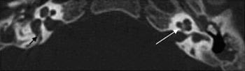
Patient A2.1 was 2.5 years old. She had been diagnosed with moderate hearing loss at the age of 12 months; auditory brainstem response (ABR) had revealed well formed waves bilaterally at 85 dB clicks. She also had otitis media with effusion. The insertion of bilateral ventilation tubes had not significantly improved the patient's hearing, and she was currently using hearing aids successfully. Her most recent audiogram revealed a 60 dB loss in the right ear and a 55 dB loss in the left. High resolution temporal bone CT demonstrated bilateral enlarged vestibular aqueducts (Figure 1), with no other inner-ear pathology.
High resolution axial computed tomography scan of patient A2.1, at the level of the vestibular aqueducts, showing enlarged vestibular aqueduct on the right side (black arrow) and normal modiolus on left side (white arrow).

Patient A2.2 was nine months old. He had been diagnosed with hearing loss at the age of three months. His ABR was similar to his older sister's, with well formed waves appearing at 85 dB. A high resolution temporal bone CT was not performed in this patient.
Family B
The parents were unrelated Jews of Moroccan descent. The family had four children, with the two eldest, both boys (B2.1 and B2.2), aged 15 and 17 years, being affected. Both had been diagnosed with distal renal tubular acidosis and treated several months after birth (Table I) and they were now well developed, with normal stature. Both had been diagnosed with bilateral moderate SNHL at the age of 24 months and had been fitted with hearing aids. The older brother (B2.1) used hearing aids until the age of 15 years, when he became completely deaf. The progression of his hearing loss was asymmetrical; he first lost serviceable hearing in his left ear and later in his right. At the age of 15 years, he underwent cochlear implantation, with excellent results.
The younger brother (B2.2) had bilateral, severe SNHL (speech reception threshold and peak twitch amplitude of 80 dB), but was able to communicate well using hearing aids (speech reception threshold of 35 dB).
Both brothers had a conductive component in the left ear of 10–30 dB, more pronounced in the low frequencies.
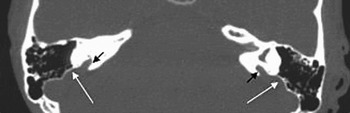
Bilateral enlarged vestibular aqueducts were observed on high resolution CT of the temporal bone in both patients at the age of 15 years, without any other inner-ear abnormalities (Figure 2).
High resolution axial computed tomography scan of patient B2.1, showing bilaterally enlarged vestibular aqueducts (black arrows) and sacs (white arrows). The enlarged endolymphatic sacs cause indentations in the mastoid.

Family C
The parents were unrelated Arab Bedouins from the south of Israel. They had three children, the youngest of whom (C2.3), aged 12 years, was affected. She had been diagnosed with distal renal tubular acidosis at three months of age following investigation for failure to thrive and vomiting. Her current height was 145 cm, similar to her unaffected mother and two sisters.
The patient had been diagnosed with hearing loss at the age of 12 months following screening, with subsequent confirmation by ABR (thresholds of 90 dB in the right ear and 75 dB in the left ear). She had worn hearing aids until the age of 10 years, but had not developed any language skills. At the age of 10, audiometry had revealed bilateral, severe, down-sloping SNHL; the speech detection threshold with hearing aids was 65 dB bilaterally.
The patient had therefore undergone cochlear implantation. Currently, she was attentive to noise and could speak simple sentences but was unable to engage in conversation.
High resolution CT, performed at the age of 10 years, had shown bilateral enlarged vestibular aqueducts.
Discussion
Since ATP6V1B1 has been recognised as the gene involved in the majority of cases of distal renal tubular acidosis and deafness,Reference Karet, Finberg, Nelson, Nayir, Mocan and Sanjad2 a total of 42 kindreds with 20 different mutations has been identified.Reference Karet, Finberg, Nelson, Nayir, Mocan and Sanjad2–Reference Ruf, Rensing, Topaloglu, Guay-Woodford, Klein and Vollmer4, Reference Borthwick, Kandemir, Topaloglu, Kornak, Bakkaloglu and Yordam10, Reference Hahn, Kang, Ha, Cheong and Choi11
The gene product of ATP6V1B1 is a B1 subunit of the H+-ATPase-driven proton pump. This H+-ATPase proton pump (also known as vacuolar or V-ATPase) is a multi-subunit complex responsible for the acidification of intracellular organelles and of luminal or interstitial spaces. It consists of two functional domains: the membrane-spanning V0 domain and a cytoplasmic V1 domain.Reference Alper1, Reference Dou, Finberg, Cardell, Lifton and Choo6, Reference Forgac12
The V1 domain is composed of eight subunit types, labelled A to H. Subunit B has two isoforms, B1 and B2.Reference Alper1 The B2 isoform is expressed ubiquitously and is encoded by the gene ATP6V1B2. The B1 isoform is expressed by ATP6V1B1 and is the subject of our study. The B1 isoform is tissue-specific, and it is expressed specifically in intercalated cells of the distal nephron in the male reproductive tract.Reference Herak-Kramberger, Breton, Brown, Kraus and Sabolic7, Reference Finberg, Wagner, Stehberger, Geibel and Lifton13 In the inner ear, it is expressed in the endolymphatic sac and in the interdental cells of the spiral limbus.Reference Karet, Finberg, Nelson, Nayir, Mocan and Sanjad2
The endolymph pH varies in different parts of the labyrinth, measuring 7.4 in the cochlea and 7.0 in the endolymphatic sac.Reference Couloigner, Teixeira, Hulin, Sterkers, Bichara and Escoubet14 Cochlear endolymph has high concentrations of K+ and low concentrations of Na+, resulting in a high positive potential across the hair cell. This would be expected to result in a passive flow of H+ from the endolymph, causing an alkaline pH. Thus, the function of H+-ATPase is probably to maintain the endolymphatic pH at 7.4 in the cochlea and at 7.0 in the endolymphatic sac. The importance of this action was supported by a study showing that specific inhibition of the pump by bafilomycin caused a rise in endolymphatic sac pH to 7.4.Reference Couloigner, Teixeira, Hulin, Sterkers, Bichara and Escoubet14
MondiniReference Mondini15 was the first to describe a large internal and external vestibular aqueduct aperture, during a temporal bone dissection in 1791. Almost 200 years later, Valvassori and Clemis,Reference Valvassori and Clemis16 using polytomograms, reported a ‘large vestibular aqueduct syndrome’ in 50 patients out of 3700 tested. Today, enlarged vestibular aqueduct, usually found bilaterally, is recognised as a part of stable, progressive and fluctuating SNHL, which may worsen with mild head trauma.Reference Levenson, Parisier, Jacobs and Edelstein17, Reference Jackler and De La Cruz18 The exact definition of enlarged vestibular aqueduct, however, varies amongst authors. Valvassori and ClemisReference Valvassori and Clemis16 defined it as a width of more than 1.5 mm in the anteroposterior dimension, whereas Jackler and de la CruzReference Jackler and De La Cruz18 define it as a diameter of more than 2.0 mm, measured midway between the common crus and the external aperture. All of our patients met the criterion of Jackler and de la Cruz.Reference Jackler and De La Cruz18
There have been two previous reports on a total of four patients with distal renal tubular acidosis and deafness associated with enlarged vestibular aqueduct.Reference Berrettini, Forli, Franceschini, Ravecca, Massimetti and Neri8, Reference Shinjo, Kaga and Igarashi9 Unfortunately, these patients' genotypes were not mentioned. We describe an additional five patients, and associate the genotype of ATP6V1B1 mutations with the finding of enlarged vestibular aqueduct in four of them. It is possible that the patients in the previous reportsReference Berrettini, Forli, Franceschini, Ravecca, Massimetti and Neri8, Reference Shinjo, Kaga and Igarashi9 had a mutation in ATP6V0A4 rather than ATP6V1B1, since three of them suffered hearing loss found later in life, which is characteristic of that mutation.Reference Stover, Borthwick, Bavalia, Eady, Fritz and Rungroj3
The vestibular aqueduct grows in a nonlinear fashion throughout embryonic life. PyleReference Pyle19 suggested that an enlarged vestibular aqueduct is probably not a developmental disorder but rather occurs during the postnatal period or in early childhood. The youngest patient in our group with enlarged vestibular aqueduct was aged two years.
Enlarged vestibular aqueduct is associated with other cochlear and vestibular anomalies, although the frequency with which these anomalies are reported has been inconsistent among studies and has changed over time.Reference Pyle19 Early reports noted a 15–25 per cent prevalence of cochlear anomalies,Reference Valvassori and Clemis16 whereas recent studies have reported a prevalence of up to 100 per cent.Reference Davidson, Harnsberger, Lemmerling, Mancuso, White and Tong20 Associated vestibular anomalies have been reported in 5 to 60 per cent of patients with enlarged vestibular aqueduct.Reference Davidson, Harnsberger, Lemmerling, Mancuso, White and Tong20
Enlarged vestibular aqueduct has been specifically associated with Pendred syndromeReference Phelps, Coffey, Trembath, Luxon, Grossman and Britton21, Reference Goldfeld, Glaser, Nassir, Gomori, Hazani and Bishara22 and with branchio-oto-renal dysplasia.Reference Chen, Francis, Ni, Cremers, Kimberling and Sato23 Both of these diseases are manifested by other anomalies of the inner ear. Pendred syndrome is a genetic disease characterised by SNHL and goitre.Reference Everett, Glaser, Beck, Idol, Buchs and Heyman24 It is caused by a mutation in the SLC26A4 (Pds) gene, which belongs to a superfamily of anion exchangers. The gene encodes the protein pendrin, originally thought to be a sulphateReference Everett, Glaser, Beck, Idol, Buchs and Heyman24 or iodide transporter,Reference Kalatzis, Sahly, El-Amraoui and Petit25 but more recently found to be a Cl−/HCO3− exchanger.Reference Dou, Xu, Wang, Smith, Soleimani and Karet26 Branchio-oto-renal dysplasia is an autosomal dominant, early developmental defect characterised by varying combinations of branchial (i.e. fistulas, sinuses and cysts), auricular (i.e. outer, middle and inner ear) and renal anomalies. It is caused by mutations in the EYA1 gene, a transcriptional co-activator.Reference Kalatzis, Sahly, El-Amraoui and Petit25
The most common radiological findings in Pendred syndrome include modiolus deficiency and vestibular enlargement.Reference Phelps, Coffey, Trembath, Luxon, Grossman and Britton21, Reference Goldfeld, Glaser, Nassir, Gomori, Hazani and Bishara22 The most common radiological findings in branchio-oto-renal dysplasia include cochlear hypoplasia (four-fifths of normal size, with only two turns), bulbous internal auditory canals, deep posterior fossa and acutely angled promontories.Reference Royaux, Suzuki, Mori, Katoh, Everett and Kohn27 By contrast, bilateral enlarged vestibular aqueduct was the only radiological pathology observed in our four patients with distal renal tubular acidosis and deafness, and in the four patients previously reported.Reference Berrettini, Forli, Franceschini, Ravecca, Massimetti and Neri8, Reference Shinjo, Kaga and Igarashi9 This difference may be explained by the different areas of expression of the proteins. Pendrin is expressed in different areas of the labyrinth: the endolymphatic duct and sac, utricle, saccule, external sulcus spiral ligament, Claudius cells, Deiters, cells and spiral ganglion.Reference Yoshino, Sato, Nakashima, Nakashima, Teranishi and Nakayama28 The EYA1 compound, implicated in the pathogenesis of branchio-oto-renal dysplasia syndrome, is a transcription factor expressed in differentiated sensory and supporting cells of the ear during early and later development.Reference Kalatzis, Sahly, El-Amraoui and Petit25 By contrast, ATP6V1B1 is expressed mainly in epithelial cells of the endolymphatic sac and interdental cells of the spiral limbus.Reference Karet, Finberg, Nelson, Nayir, Mocan and Sanjad2, Reference Dou, Finberg, Cardell, Lifton and Choo6
Several genetic studies have been performed in patients with enlarged vestibular aqueduct.
Usami et al. Reference Usami, Abe, Weston, Shinkawa, Van Camp and Kimberling29 screened for mutations in the SLC26A4 gene in six families with congenital, nonsyndromic, high-frequency, fluctuating, sometimes progressive SNHL and with high resolution CT confirmed enlarged vestibular aqueduct. One patient had a history of vertigo; none had a Mondini malformation. Four of the six families were found to be homozygous or compound heterozygous for mutations.
Tsukamoto et al.,Reference Tsukamoto, Suzuki, Harada, Namba, Abe and Usami30 in a Japanese study, screened for SLC26A4 gene mutations in 10 families with Pendred syndrome and 32 families with bilateral SNHL associated with enlarged vestibular aqueduct. None of their patients had the Mondini malformation. Causative mutations were identified in 90 per cent of the typical Pendred syndrome families and in 78 per cent of those with SNHL and enlarged vestibular aqueduct. These authors noted that the same combination of mutations resulted in a variable phenotypic expression, suggesting that these two conditions are part of a continuous spectrum of disease.
In contrast, Pryor et al. Reference Pryor, Madeo, Reynolds, Sarlis, Arnos and Nance31 found that all 11 of their patients with Pendred syndrome had two mutant SLC26A4 alleles, whereas all 18 of their patients with nonsyndromic enlarged vestibular aqueduct had either one or no SLC26A4 mutant alleles. They concluded that the two entities are distinct clinically and genetically.
Other studies from KoreaReference Park, Lee, Jin, Lee, Go and Jang32 and TaiwanReference Wu, Yeh, Chen and Hsu33 found a high percentage of SLC26A4 mutations in patients with enlarged vestibular aqueduct (92 and 87 per cent, respectively), although there were some patients with enlarged vestibular aqueduct who did not have SLC26A4 mutations. We speculate that some of these patients may have had an ATP6V1B1 mutation.
• Primary distal renal tubular acidosis with sensorineural hearing loss (SNHL) is a rare autosomal recessive disease, usually caused by mutations in the ATP6V1B1 gene
• The aim of this study was to characterise the phenotype of this disease, with emphasis on auditory findings, in a cohort of Israeli children
• Several types of mutations in the ATP6V1B1 gene may cause distal renal tubular acidosis and SNHL
• A bilaterally enlarged vestibular aqueduct was the only radiological abnormality in the inner ear in all cases
Idiopathic enlarged vestibular aqueduct without kidney involvement, caused by an ATP6V1B1 gene mutation, can be explained in two ways. Both alleles may be mutated, but the mutation causes only a reduction (not loss) of function, so patients have hearing loss without kidney problems. Alternatively, a mutation may be present in only one allele, causing enlarged vestibular aqueduct but not distal renal tubular acidosis, due to very low penetrance (similar to the finding of Pryor et al. Reference Pryor, Madeo, Reynolds, Sarlis, Arnos and Nance31 for SLC26A4).
Several theories have been proposed to explain the progression of hearing loss observed in cases of enlarged vestibular aqueduct. The two main theories involve reflux of cerebrospinal fluid through the abnormal vestibular aqueduct,Reference Levenson, Parisier, Jacobs and Edelstein17 or intracochlear membrane rupture.Reference Jackler and De La Cruz18 Both theories are based on the finding that minor head trauma can cause sudden worsening in hearing.
We propose a different theory. Both pendrin and the B1 subunit of apical H+-ATPase are found not only in the endolymphatic sac but also in the cochlea, and both have a metabolic function. Other proteins may compensate for the absence of pendrin and B1 H+-ATPase subunit under normal conditions but not under stress, e.g. following mild trauma or viral infection. Accordingly, mice that lack ATP6V1B1 do not develop deafness,Reference Dou, Finberg, Cardell, Lifton and Choo6 apparently because the mouse has a compensatory system in the inner ear. Our theory might better account for the finding that, in patients with enlarged vestibular aqueduct, sudden deterioration in hearing is often related to a viral infection.Reference Lin, Lin, Kao and Wu34
Conclusion
A mutation in the gene encoding ATP6V1B1, a subunit of apical H+-ATPase, causes distal renal tubular acidosis and deafness. The hearing loss is asymmetrical, bilateral, moderate to severe and progressive. The radiological finding of enlarged vestibular aqueduct on high resolution CT and MRI should prompt renal function tests as well as blood pH analysis.
Patients with distal renal tubular acidosis and SNHL, like those with other progressive cochlear disorders, are expected to achieve good hearing with hearing aids and cochlear implants. They should be counselled regarding the danger of minimal head trauma.