INTRODUCTION
Visceral leishmaniasis (VL) is a vector-borne neglected disease, caused mainly by Leishmania donovani and Leishmania infantum chagasi. These parasite species infect macrophage-rich internal organs such as the spleen, liver and bone marrow causing a deadly disease that affects over three to five hundred thousand people each year (World Health Organization, 2010). Available antileishmanial drugs are costly, toxic and may be ineffective against emerging drug-resistant strains.
Thus, Leishmania infection is currently an unmet clinical need, and research for prophylactic treatments is urgent and remains critical. Considerable progress has been made in identifying a large number of Leishmania antigens, including whole parasite antigens (Mayrink et al. Reference Mayrink, Dos Santos, Peixoto De Toledo, Dabés Guimarães, Lins Machado-Coelho, Genaro and Da Costa2002). Purified proteins (Cardoso et al. Reference Cardoso, Da Silva, Da Costa, Mayrink, Melo, Marques Michalick, Willi Liu, Fujiwara and Nascimento2003) and DNA encoding immunogenic epitopes (Melby et al. Reference Melby, Yang, Zhao, Perez and Cheng2001) have been investigated as potential vaccine candidates. These antigens have been found to have a limited ability to induce protection against parasite infection even after promoting a strong Th1 immune response, which supports the hypothesis that failure of some vaccines may not only be related to the immunity magnitude induced by vaccination, but also to the ability to induce other T cells subsets and in controlling disease associated responses (Sánchez-Sampedro et al. Reference Sánchez-Sampedro, Gómez, Mejías-Pérez, Sorzano and Esteban2012; Majumder et al. Reference Majumder, Bhattacharjee, Chowdhury, Majumdar and Majumdar2014).
For many years, we have used the intranasal and oral routes to administer whole antigen from Leishmania amazonensis (LaAg) in the absence of adjuvants providing a partial protective immune response against experimental cutaneous leishmaniasis in mice (Pinto et al. Reference Pinto, De Mello Cortezia and Rossi-Bergmann2003, Reference Pinto, Pinheiro, Rayol, Larraga and Rossi-Bergmann2004; Pinheiro et al. Reference Pinheiro, Pinto, de Matos Guedes, Filho, de Mattos, Saraiva, de Mendonça and Rossi-Bergmann2007; Leonel et al. Reference Leonel, Guedes, Lilian, Chaves, Cláudio, Gomes, Nosanchuk and De Simone2014). Adjuvants have been tested in both first and second generations of experimental vaccines against leishmaniasis, increasing the specific Th1 immune response. Promising results have been acquired, especially when considering that protein antigens on their own cannot induce significant immune response, requiring the addition of an adjuvant system to ensure appropriate antigen delivery and concomitant immune activation (Srivastava et al. Reference Srivastava, Misra, Sharma, Srivastava, Naik and Dube2003; Khalil et al. Reference Khalil, Musa, Modabber and El-Hassan2006). Furthermore, the use of adjuvants may contribute to the lower costs for vaccine production, due to the lower Ag concentration/booster required.
CAF01 is a novel two-component liposomal adjuvant system composed of a cationic liposome vehicle [dimethyldioctadecylammonium (DDA)] stabilized with a glycolipid immunostimulator [trehalose 6,6-dibehenate (TDB)], which is a synthetic analogue of mycobacterial cord factor. Formulations using CAF01 adjuvant have been evaluated in several experimental vaccine candidates, as example against Tuberculosis, HIV, Chlamydia and Influenza (Olsen et al. Reference Olsen, Theisen, Christensen, Follmann and Andersen2010; Fomsgaard et al. Reference Fomsgaard, Karlsson, Gram, Schou, Tang, Bang, Kromann, Andersen and Andreasen2011; Martel et al. Reference Martel, Agger, Poulsen, Jensen, Andresen, Christensen, Nielsen, Blixenkrone-Møller, Andersen and Aasted2011; Pinto et al. Reference Pinto, Salanti, Joergensen, Dahlbäck, Resende, Ditlev, Agger, Arnot, Theander and Nielsen2012; Van Dissel et al. Reference Van Dissel, Joosten, Hoff, Soonawala, Prins, Hokey, O'Dee, Graves, Thierry-Carstensen, Andreasen, Ruhwald, de Visser, Agger, Ottenhoff, Kromann and Andersen2014), and also in Phase 1 clinical trials, combined with the tuberculosis fusion antigen Ag85B-ESAT6 (H1) (NCT ID: NCT00922363) and an HIV-1 peptide mix (NCT ID: NCT01141205; NCT01009762), respectively (Fomsgaard et al. Reference Fomsgaard, Karlsson, Gram, Schou, Tang, Bang, Kromann, Andersen and Andreasen2011; Van Dissel et al. Reference Van Dissel, Joosten, Hoff, Soonawala, Prins, Hokey, O'Dee, Graves, Thierry-Carstensen, Andreasen, Ruhwald, de Visser, Agger, Ottenhoff, Kromann and Andersen2014). In addition, CAF01 adjuvant formulations can be administrated by the parenteral (Dietrich et al. Reference Dietrich, Andreasen, Andersen and Agger2014) or mucosal route (Rosenkrands et al. Reference Rosenkrands, Vingsbo-Lundberg, Bundgaard, Lindenstrøm, Enouf, van der Werf, Andersen and Agger2011), which provides an advantage over other adjuvants, which are usually limited to a single administration route. Immunologically, CAF01 can induce a strong CD4+ T-cell response characterized by a mixed Th1/Th17 profile and pro-inflammatory cytokine production, such as IFN-γ, IL-12, IL-17 and TNF-α (Rosenkrands et al. Reference Rosenkrands, Vingsbo-Lundberg, Bundgaard, Lindenstrøm, Enouf, van der Werf, Andersen and Agger2011; Kamath et al. Reference Kamath, Mastelic, Christensen, Rochat, Agger, Pinschewer, Andersen, Lambert and Siegrist2012). Furthermore, CAF01 has been shown to increase humoral and cell-mediated memory immune responses (Agger et al. Reference Agger, Rosenkrands, Hansen, Brahimi, Vandahl, Aagaard, Werninghaus, Kirschning, Lang, Christensen, Theisen, Follmann and Andersen2008; Lindenstrøm et al. Reference Lindenstrøm, Agger, Korsholm, Darrah, Aagaard, Seder, Rosenkrands and Andersen2009; Rosenkrands et al. Reference Rosenkrands, Vingsbo-Lundberg, Bundgaard, Lindenstrøm, Enouf, van der Werf, Andersen and Agger2011).
The development of effective needle-free vaccine delivery strategies is recognized as being important for increasing vaccine coverage in developing countries and thus increasing global health. In this work, we attempted to optimize the airway vaccination strategy against VL by the intranasal delivery of the LaAg vaccine associated with the CAF01 adjuvant, which was used in this context for the first time.
MATERIALS AND METHODS
Animals
BALB/c mice were originally purchased from Jackson Laboratory (Bar Harbor, Maine). They were bred and maintained at our own facilities using sterilized bedding, filtered water and pelleted food. For each experiment, eight to ten female mice (4–6 weeks old) were used. The experimental protocols were approved by the Ethical Committee for Experimental Animal Use established in the Universidade Federal do Espírito Santo under referential number: 014/2011.
Parasites and antigens
L. (infantum) chagasi strain MHOM/BR/1975/PP75 and L. amazonensis strain MHOM/BR/75/Josefa promastigotes were cultured at 26 °C in GRACE'S medium, pH 7·2, supplemented with 10% heat-inactivated fetal bovine serum, 2 mm L-glutamine, 25 mm HEPES and 20 µg mL−1 of gentamicin. For both L. infantum chagasi (LcAg) and L. amazonensis (LaAg) antigens, late-log-phase culture promastigotes were centrifuged, washed five times in phosphate-buffered saline (PBS) and disrupted by ten rounds of freezing and thawing. The protein content was determined by the Lowry method and kept at –70 °C until use. CAF01 adjuvant was kindly donated by Statens Serum Institut (SSI, Copenhagen, Denmark). For vaccination, the adjuvant was reconstituted by the addition of LaAg until the concentration use and given by nasal instillation in 20 µL.
Vaccination and parasite challenge
Mice were vaccinated by intranasal route twice, with a 1 week interval, in the following groups: group I (PBS); group II (25 µg of whole LaAg antigens); group III (25 µg of LaAg associated with 150 µg of CAF01 adjuvant), and group IV (150 µg of CAF01 adjuvant), in a total volume of 20 µL of PBS. For the booster immunization, mice received the same protocol. One week after the booster, the animals were infected by the intravenous route with 107 L. infantum chagasi promastigotes at the stationary phase of growth, as described in Gomes et al. (Reference Gomes, Pinto, Melo, Lima, Larraga, Lopes and Rossi-Bergmann2007).
Parasite burden
On day 15 of infection, the parasite burden in each liver and spleen was individually determined by Limiting Dilution Assay, as described in (Marques-Da-Silva et al. Reference Marques-Da-Silva, Coelho, Gomes, Vilela, Masioli, Tavares, Fernandes, Afonso and Rezende2005). Briefly, each organ was weighted and homogenized in GRACE'S medium, supplemented with 10% heat-inactivated fetal bovine serum. Serial dilutions of single-cell suspensions from each animal were cultured for 10 days at 26 °C. The original number of parasites in each organ was calculated from the reciprocal of the highest dilution containing promastigotes.
Production of cytokines and cell proliferation analysis
Splenocyte suspensions from each mouse were prepared and incubated at 37 °C in 24-well flat-bottomed plates at 5 × 10⁶ cells mL−1 in dulbecco's modified eagle's medium (DMEM) supplemented with 10% heat-inactivated fetal bovine serum, and 50 µ m 2-mercaptoetanol in the presence of LcAg antigen (50 µg mL−1) or Con A (10 µg mL−1) for 72 h. Unstimulated cells were used as negative control. The IL-10 and IFN-γ production were determined by enzyme-linked immunosorbent assay (ELISA) assay following the manufacturer's instructions (R&D Systems, Minneapolis, USA) in the cell culture supernatants.
Proliferation analyses were performed using splenocytes individually labelled with 5 µ m of carboxyfluorescein succinimidyl ester (CFSE), and cultured in 96-well plates at 5 × 10⁶ cells mL−1 with LcAg (50 µg mL−1) or Con A (10 µg mL−1) for 72 h. FACS analysis using the FlowJo Proliferation Platform (software v. 9·7·7) was employed to assess proliferation by CFSE dye dilution. Unlabelled cells and unstimulated CFSE-labelled cells were used as controls.
Nitrite production
On day 15 of infection, splenocytes were re-stimulated individually in vitro with LcAg (50 µg mL−1) for 72 h and the nitrite production was measured in the supernatants using the Griess method (Green et al. Reference Green, Wagner, Glogowski, Skipper, Wishnok and Tannenbaum1982). Briefly, 50 µL of cell culture supernatants were mixed with 50 µL of Griess reagent (1% sulphanilamide and 0·1% N-1-naphthylethylenediamin dihydrochloride in 2·5% o-phosphoric acid) and incubated at room temperature for 10 min. The nitrite concentrations were determined at 540 nm wavelength using a standard sodium nitrite curve.
Parasite-specific antibodies
Parasite-specific IgG, IgG1 and IgG2a levels were measured in sera within a given group by direct ELISA using LcAg-coated microplates, as described by Gomes et al. (Reference Gomes, Pinto, Melo, Lima, Larraga, Lopes and Rossi-Bergmann2007). Briefly, 96-well ELISA plates were coated with soluble antigens of L. chagasi (50 µg mL−1) for 18 h at 4 °C. Blocking was done using 5% of heat inactivated fetal calf serum (HIFCS) in PBS following three washes. Serum of each animal was individually added at a ratio of 1:400 for 2 h at room temperature. The anti-mouse IgG, IgG1 or IgG2a biotinylated was added for 1 h at 37 °C. Plates were further incubated with streptavidin-conjugated peroxidase for 1 h at room temperature, followed by 100 µL of 3,3′,5,5′-tetramethylbenzidine (TMB) substrate addition. The antibody concentrations were determined at 450 nm wavelength.
Statistics
Data were statistically analysed using the GraphPad Prism software version 6.0 for Mac. Means of normally distributed variables were compared by analysis of variance (ANOVA) simple factorial test and by one-way ANOVA–Tukey's honestly significant difference (Tukey's HSD) post hoc and Spearman's rank-correlation method. Data were considered significantly different when p < 0·05.
RESULTS
LaAg/CAF01 association enhances the protective immune response against parasite challenge
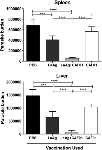
Immunized mice and control groups were challenged with L. infantum chagasi promastigotes 1 week after booster and euthanized 15 days post infection (dpi) corresponding with the spleen and liver parasitism peak, in order to assess protective immune response against the parasite. LaAg/CAF vaccinated–infected mice exhibited a significant parasite burden reduction in both liver and spleen compared with PBS, CAF01 and LaAg vaccinated mice (Fig. 1). A similar decrease of splenic and liver parasite burden was observed in LaAg vaccinated mice compared with the PBS and CAF01 alone groups. In addition, the control groups (PBS and CAF01) failed to control infection in both organs, presenting similar parasite burden levels (Fig. 1).
Effect of vaccination in parasite burden control. Mice received 2 intranasal doses of (LaAg) (25 µg) or CAF (150 µg), either alone or in combination. The negative control group received PBS (20 µL). One week after booster, they were infected with 107 Leishmania infantum chagasi promastigotes and 15 dpi, the parasite burden was individually measured in both liver and spleen. Results are represented as arithmetic means ± s.d. from 3 combined experiments. Abbreviations: LaAg, Leishmania amazonensis antigens; dpi, days post infection; PBS, phosphate-buffered saline. (*P < 0·05, ***P < 0·001, ****P < 0·0001).

LaAg/CAF01 association leads to increased antigen-specific immune response in immunized mice
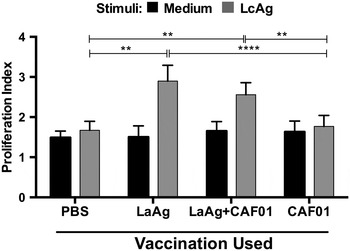
To further evaluate the vaccine-specific cell-mediated immune responses we performed an experiment to assess the lymphoproliferative response to LcAg antigen recall, using CFSE-labelled splenocytes from vaccinated-infected mice. Under this condition, cells from both LaAg and LaAg/CAF01 groups exhibited significantly higher proliferation upon antigen recall than the PBS and CAF01 vaccinated groups, but not compared with each other (Fig. 2). Furthermore, no significant difference was observed in the lymphoproliferative capacity of PBS and CAF01 cells (Fig. 2).
Effect of vaccination in the parasite-specific lymphoproliferations. Mice were vaccinated and infected as described in Fig. 1. On day 15 of infection, their spleen cells were labelled with CFSE and cultured at 5 × 10⁶ cells mL−1 in the absence (Medium) or presence of 50 µg mL−1 of L. infantum chagasi (LcAg) for 96 h. The proliferation index was calculated by the ratio of the total number of divisions/the number of divided cells using the FlowJo software after flow cytometry acquisition. Results were expressed as arithmetic means ± s.d. from 3 combined experiments. Abbreviation: CFSE, carboxyfluorescein succinimidyl ester. (**P < 0·01, ****P < 0·0001).

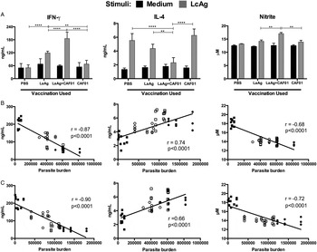
To evaluate the possible molecular mechanisms involved in protection after administration of single LaAg or in association with a CAF01 adjuvant, we investigated the IFN-γ, IL-4, IL-10 and nitrite production in the supernatants of splenocytes from vaccinated–infected mice stimulated in vitro with parasite antigens. As shown in Fig. 3A, cells from LaAg/CAF01 vaccinated mice produced significantly more IFN-γ than the PBS, CAF01 and LaAg vaccinated groups. LaAg vaccinated splenocytes produced higher IFN-γ amounts compared with the PBS and CAF01 groups. Only LaAg/CAF01 intranasal vaccinated splenocytes showed a significant increase in nitrite production and also a decrease in IL-4 production when compared with the PBS, CAF01 and LaAg vaccinated groups (Fig. 3A). No IL-10 production was detected in the supernatants of any evaluated groups in response to parasite antigens stimulation (data not shown). As a complement to these findings, a negative correlation was observed between IFN-γ or nitrite production and parasite burden in both spleen and liver, indicating that IFN-γ and nitrite levels are correlating with reduced infection burden. On the other hand, a positive correlation between IL-4 production and parasite load increase was observed, suggesting its association with the parasitism (Fig. 3B and C).
Effect of vaccination in parasite-specific cytokine and nitrite production. Mice were vaccinated and infected as described in Fig. 1. On day 15 of infection, their spleen cells were cultured at 5 × 10⁶ cells mL−1 in the absence (Medium) or presence of 50 µg mL−1 of LcAg for 72 h. (A) IFN-γ, IL-4 and nitrite amounts were determined in the culture supernatants by ELISA. Results are represented as arithmetic means ± s.d. from 3 combined experiments. (B) Correlation between IFN-γ, IL-4, or nitrite production and parasite burden in liver or (C) spleen. The correlation graph shows a fit line with a confidence curve. r = correlation coefficient; P = significance level. Each point represents one animal. PBS (●), LaAg (○), CAF (⩽), or LaAg/CAF (■). The horizontal bars represent the median values. Abbreviations: LcAg, L. infantum chagasi antigens; ELISA, enzyme-linked immunesorbent assay; PBS,. phosphate-buffered saline; LaAg, Leishmania amazonensis antigens. (**P < 0·01, ****P < 0·0001)

Specific antibodies elicited after immunization
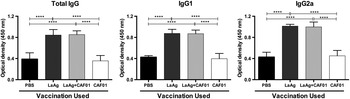
In order to analyse specific humoral immunity against parasites, we evaluated the total production of antileishmania IgG, IgG1 and IgG2a antibody isotypes in sera from all vaccinated groups. Total IgG, IgG1 and IgG2a isotype levels in the LaAg and LaAg/CAF01 vaccinated groups were significantly higher than those obtained from the PBS or CAF01 immunized groups. Moreover, no significant difference in IgG or its subtypes was observed between LaAg or LaAg/CAF01 vaccinated groups (Fig. 4).
Effect of vaccination in antibody production. BALB/c mice were vaccinated and infected as described in Fig. 1. On day 15 of infection, the animals were bled and total IgG, IgG1 and IgG2a analysed in the serum by ELISA assay. Results are represented as arithmetic means ± s.d. from 3 combined experiments. Abbreviation: ELISA, enzyme-linked immunesorbent assay. (****P < 0·0001).

DISCUSSION
In prior studies, the immunomodulatory and biosafety properties of the CAF01 adjuvant have shown an increased competence to induce both immunogenic and protective immune responses against bacterial and viral infections, which in some cases is associated with a shift to a Th1-type immune response (Fomsgaard et al. Reference Fomsgaard, Karlsson, Gram, Schou, Tang, Bang, Kromann, Andersen and Andreasen2011; Dietrich et al. Reference Dietrich, Andreasen, Andersen and Agger2014; Van Dissel et al. Reference Van Dissel, Joosten, Hoff, Soonawala, Prins, Hokey, O'Dee, Graves, Thierry-Carstensen, Andreasen, Ruhwald, de Visser, Agger, Ottenhoff, Kromann and Andersen2014). For many years, our group has proposed the use of mucosal vaccines against experimental cutaneous and VL (Pinto et al. Reference Pinto, De Mello Cortezia and Rossi-Bergmann2003, Reference Pinto, Pinheiro, Rayol, Larraga and Rossi-Bergmann2004; Gomes et al. Reference Gomes, Pinto, Melo, Lima, Larraga, Lopes and Rossi-Bergmann2007; De Oliveira Gomes et al. Reference De Oliveira Gomes, Da Silva Costa Souza, De Matos Guedes, Lopes and Rossi-Bergmann2011). In particular, the adjuvant-free crude LaAg antigens given by intranasal route has shown promising results for protection against experimental cutaneous leishmaniasis caused by L. amazonensis and Leshmania brasiliensis (Chaves et al. Reference Chaves, Claudio, Gomes, De-simone, Rossi-bergmann and De Matos2015; Da Silva-Couto et al. Reference Da Silva-Couto, Ribeiro-Romão, Saavedra, da Silva Costa Souza, Moreira, Gomes-Silva, Rossi-Bergmann, Da-Cruz and Pinto2015). Up to now, however, it had not been used against VL, the most important clinical form of the disease. Here, we extended these findings by demonstrating for the first time the feasibility of LaAg intranasal administration against VL by inducing partial protection in BALB/c mice challenged with L. (infantum) chagasi. The specific immune response given by LaAg vaccination has been critically dependent upon the mucosal route of immunization, as previously observed in murine models infected with L. amazonensis. (Pinto et al. Reference Pinto, Pinheiro, Rayol, Larraga and Rossi-Bergmann2004; Pinheiro et al. Reference Pinheiro, Pinto, Lopes, Guedes, Fentanes and Rossi-Bergmann2005). This supports the mucosal ability to induce tolerance to Th2 response-associated allergens and other disease-promoting antigens (Mestecky et al. Reference Mestecky, Moldoveanu and Elson2005; Takagi et al. Reference Takagi, Hiroi, Yang, Tada, Yuki, Takamura, Ishimitsu, Kawauchi, Kiyono and Takaiwa2005), but also to elicit active systemic immunity against pathogens (Fouda et al. Reference Fouda, Amos, Wilks, Pollara, Ray, Chand, Kunz, Liebl, Whitaker, Carville, Smith, Colvin, Pickup, Staats, Overman, Eutsey-Lloyd, Parks, Chen, Labranche, Barnett, Tomaras, Ferrari, Montefiori, Liao, Letvin, Haynes and Permar2013; Carapau et al. Reference Carapau, Mitchell, Nacer, Shaw, Othoro, Frevert and Nardin2013; Wahid et al. Reference Wahid, Fresnay, Levine and Sztein2015). In the present study, CAF01 enhanced the protective effect of LaAg vaccination, as shown by the decreased parasitism in the livers and spleens of vaccinated mice. This finding supports previous reports, which suggest that protein antigen formulations offer an advantage in safety, but a low immunogenicity as compared with protein alone, thus requiring the help from adjuvants (Pinheiro et al. Reference Pinheiro, Pinto, de Matos Guedes, Filho, de Mattos, Saraiva, de Mendonça and Rossi-Bergmann2007). CAF01 administration has shown the ability to prime pro-inflammatory cytokine production (Rosenkrands et al. Reference Rosenkrands, Vingsbo-Lundberg, Bundgaard, Lindenstrøm, Enouf, van der Werf, Andersen and Agger2011; Kamath et al. Reference Kamath, Mastelic, Christensen, Rochat, Agger, Pinschewer, Andersen, Lambert and Siegrist2012) and also has displayed safety/biocompatibility advantages when used in vaccine formulation, especially for humans (Fomsgaard et al. Reference Fomsgaard, Karlsson, Gram, Schou, Tang, Bang, Kromann, Andersen and Andreasen2011; Van Dissel et al. Reference Van Dissel, Joosten, Hoff, Soonawala, Prins, Hokey, O'Dee, Graves, Thierry-Carstensen, Andreasen, Ruhwald, de Visser, Agger, Ottenhoff, Kromann and Andersen2014).
Unlike cutaneous infection caused by Leshmania major, the VL outcome is determined by the magnitude/quality of the parasite-specific Th1 immune response, regardless of the Th2 immune response or Th1/Th2 cytokine production balance (De Oliveira Gomes et al. Reference De Oliveira Gomes, Schwedersky, De-Melo, Da Silva Costa Souza, De Matos Guedes, Lopes and Rossi-Bergmann2012; Yadav et al. Reference Yadav, Amit, Chaudhary, Chandel, Mahantesh, Suman, Singh, Dikhit, Ali, Rabidas, Pandey, Kumar, Das and Bimal2015). After specific antigen recall, LaAg and LaAg/CAF01 vaccinated mice produced significantly more IFN-γ than control groups. However, only the LaAg/CAF01 vaccination decreased the production of IL-4, which, together with IL-10, has been associated with disease severity during VL and establishment of chronic infection (Oliveira et al. Reference Oliveira, Costa, Chavez-Fumagalli, Valadares, Duarte, Costa, Martins, Gomes, Melo, Soto, Tavares and Coelho2012; Singh and Sundar, Reference Singh and Sundar2014). Since IFN-γ-activated macrophages are necessary for nitric oxide production and Leishmania intracellular killing, its production has been considered one of the main factors in acquiring immunity against infection with viscerotropic and non-viscerotropic Leishmania species (Singh and Sundar, Reference Singh and Sundar2014). To investigate if the higher IFN-γ production observed in LaAg/CAF01-vaccinated mice was associated with immune response activation, nitrite production (used as an indirect marker of nitric oxide production) from Ag-stimulated splenocytes was analysed. Our results showed that the nitrite levels run in direct correlation with IFN-γ produced after LaAg/CAF01 vaccination, suggesting that infection control could be mediated by a nitric oxide-dependent pathway (Iniesta et al. Reference Iniesta, Gómez-Nieto, Molano, Mohedano, Carcelén, Mirón, Alonso and Corraliza2002; Yadav et al. Reference Yadav, Amit, Chaudhary, Chandel, Mahantesh, Suman, Singh, Dikhit, Ali, Rabidas, Pandey, Kumar, Das and Bimal2015). In addition, the failure to control disease presented by the control groups or even after LaAg vaccination, may be related to the high IL-4 levels or lower IFN-γ production as observed. Splenocytes from LaAg/CAF01 and LaAg vaccinated mice presented increased lymphoblastic activity in response to Leishmania antigen stimulation, suggesting that both may preferably prime a cellular immune response with a high production of IFN-γ, which has been related to resistance to visceral and cutaneous leishmaniasis (De Oliveira Gomes et al. Reference De Oliveira Gomes, Schwedersky, De-Melo, Da Silva Costa Souza, De Matos Guedes, Lopes and Rossi-Bergmann2012; Nico et al. Reference Nico, Gomes, Alves-Silva, Freitas, Morrot, Bahia, Palatnik, Rodrigues and Palatnik-de-Sousa2014).
Levels of IgG2a and IgG1 isotypes are used indirectly as markers of Th1 and Th2 immune response, respectively. In BALB/c mice, the Th2-dependent production of IgG1 antibodies is associated with disease progression due to some Leishmania species, including L. amazonensis (Pereira and Alves, Reference Pereira and Alves2008), but this is not confirmed in L. infantum or L. donovani (Ghosh et al. Reference Ghosh, Zhang and Matlashewski2001; Garg and Dube, Reference Garg and Dube2006). Our results showed no significant difference in the total IgG, IgG2a or IgG1 levels between the LaAg/CAF01 and LaAg-vaccinated groups, suggesting that the CAF01 adjuvant did not directly participate in the antibody production after vaccination. Also, we did not find differences between IgG2a and IgG1 antibody levels in the immunized groups, indicating that LaAg or CAF01 vaccination did not contribute directly to a humoral immune response correlated with cytokine production.
The results herein support a model whereby the well-established formulations of intranasal delivery could be improved by association with a promising adjuvant (CAF01), predicted to target an immune response against VL
FINANCIAL SUPPORT
This work was financially supported by Conselho Nacional de Desenvolvimento Científico e Tecnológico-CNPq (grant number 482167/2013-0) and Fundação de Amparo à Pesquisa do Estado do Espírito Santo – FAPES (grant number 54685532/2011).