Introduction
Lichenicolous fungi are inconspicuous species that grow exclusively on lichens and develop diverse degrees of specificity and parasitic behaviour towards their hosts. They are generally host-specific parasites, but broad-spectrum pathogens, saprotrophs or commensals are not rare. Over 1800 species have been described throughout the Ascomycota and Basidiomycota, and it is estimated that c. 3000 species still need to be described (Lawrey & Diederich Reference Lawrey and Diederich2003, Reference Lawrey and Diederich2016).
Among the lichens collected from the southern subpolar region by Henry Imshaug and co-workers and housed in the herbarium of Michigan State University (MSC; Fryday & Prather Reference Fryday and Prather2001), there is a single collection of an endohymenial species with more or less spherical asci and hyaline, muriform ascospores that we provisionally assign here to the genus Arthothelium A. Massal. The genus Arthothelium is a heterogeneous group of c. 121 more or less lichen-forming species (Kirk et al. Reference Kirk, Cannon, Minter and Stalpers2008), but no lichenicolous species are known (Lawrey & Diederich Reference Lawrey and Diederich2016). As we are unaware of any other endohymenial species of Arthoniaceae producing muriform ascospores, we describe it here as a species new to science.
Material and Methods
Specimens were studied using an Olympus SZX12 stereomicroscope and an Olympus BX51 compound microscope. Hand-cut sections were investigated by light microscopy on material mounted in water, 5% KOH (K) and Lugol’s reagent (1%) without (I) or with (K/I) pre-treatment with K. Measurements of asci and ascospores refer to material examined in water.
Arthothelium hymeniicola Ertz & Fryday sp. nov.
MycoBank No.: MB 817356
Endohymenial species of Arthothelium reduced to±spherical, 8-spored asci, 35–40×24–30 µm, containing hyaline, muriform, 4–6×1-septate ascospores, 14–20×6–8 µm.
Type: New Zealand, Campbell Island, north of Beeman Station, on branch of Dracophyllum scrub, on a Bacidia sp., 22 December 1969, R. C. Harris 4386 (MSC0102168—holotype!).
(Fig. 1)
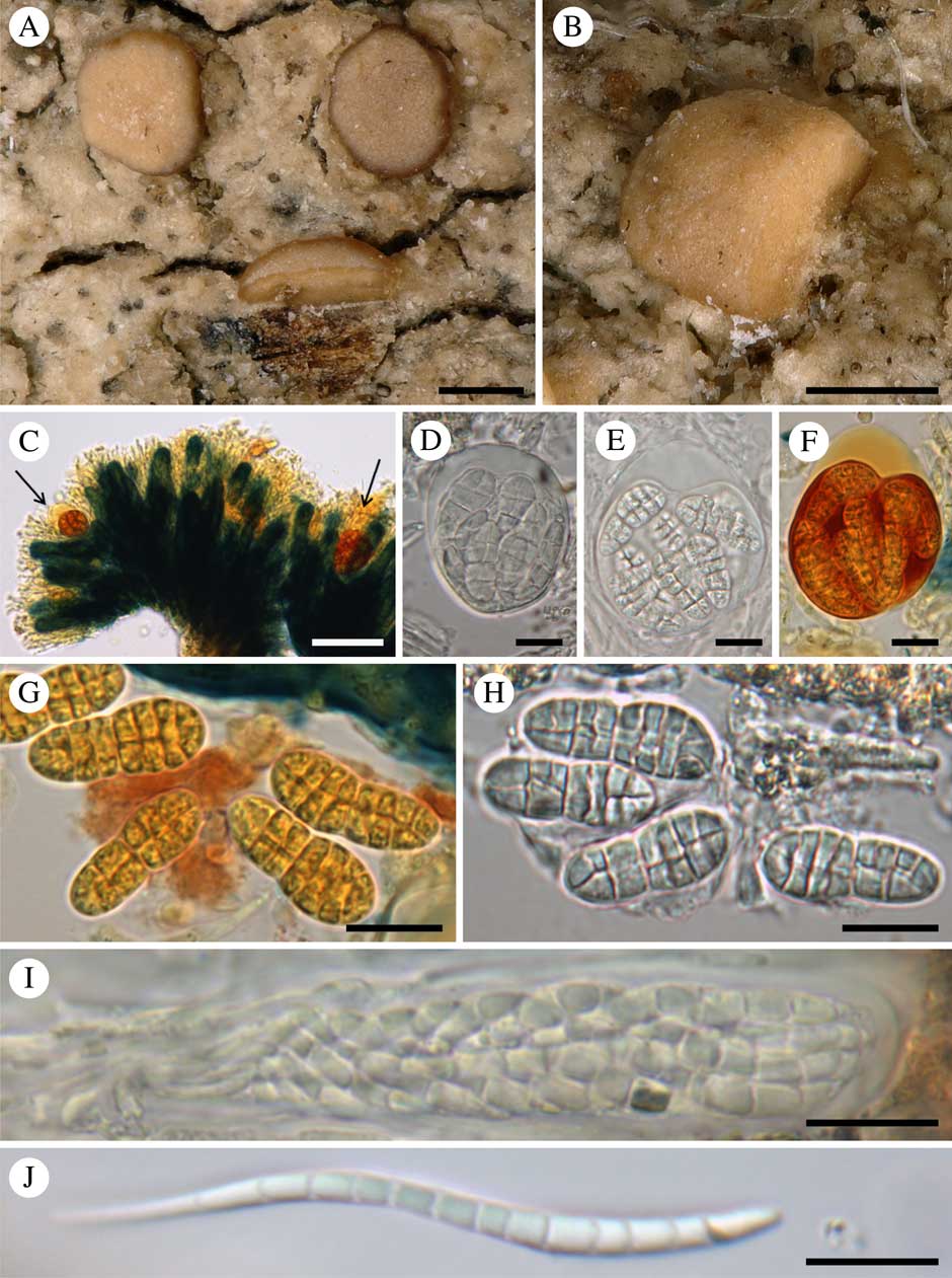
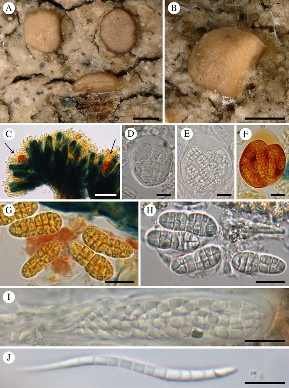
Arthothelium hymeniicola and its host lichen (Bacidia sp.) (holotype). A, apothecia of the host lichen: the lowermost apothecium, for which a large part was removed, was examined under the microscope and found to be infected by A. hymeniicola, although no visible sign of infection was evident to the naked eye; the two apothecia situated above look healthy but they are probably also infected. B, macroscopical overview of an apothecium of the host lichen which was infected with A. hymeniicola, with the right-hand part removed; no visible sign of infection was evident to the naked eye. C, cross-section of the hymenium of the Bacidia sp. in KI, showing two KI+ orange-red asci of A. hymeniicola (arrows) in the upper part of the hymenium and many KI+ blue asci of the host lichen. D–F, asci of A. hymeniicola (D, in water; E, in KOH; F, in KI). G & H, ascospores of A. hymeniicola (G, in KI; H, in water). I, ascus of the host lichen (in water). J, ascospore of the host lichen. Scales: A & B=250 µm; C=50 µm; D–J=10 µm. In colour online.
Thallus absent. Lichenicolous, non-lichenized, in hymenium of a Bacidia sp.
Ascomata absent. Asci loosely aggregated in the upper third of the hymenium of the host that is not modified in shape or colour by the infection, ±spherical, 35–40×24–30 µm, with a very short, almost inconspicuous foot, 8-spored, wall thicker in upper part, c. 5 µm thick, I−, KI+ orange (internal plasma I+ red, KI+ red), with a small, often inconspicuous ocular chamber. Ascospores hyaline, ovoid-ellipsoid, muriform, with 4–6 transverse septa and one longitudinal septum, I+ orange, KI+ orange, 14–20×6–8 µm (in water), without a distinct gelatinous sheath. Paraphysoids not seen.
Conidiomata not seen.
Host (Bacidia sp.). Thallus greyish to pale brown or cream, thin, rimose.
Apothecia round, constricted at the base, 0·3–0·8 mm diam., at first flat with a thin pale brownish to cream margin, often becoming slightly convex and immarginate; hymenial disc cream to pale brown, sometimes paler than the margin. True exciple colourless. Hymenium 70–85 µm high, colourless; epihymenium yellowish, K−, often covered by a thin layer of pruina with colourless crystals dissolving in K. Hypothecium colourless or yellowish; paraphyses 1·0–1·5 µm wide, irregularly branched, apices not distinctly widening. Asci narrowly clavate, 8-spored, 68–76×10–13 µm, I+ blue becoming brownish, KI+ blue. Ascospores hyaline, tapered at one end, slightly to strongly sigmoid, without gelatinous sheath, 8–13-septate, 55–57×3–5 µm.
Distribution and ecology. Known only from the type collection from Campbell Island, on a branch of a Dracophyllum shrub, and strictly confined to the hymenia of its host (an apparently undescribed Bacidia sp.).
Notes. The absence of ascomatal structure renders the generic placement of the new species difficult. The short subspherical asci with a thick upper wall and the parasitic lifestyle on other ascomycetes are reminiscent of species of Arthoniaceae (Arthoniomycetes) and of Myriangiales (encompassing Cookellaceae; Dothideomycetes). We decided to tentatively place the new species in the family Arthoniaceae and to describe it in the genus Arthothelium s. lat. for the reasons discussed hereafter.
The family Arthoniaceae is well known for including taxa with reduced or absent ascomatal structures. It also includes many lichenicolous fungi. Among them, Arthonia intexta Almq. is a notable example of an endohymenial parasite that forms only asci and a few interascal filaments intermixed with those of the host. However, contrary to our new species, A. intexta differs by having only transversely 2–3(–4)-septate ascospores, narrower asci (10–20 µm wide) generally having a KI+ blue ring-structure and different host species belonging to the genus Lecidella (e.g. Hertel Reference Hertel1969; Triebel Reference Triebel1989; Grube Reference Grube2007; Sussey Reference Sussey2012). Many other lichenicolous Arthonia species are strictly or facultatively parasitic on the hymenia of their host lichen but all produce distinct ascomatal structures (e.g. Arthonia apotheciorum (A. Massal.) Almq., A. clemens (Tul.) Th. Fr., A. glacialis Alstrup & E. S. Hansen, A. haematommatum Kalb & Hafellner, A. lecanoricola Alstrup & Olech, A. lecanorina (Almq.) R. Sant., A. obscurior Triebel, A. protoparmeliopseos Etayo & Diederich, A. sherparum Grube & Matzer, A. subfuscicola (Linds.) Triebel and A. varians (Davies) Nyl.) and non-muriform ascospores (Triebel Reference Triebel1989; Kalb et al. Reference Kalb, Hafellner and Staiger1995; Grube & Matzer Reference Grube and Matzer1997; Alstrup & Hansen Reference Alstrup and Hansen2001; Grube Reference Grube2007; Coppins & Aptroot Reference Coppins and Aptroot2009; Etayo & Diederich Reference Etayo and Diederich2009), whereas a tendency for reduced ascomatal structures is observable in Arthonia varians (=A. glaucomaria) (Hertel Reference Hertel1969; Triebel Reference Triebel1989). The genus Arthonia was formerly separated from Arthothelium by ascospore septation: transversely septate in Arthonia and muriform in Arthothelium (e.g. Santesson Reference Santesson1952; Makhija & Patwardhan Reference Makhija and Patwardhan1995; Grube & Giralt Reference Grube and Giralt1996; Sundin & Tehler Reference Sundin and Tehler1998; Sundin Reference Sundin1999; Grube Reference Grube2007). However, in recent molecular studies Arthonia and Arthothelium were recovered as polyphyletic with different lineages including species of both genera (Ertz & Tehler Reference Ertz and Tehler2010; Frisch et al. Reference Frisch, Thor, Ertz and Grube2014; Van den Broeck & Ertz Reference Van den Broeck and Ertz2016). Moreover, the lichenicolous lifestyle has evolved several times in the Arthoniaceae (Diederich et al. Reference Diederich, Lawrey, Sikaroodi, van den Boom and Ertz2012; Frisch et al. Reference Frisch, Thor, Ertz and Grube2014) but too few species of Arthoniaceae were included in molecular phylogenies for a reassessment of generic delimitations of the lichenicolous taxa. Therefore, we decided to describe our new species in the very heterogeneous genus Arthothelium pending a molecular taxonomic revision of the lichenicolous taxa of Arthoniaceae. The placement in Arthothelium must be considered provisional because the type species of Arthothelium (A. spectabile A. Massal.) is a lichenized species forming distinct ascomata. It must be noted that Cryptothecia Stirt. is a genus of Arthoniaceae with muriform ascospores that does not form ascomatal structures, the asci being loosely scattered in the thallus (Grube Reference Grube1998). However, no lichenicolous/parasitic species of Cryptothecia are known and the asci are of a different type (Grube Reference Grube1998, fig. 3). Stirtonia A. L. Sm. is another genus of Arthoniaceae that does not form ascomatal structures, but species of this genus differ by having transeptate ascospores and a lichenized habit. Lichenicolous Arthoniales with globose to subglobose asci and muriform ascospores are also found in the genera Paradoxomyces Matzer and Trichophyma Rehm (Matzer Reference Matzer1996, who questioned the placement of the latter genus in either Arthoniales or Myriangiales). Both genera differ from Arthothelium notably by ascomata having a true exciple. Another Arthoniales known to occur on Bacidia is Opegrapha diffracticola (Harris & Ladd Reference Harris and Ladd2007) but this species has very different ascomata, asci and ascospores.
We also considered a possible placement of our new species in the Myriangiales because similar asci and ascospores are found in some taxa. This order is characterized by pulvinate, irregular ascostromata in which asci are irregularly arranged in one or more layers in locules, each locule containing single or multiple asci (Hyde et al. Reference Hyde, Jones, Liu, Ariyawansa, Boehm, Boonmee, Braun, Chomnunti, Crous and Dai2013). However, most species are parasitic or saprobic on plants (Hyde et al. Reference Hyde, Jones, Liu, Ariyawansa, Boehm, Boonmee, Braun, Chomnunti, Crous and Dai2013) and no lichenicolous species are known from the order (Lawrey & Diederich Reference Lawrey and Diederich2016) making a placement of our new species in this order unlikely. However, parasites on fungi are known in the family Cookellaceae which was placed in Myriangiales by von Arx (Reference von Arx1963) but now considered as Dothideomycetes of uncertain order by some authors (e.g. Eriksson Reference Eriksson2005; Lumbsch & Huhndorf Reference Lumbsch and Huhndorf2010; Hyde et al. Reference Hyde, Jones, Liu, Ariyawansa, Boehm, Boonmee, Braun, Chomnunti, Crous and Dai2013). Unfortunately, no molecular data are available for this poorly known family which includes only 13 species in three genera (Cookella Sacc., Uleomyces Henn. and Pycnoderma Syd. & P. Syd.) (Kirk et al. Reference Kirk, Cannon, Minter and Stalpers2008). Cookella differs from our new species by having dark brown ascospores, whereas, according to Hyde et al. (Reference Hyde, Jones, Liu, Ariyawansa, Boehm, Boonmee, Braun, Chomnunti, Crous and Dai2013) the differences between Pycnoderma and Uleomyces, the species of which are mainly hyperparasites of leaf fungal parasites (Hennings Reference Hennings1895; von Arx Reference von Arx1963), refer to their ascomatal structure: superficial, cushion or disc-shaped ascostromata in Uleomyces and superficial, disc-shaped thyriothecia in Pycnoderma. However, the asci and ascospores of several members of these two genera are similar to our new species (e.g. Pycnoderma bambusinum Syd. & P. Syd., P. congestum (Syd.) Arx, Uleomyces brenesii (Petr.) Arx, U. comedens Syd., U. struthanthi G. Arnaud and U. wellmanii Jenkins & Limber) so that a placement here might be considered, especially as these species do not form (pseudo)paraphyses. However, these fungi differ from our new species by the formation of ascostromata on fungal leaf parasites (von Arx Reference von Arx1963) and lichenicolous species are not known from the Cookellaceae. It must be noted that, interestingly, Grube (Reference Grube1998, p. 377) mentioned that members of the Cookellaceae have some similarities with Arthothelium, Stirtonia and Cryptothecia species, which highlights the difficulties we encountered placing a species not developing ascomatal structures in either Arthoniaceae or Cookellaceae.
As a conclusion, our new species could be placed in the Arthoniaceae or the Cookellaceae, but because the former family is the only one known to include lichenicolous species, and as some of these species are known to have a tendency for reduced ascomatal structures, a placement in the Arthoniaceae was preferred. The placement in Arthothelium should, however, be considered provisional.