Introduction
Millsite, CuTeO3·2H2O, was identified as a new mineral at the Natural History Museum in London, UK (henceforth NHM) after two specimens, each containing a different blue-coloured mineral were donated to the museum by a member of the public. The specimens were obtained by the donor during a field excursion to the Oppdal area in Norway many years earlier, who was told during the excursion that the blue material on the specimens was rare and might be a new mineral.
Curatorial protocol at the NHM strives for the systematic verification using both EDX and XRD analysis of newly obtained unusual or potentially significant specimens. To the surprise of one of us (M.S.R.) this analysis revealed that one of the blue minerals on the specimens was the rare tellurite teineite, and the other was indeed potentially new, namely millsite. Here we report the structural, chemical and optical characterization of millsite.
Millsite is named after Dr. Stuart J. Mills (b. 1982), currently Senior Curator of Geosciences at Museums Victoria, Australia. He has published extensively on new minerals and has focused particularly in recent years on the secondary mineralogy and crystal chemistry of tellurium bearing oxysalts (e.g. Mills and Christy, Reference Mills and Christy2013, Christy and Mills, Reference Christy and Mills2013; Christy et al., Reference Christy., Mills and Kampf2016; Fornadel et al., Reference Fornadel, Spry, Haghnegahdar, Schauble, Jackson and Mills2017). The new mineral and its name, pronounced (‘mills-ite’), have been approved by the Commission on New Minerals, Nomenclature and Classification of the International Mineralogical Association (IMA2015-086). Holotype material is registered as BM 2011,243, housed at the NHM and comprises the original specimen, a polished probe block and two single crystal mounts.
The discovery of millsite highlights the importance of a well-resourced museum, with a systematically maintained reference collection, as well as the investment of time in routine and speculative identification of mineral species.
Occurrence
After the investigation of millsite had begun, it was established more specifically that the specimens being studied were from Gråurdfjellet in Oppdal kommune, Norway (62°29'11”N, 9°29'25''E) and were almost certainly part of a find made in 1991 by Kjell Arve Isbrekken which was significant for containing gold and tellurium minerals and a supposed new mineral. It also transpired that a sample of the new mineral had been obtained by one of the authors (FM) from K.A. Isbrekken (specimen KAI-93). The crystal structure of this sample was determined by single-crystal XRD using AgKα radiation (Mo, F., Larsen, F.K., Mathiesen, R., Måseide, K. and Overgaard, J. (Reference Mo, Larsen, Mathiesen, Måseide and Øvergaard2000) Unpublished results). The crystallographic data showed unequivocally that the new mineral is distinct from teineite and likely to be a new mineral. This new mineral is millsite and it is essentially the same as the material donated recently to the NHM via M.S.R.
Millsite occurs as part of an unusual suite of secondary copper minerals found within a granular quartz and chlorite/muscovite matrix. Traces of primary ore minerals were identified within the matrix when the specimen was split for analysis. These have been shown to consist of digenite/chalcocite, hessite and small specks of native gold. Uraninite, monazite, dravite and euxenite have also been reportedly identified within the matrix (http://www.mindat.org retrieved 05/2016) but were not observed during this study. The secondary phases present occur in vughs, cracks and interstices between the granular quartz, and aside from millsite include teineite, malachite, U-rich mcalpineite, schmitterite, a copper sulfate (probably brochantite), goethite and an amorphous, heterogeneous Cu–Te–Si gel-like component with variable chemistry.
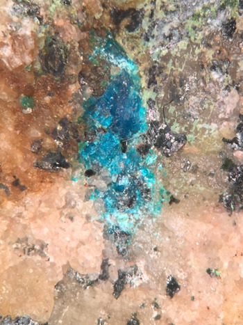
On the type specimen, BM 2011,243 (Fig. 1), millsite occurs in one significant cavity that is completely filled by the mineral. Millsite and teineite occur separately on each specimen from Gråurdfjellet in the NHM, other examples from the investigation by Mo et al., (2000) show that the two polymorphs can occur together, intergrown on the same specimen. The geological origin of the millsite-bearing specimens and the entire suite including primary tellurides and gold is unclear. It is reported anecdotally that all the known specimens were extracted from a single boulder, rather than from an in situ geological unit. Recent trips to the area have failed to find any remains of the boulder or any signs of similar mineralization (R. Kristiansen, pers. comm. to M.S.R., 2015). It is possible that the boulder is a glacial erratic from an as yet unidentified Cu–Au–Ag–Te deposit.
Holotype millsite BM2011,243. Width of field of view = 6 mm. All the blue phase is millsite.

Millsite and other secondary minerals from the boulder probably formed through the oxidation of primary sulfides and tellurides under near-surface oxidizing conditions in the presence of water. Such an origin is consistent with the secondary tellurium mineralization observed at Otto Mountain and Centennial Eureka mine where similar phases are observed (Kampf et al., Reference Kampf, Housley, Mills, Marty and Thorne2010; Marty et al., Reference Marty, Kampf, Housley, Mills and Weiβ2010; Housley et al., Reference Housley, Kampf, Mills, Marty and Thorne2011).
Physical and optical properties
Millsite occurs on the holotype as cyan blue (HTML 0099CC) to pale royal blue (HTML 0066FF) transparent crystalline grains infilling cavities, fractures and interstices in a granitic matrix. On this specimen the millsite fills a cavity 5 mm × 2 mm × 2 mm in size. Since approval of millsite as a new species, numerous larger specimens have been tentatively identified in private collections and examples can be seen on http://www.mindat.org (retrieved 05/2016). The largest of these aggregates is ~20 mm × 15 mm × 5 mm, with distinct crystals in the mm range. It is of significant note that even though there is a subtle colour difference between teineite (darker royal blue) and millsite on the two specimens at the NHM, the authors do not consider that this is an infallible way to distinguish the two phases. It is clear that this feature has been used by collectors to distinguish the two phases, but tests by one of the authors (MSR) and other researchers have shown that specimens having a cyan colour are still teineite. Not enough of the specimens now known to exist were available to the authors to conclusively determine reliable differences in colour.
There are no discrete terminated crystals and so forms and twinning are difficult to assess macroscopically. Tiny broken fragments show perfect cleavage and appear platy on (100). Millsite is transparent with a vitreous lustre and is non-fluorescent in both shortwave and longwave ultraviolet light. Millsite has a pale green streak. Hardness and density could not be measured due to paucity of sample, but using the determined empirical formula and volume the calculated density is 3.963 g cm–3.
The mineral is optically biaxial (+) with optical constants measured in white light: α = 1.756(5), β = 1.794(5) and γ = 1.925calc. Due to the reaction of millsite with index liquids with n > 1.8, γ was calculated based on α, β and 2V. The 2V angle was determined by conoscopic observation on a spindle stage as 60(1)° and obvious r < v dispersion was observed. The optical orientation is Y = b, X ^ a = 41° in obtuse β; one optical axis ≈ ⊥ to (001). The crystal studied is weakly pleochroic: X ≈ Z > Y, greenish blue.
The Gladstone-Dale compatibility index, 1–(Kp/Kc) as defined by Mandarino (Reference Mandarino1981), provides a measure of the consistency among the average index of refraction, calculated density and chemical composition. For millsite of end-member CuTeO3·2H2O composition the compatibility index is 0.0095, indicating ‘superior’ agreement among these data. Based on the empirical composition the compatibility index is 0.056, indicating ‘good’ agreement.
Chemical composition
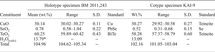
A small fragment of holotype specimen BM 2011,243 was removed, mounted in epoxy and polished using aluminium oxide. The polished block was carbon-coated for analysis using a Cameca SX100 electron microprobe at the NHM. The Cameca microprobe was operated in wavelength-dispersive mode at 20 kV, a probe current of 20 nA and a beam diameter of 10 µm. Only Cu, Te and Se were detected, although S, Fe, Co, Ni, Zn, Ag and U were all sought but found to be below detection limits. Table 1 summarizes the microprobe data. The anhydrous empirical formula (O = 3) is Cu0.99(Te0.98Se0.02)O3, which is consistent with the hydrous end-member formula determined in the crystallographic study, CuTeO3·2H2O. The ideal formula requires 28.91 wt.% CuO, 58.00 wt.% TeO2 and 13.09 wt.% H2O and is identical to the composition of teineite.
Electron microprobe analyses of millsite from holotype specimen BM 2011,243 and cotype specimen KAI-93.

* The H2O content is that calculated on the basis of the structure determination.
S.D. – Standard deviation.
A small piece of the cotype specimen KAI-93 was embedded in a drop of adhesive on a microscope glass slide and polished with 1 µm diamond powder. Quantitative element analyses were carried out using a JEOL JXA-8500 F electron microprobe (field-emission electron gun) equipped with a high-resolution wavelength X-ray dispersive spectrometer. A teineite standard was used for CuO and TeO2, and for SeO2 a Se metal standard. Raw data were reduced to weight percentages using the ZAF algorithm. The analyses were done using 15 kV accelerating voltage and an electron beam current of 20 nA with a defocused probe size of 10 μm. Selenium was measured as a trace element, and the beam was defocused to avoid beam damage to the specimen and limit the charging effects because of long counting times on Se (300 s). A further reason for employing such mild conditions is that several previous electron microprobe analyses implied a structure with only one water molecule, at variance with the X-ray crystal structure, and indicating the loss of ~1 H2O per formula unit (pfu) due to the charging and local heating of the sample under the impact of the electron beam. For the elements Cu and Te counting times of 10 s were adequate. The spectra showed weak emission lines for SeLα1,2 and SeLβ1. As the latter is well resolved from any Te lines we believe that the sample does contain a small amount of Se, presumably as a substitute for Te. The results from 5 analyses are given in Table 1.
There was not enough material on the holotype specimen to perform CHN analysis or directly quantify water content by any other means other than structure refinement. However, both crystal structure determinations demonstrate clearly that there are two H2O pfu. These two non-equivalent H2O groups form part of the octahedral coordination of Cu and are not ‘zeolitic’, but a fixed and structurally well-defined feature. Consequently, the H2O content of millsite correlates with the Cu content, and as such can be inferred from the stoichiometry of the anhydrous empirical formula.
Raman spectroscopy
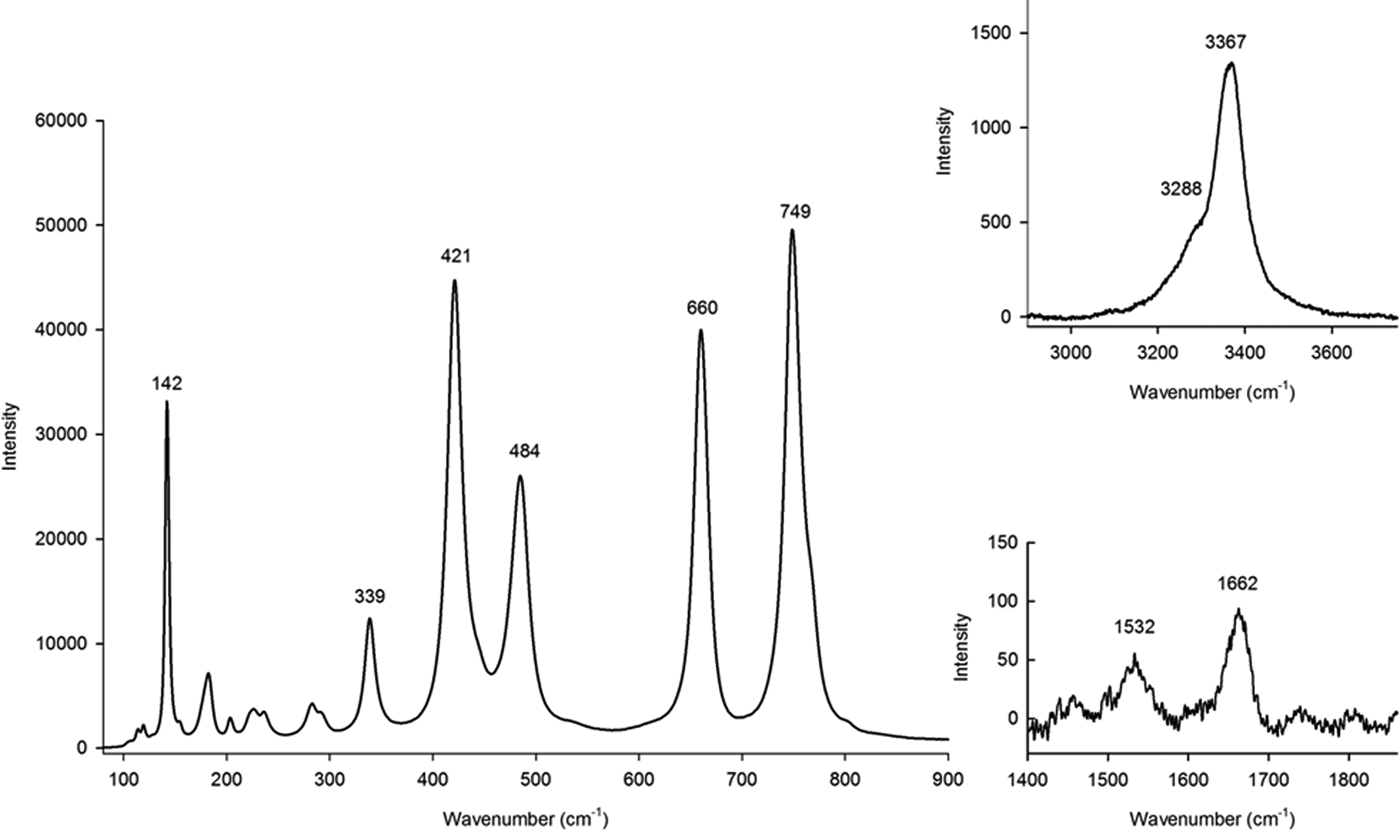
An unpolarized Raman spectrum was obtained for a crystal of millsite (30 µm × 75 µm × 115 µm) removed from the holotype specimen 2011,243. Excitation induced by the 473 nm line of a 50 mW Cobalt Blues TM laser focused down to a 10 µm spot. The spectrum is shown in Fig. 2. Peaks in the 100–1000 cm–1 range are assigned by comparison with the Raman spectrum of teineite (Frost and Keeffe, Reference Frost and Keeffe2009). The 749 cm–1 band and a shoulder are assigned to symmetric and antisymmetric modes of the TeO3 group; a band at 339 cm–1 is assigned to the TeO3 bending mode; the 484 cm–1 band is a possible H2O libration mode. Two low-intensity but distinct bands occur at 1532 and 1662 cm–1 and are attributed to the bending modes of two non-equivalent H2O molecules (see Crystal structure). Two bands at 3288 cm–1 and 3367 cm–1 are due to O–H stretching modes of H2O molecules.
Raman spectrum of millsite, showing O‒H vibrational modes in the insets.

Crystal structure
The structure of a small prismatic crystal (70 µm × 40 µm × 30 µm) from the holotype specimen BM 2011,243 was determined. This crystal was mounted on an XcaliburE four-circle diffractometer equipped with an Eos CCD detector (Rigaku Oxford Diffraction) at the Natural History Museum, London. Graphite-monochromated MoKα X-rays were used at 45 kV and 40 mA. Reflection intensities were corrected for Lorentz-polarization and absorption effects, and converted to structure factors using CrysalisPro (®Rigaku Oxford Diffraction). A whole sphere of data was collected to 60°2θ with 99.9% completeness. Structure solution by direct methods and refinement by least-squares were carried out using SHELX (Sheldrick, Reference Sheldrick2008) within the WinGX environment (Farrugia, Reference Farrugia1999). Neutral scattering factors for all atoms were taken from the International Tables for Crystallography Volume C (Wilson, Reference Wilson1992).
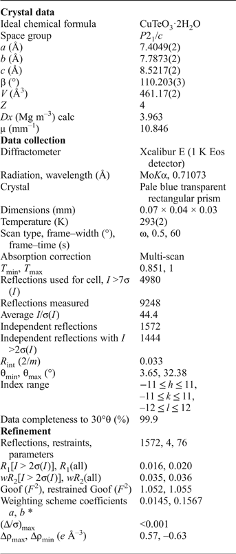
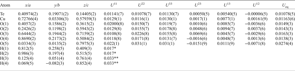
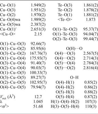
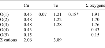
Systematic absences are consistent with space group P21/c. Structure solution using direct methods (SHELX, Sheldrick, Reference Sheldrick2008) located the Cu and Te atoms (one of each). All five O atoms of the asymmetric unit were found after 10 cycles of least-squares refinement. All four H atoms of the asymmetric unit were found in subsequent stages of weighted least-squares isotropic refinement, and their coordinates refined with their U iso values set to 1.2 times those of the oxygen atoms to which they are bonded. A soft restraint on O–H bond lengths was used. All other atoms were then refined anisotropically. Information relating to the data collection and structure refinement is summarized in Table 2. Atom coordinates and displacement parameters are given in Table 3. Key interatomic distances, angles and polyhedral parameters are given in Table 4. Bond-valence calculations are summarized in Table 5. A crystallographic information file is provided as Supplementary material.
Summary of information relating to data collection and structure refinement of millsite from holotype specimen BM 2011,243.

Atom coordinates, anisotropic and equivalent-isotropic displacement parameters (Å2) for millsite from holotype specimen BM 2011,243.

*Value coupled to 1.2 × refined U eq of O(4). **Value coupled to 1.2 × refined U eq of O(5). See text for explanation.
Interatomic distances (Å), bond angles (°) and polyhedral parameters for millsite.

wa = oxygen of a H2O molecule.
Bond valences and bond-valence sums (BVS, valence units) for millsite.

Notes: Atoms O(3) and O(4) each receive a hydrogen bond. Atom O(2) receives two hydrogen bonds. Atoms O(4) and O(5) belong to H2O molecules. Values calculated using the parameters for the TeIV–O bond of Mills and Christy (Reference Mills and Christy2013). CuII–O bond valences were calculated using the parameters of Brown and Altermatt (Reference Brown and Altermatt1985).
* This is a secondary bond in the sense of Mills and Christy (Reference Mills and Christy2013).
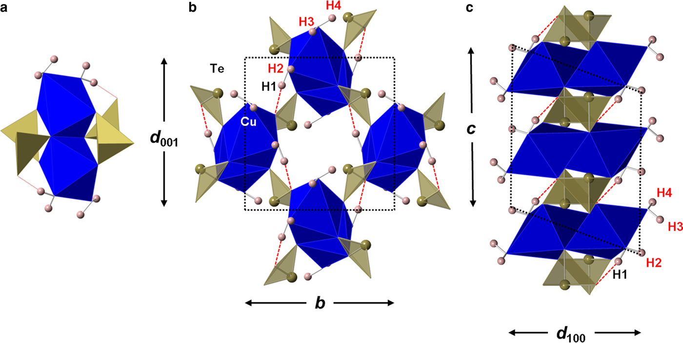
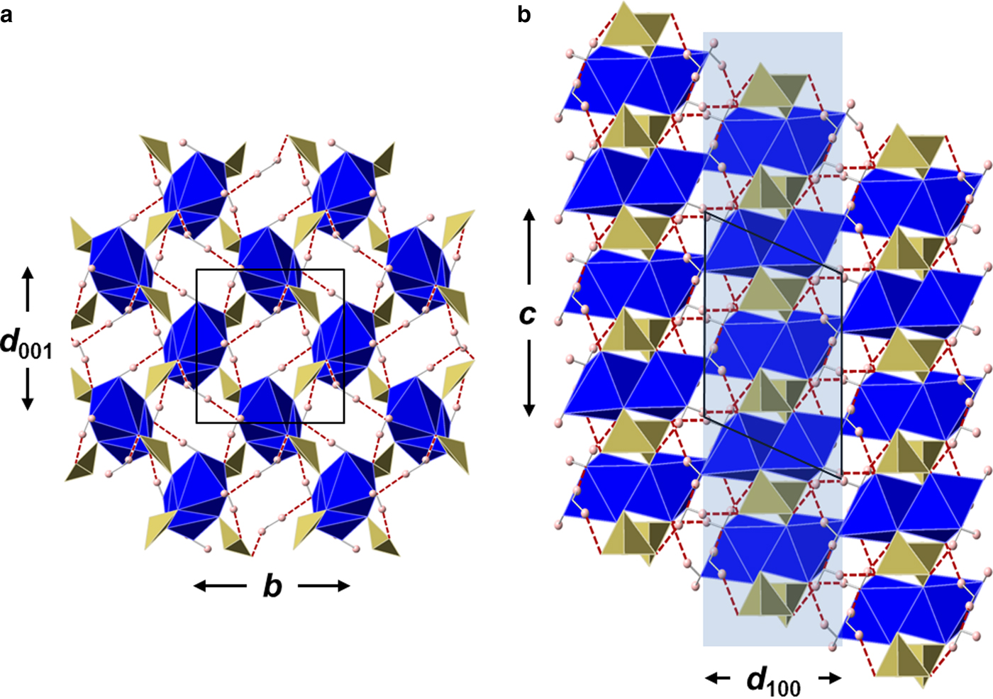
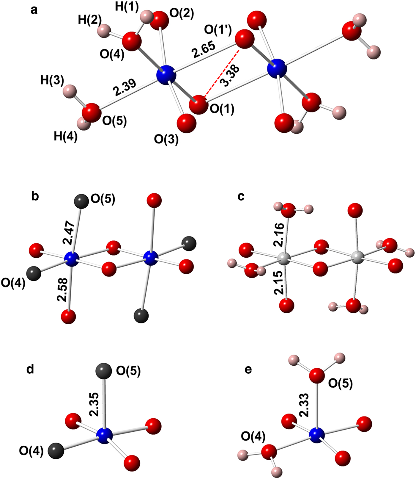
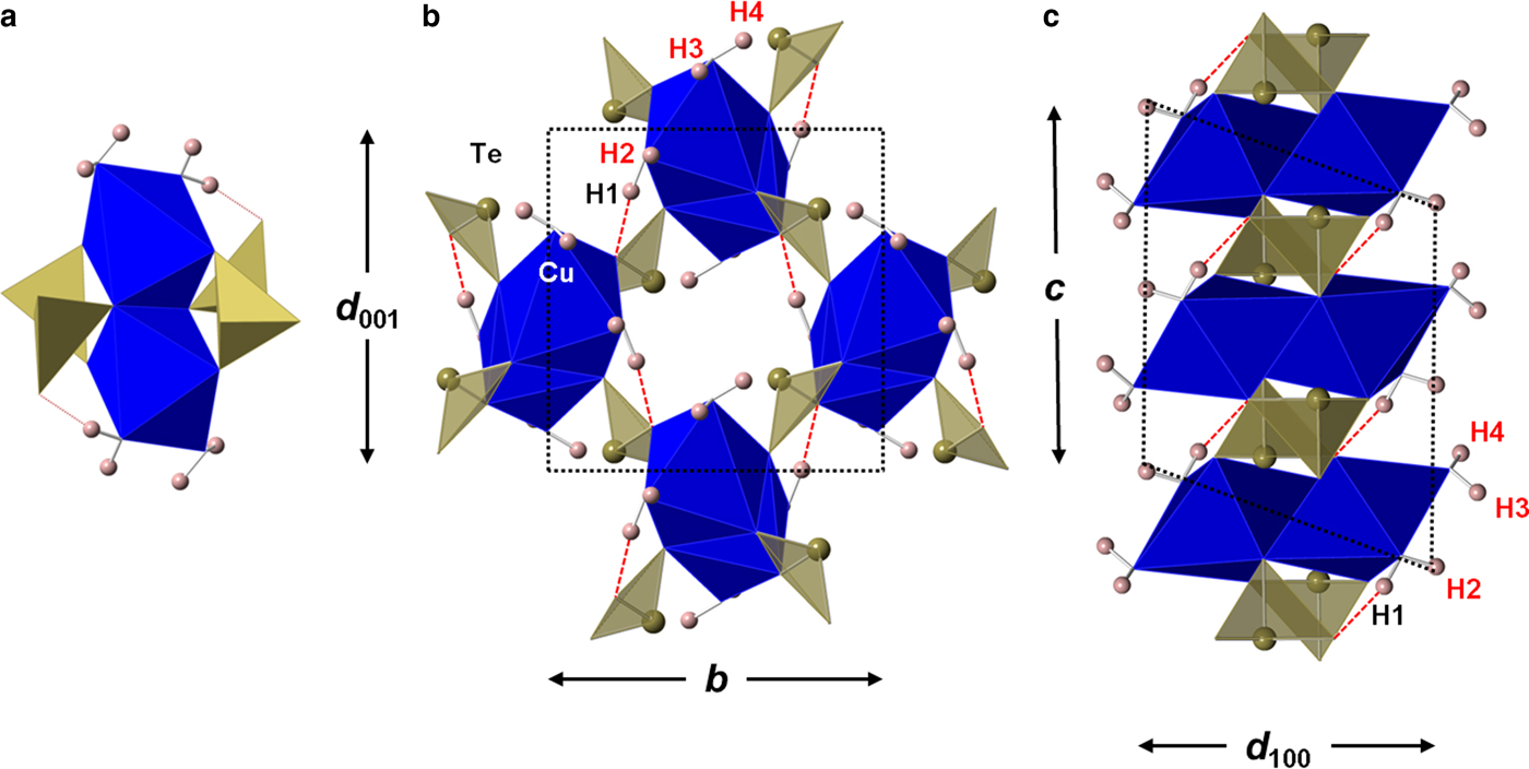
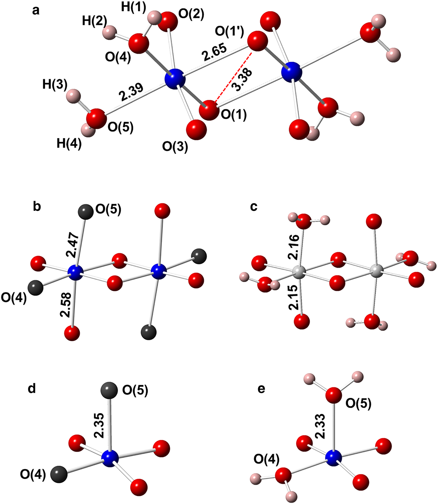
Structural features of millsite are shown in Figs 3–7. All atoms lie on general positions (4e). Cu forms four short, nearly coplanar, Cu–O bonds at 1.95–1.99 Å, one of which Cu–O(4) (1.99 Å) is to an H2O molecule. In addition to these short bonds there are two much longer bonds Cu–O(5)(H2O) at 2.387(2) Å and Cu–O(1') at 2.651(3) Å. These two long bonds lie trans with respect to the strongly-bonded planar CuO3(H2O) grouping. Thus, Cu2+ in millsite has a highly Jahn-Teller-distorted (prolate) octahedral coordination. A pair of CuO4(H2O)2 octahedra share a long edge (3.38 Å) to form a centrosymmetric Cu2O6(H2O)4 dimer (Figs 3 and 4). It is conceivable that the long Cu–O(5)(H2O) bond may be more easily broken than the stronger Cu–O(4)(H2O) bond, leading to the loss of ~1 H2O pfu during electron microprobe analysis mentioned above.
(a) The fundamental structural unit of millsite in which each Cu2O6(H2O)4 dimer is decorated with four TeO3 groups which connect to four adjacent dimers to form the heteropolyhedral sheet; (b) and (c) different views of the intersheet connectivity of millsite. H(1) forms an intrasheet hydrogen bond shown here as a dashed red line. Hydrogen atoms H(2,3,4) form intersheet hydrogen bonds.
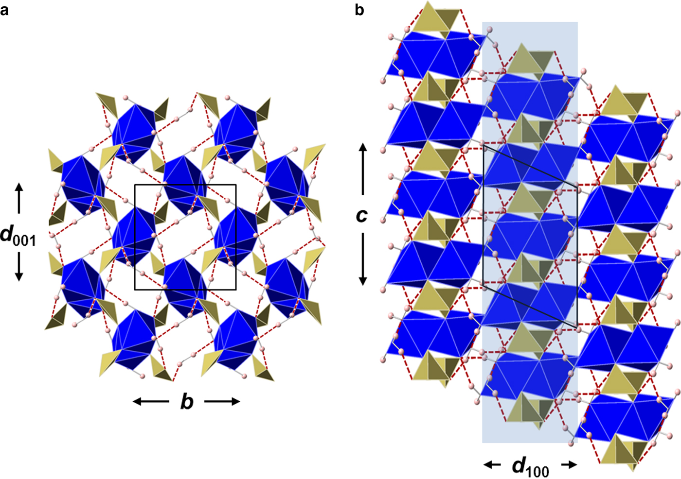
The extended millsite topology showing channels and intersheet connectivity, with a single heteropolyhedral sheet highlighted in (b). Hydrogen bonds shown as dashed red lines.
The  $M_2^{{\rm 2 +}} {\rm O}_6\left( {{\rm H}_2{\rm O}} \right)_4$ dimer units in (a) millsite, (b) monoclinic CuSeO3·2H2O, (c) ahlfeldite, (d) teineite and (e) chalcomenite. Only millsite has a sheet topology, whereas the others are frameworks. Teineite and chalcomenite do not have dimer units. Instead they have single CuO3(H2O)2 groups coordinated to three Te atoms. Water oxygens of teineite and chalcomenite are shown as black spheres. All distances are in Å.
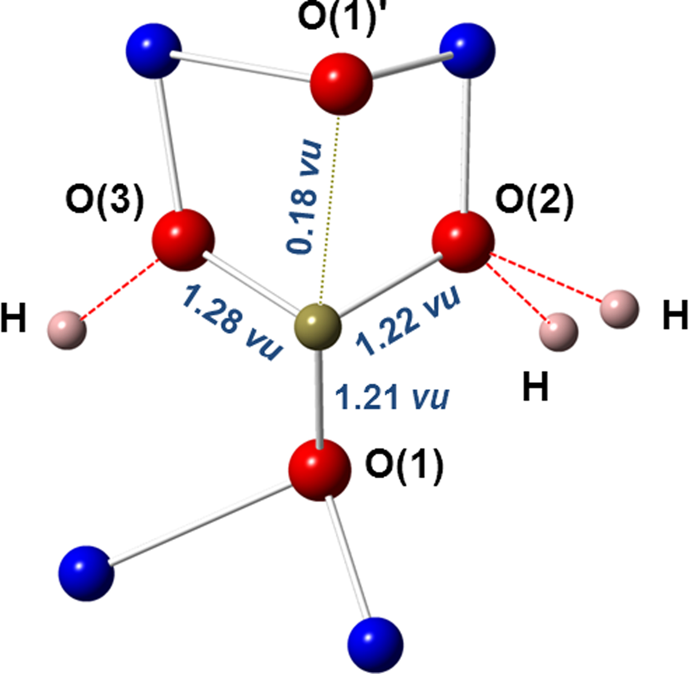
The bonding environment and bond valences of the TeO3 group of millsite. A fourth secondary Te−O(1)' bond contributing 0.18 vu lies on the lone-pair side of the Te atom.

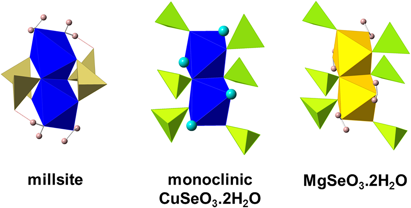
The fundamental structural units of millsite and the related structures of monoclinic CuSeO3·2H2O and synthetic MgSeO3·2H2O. Water oxygens of monoclinic CuSeO3·2H2O are shown as light blue spheres. CuO4(H2O)2 octahedra = blue, MgO4(H2O)2 octahedra = yellow, TeO3 = brown and SeO3 = green.
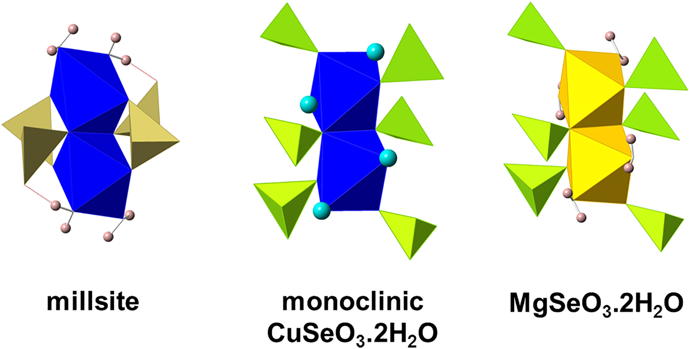
The Cu2O6(H2O)4 dimer is decorated with four TeO3 groups and it is terminated by a pair of H2O molecules at each end (Fig. 4), which prevent polymerization in those directions. Dimers are connected to each other via TeO3 groups and hydrogen bonding. The dimer−TeO3 connectivity defines a hetero-polyhedral sheet (100); the only connections between these sheets perpendicular to (100) are via hydrogen bonds. This topology underlies the (100) cleavage of millsite (Fig. 4b). The unit-cell volume of millsite is ~2.5% smaller than that of teineite, implying a denser packing of the sheet structure in the former.
The coordination of Te4+ is strongly asymmetric. Te lies at one apex of a TeO3 trigonal pyramid and forms three Te–O bonds at 1.86−1.88 Å, with three very similar bond angles at 94−99°. TeO3 groups are not polymerized.
As part of the mineral characterization supporting the first proposal submitted to the IMA in 2001 (IMA2001-000), a structure determination was carried out on a large crystal (415 µm × 180 µm × 130 µm) taken from the cotype specimen KAI-93. A 4-circle Huber diffractometer equipped with a point detector was used with graphite-monochromated AgKα radiation (λ = 0.56089 Å). A total of 4953 intensities which included reflections to a resolution of 0.885 Å–1 were collected, corrected for Lorentz-polarization, absorption and anomalous dispersion, and averaged to yield a total of 2253 reflections, R int = 0.0159. Refinement of all atoms converged at R 1(F) = 0.0236 and R w(F 2) = 0.0528 for all 2253 F, Goof = 1.154. A full structure report has not been published. To assess the agreement between the two structure determinations we have compared the nine (Cu, Te)–O distances and thirteen O–(Cu, Te)–O angles. The maximum and average differences in distances are 0.007 Å and 0.003 Å, respectively; for the angles the maximum and average differences are 0.22o and 0.09o, respectively. The largest differences are comparable to 2σ. On the basis of the crystallographic results, as well as on observations of other physical properties, it is clear that the crystals from specimens BM 2011,343 and KAI-93 are both millsite.
Hydrogen bonding in millsite
Hydrogen bonding is a structurally significant component of millsite (Figs 3 and 4). The refined positions of the four non-equivalent H atoms found in the structure determination allow hydrogen acceptors to be identified. Oxygens O(3) and O(4) each receive a hydrogen bond and O(2) receives two hydrogen bonds (one from each H2O molecule). The H(1) atom forms a hydrogen bond within its heteropolyhedral sheet. Atoms H(2,3,4) form hydrogens bonds between dimers of adjacent heteropolyhedral sheets.
Atoms O(2) and O(3) are under-bonded at 1.78 and 1.84 vu, respectively. Hydrogen bonds contribute additional bond valence to these two atoms. O(4) is different: it is part of a H2O molecule bonded to Cu at 1.99 Å and receives 0.43 vu from the Cu–O(4) bond. It also receives a hydrogen bond [O(4)···O(5) = 2.79 Å]. A very similar arrangement occurs in chalcomenite, for which the O···O distance is 2.91 Å. Robinson et al. (Reference Robinson, Sen Gupta, Swihart and Houk1992) calculate that the corresponding hydrogen bond in chalcomenite contributes 0.15 vu to the H2O oxygen. If a similar contribution is made to O(4) in millsite, then its bond-valence sum is 0.58 vu, implying a total value (with ~0.8 vu from each hydroxyl bond) of ~2.2 vu. This compares with 2.19 vu in chalcomenite.
We note that synthetic MgSeO3·2H2O, which is isostructural with monoclinic CuSeO3·2H2O (see Discussion), also has a hydrogen bond to the MgO4(H2O)2 group. However, the bond-valence of the Mg–O bond is 0.27 vu rather than 0.43 vu of the corresponding Cu–O bond in millsite and monoclinic CuSeO3·2H2O. Thus, it appears that ‘over-bonding’ of this H2O oxygen is characteristic of the CuO4(H2O)2 group, and a consequence of the unusual coordination.
Powder X-ray diffraction
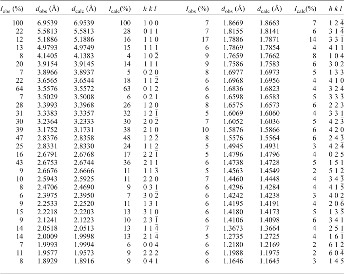
Due to the very small quantities of millsite available from specimen BM 2011,243, it was not possible to collect powder X-ray diffraction data for this sample. However, powder diffraction was carried out successfully on a small sample from the cotype specimen KAI-93 using synchrotron radiation with λ=0.79948 Å at the Swiss-Norwegian Beamlines, ESRF. Table 6 lists hkl, d hkl and I/I o values for millsite from sample KAI-93. Unit-cell parameters derived from the Rietveld refinement are shown in Table 7. Rietveld refinement of the powder data confirmed the identity of the powder as millsite. For small amounts of sample, a synchrotron beam provides a spatial (2θ) resolution of the diffraction peaks (very narrow, highly symmetric peak-shapes) that is superior to what can be achieved with radiation from a standard X-ray source.
Powder X-ray diffraction data for millsite sample KAI-93. Reflections with I/I max <5% are omitted.

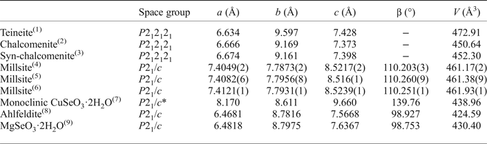
Unit-cell parameters of millsite and related phases.

(1) Effenberger (Reference Effenberger1977); (2) Pasero and Perchiazzi (Reference Pasero and Perchiazzi1989); (3) Robinson et al. (Reference Robinson, Sen Gupta, Swihart and Houk1992); (4) This study, holotype specimen BM 2011,243; (5) KAI-93, Mo et al. (2000); (6) This study, powder-diffraction of cotype specimen KAI-93; (7) Luo et al. (Reference Luo, Chen and Ma1984); (8) Larranaga et al. (Reference Larranaga, Mesa, Pizarro, Pena, Chapman, Arriortua and Rojo2005); (9) Johnston and Harrison (2001). *Transformed from the reported P21/n cell by ( $\bar 1$00/0
 $\bar 1$0/101).
Discussion
Millsite is a new mineral from Gråurdfjellet in Oppdal, Norway and is a monoclinic polymorph of teineite (orthorhombic, space group P212121). Chalcomenite, the selenite analogue of teineite also has a polymorph, monoclinic CuSeO3·2H2O (space group P21/c). However, no known tellurites or selenites are isostructural with millsite. Ahlfeldite NiSeO3·2H2O (Larranaga et al., Reference Larranaga, Mesa, Pizarro, Pena, Chapman, Arriortua and Rojo2005) is the Ni-analogue of monoclinic CuSeO3·2H2O; there are also synthetic Mg-, Zn- and Cd-analogues of monoclinic CuSeO3·2H2O (Johnson and Harrison, Reference Johnson and Harrison2001). The M 2+ coordination polyhedra of millsite, monoclinic CuSeO3·2H2O and MgSeO3·2H2O are shown in Fig. 7. Copper is five-coordinated in teineite and chalcomenite, forming single CuO3(H2O)2 square-pyramids that are not dimerized. In contrast, the M 2+ cation is octahedrally-coordinated in millsite, monoclinic CuSeO3·2H2O, ahlfeldite and synthetic MgSeO3·2H2O, with two octahedra sharing an edge to form an  $M_2^{{\rm 2 +}} {\rm O}_6\left( {{\rm H}_2{\rm O}} \right)_4$ dimer. Millsite is unique in having a different connectivity of its dimer: its two CuO4(H2O)2 octahedra are connected via the long O(1)−O(1') shared edge (3.38 Å), whereas in monoclinic CuSeO3·2H2O the connection is via a short shared edge (2.69 Å). The Cu−Cu distances in millsite and monoclinic CuSeO3·2H2O dimers are 3.21 Å and 2.98 Å, respectively.
The fundamental structural units of millsite, monoclinic CuSeO3·2H2O and synthetic MgSeO3·2H2O are shown in Fig. 7. The units of monoclinic CuSeO3·2H2O and synthetic MgSeO3·2H2O (and ahlfeldite) are isostructural and have six SeO3 groups decorating their Cu2O6(H2O)4 dimers. This arrangement differs from the four TeO3 groups decorating the dimer of millsite. In millsite, each TeO3 group has two Te–O bonds to the same dimer and a third bond to a second dimer, whereas in monoclinic CuSeO3·2H2O, ahlfeldite and MgSeO3·2H2O each Se atom forms a single Se–O bond to three different dimers. The different configuration of the dimer and its bonding to four TeO3 group leads to millsite having a sheet topology which is unique among tellurites and selenites.
Unit-cell parameters of these various related tellurites and selenites are shown in Table 7. Although millsite has the same space group as related monoclinic selenites (P21/c), it has distinctive unit-cell parameters. It is clearly distinguished from teineite by its unit cell, space group and the values of OH-stretching bands of water molecules in vibrational spectra (3288 cm–1/3367 cm–1 in millsite and 3040 cm–1/3495 cm–1 in teineite). While other M 2+ analogues with the teineite and monoclinic CuSeO3·2H2O structures may exist, millsite may be unique to Cu-bearing examples. The existence of a selenite analogue of millsite is a distinct possibility, and all the known species are worthy of consideration for a group nomenclature.
Acknowledgements
The NHM authors thank Roy Kristiansen, Henrik Friis (University of Oslo) and Knut Edvard Larsen for informing them about the history of the initial discovery of the material, the earlier IMA proposal and putting them in touch with the Norwegian authors. Tony Wighton of the NHM is thanked for specimen preparation for chemical analysis. The Norwegian authors are indebted to John Walmsley of NTNU for preparing samples for chemical analysis, and to Randi and Sigmund Rise of Oppdal for sharing with us mineral samples and their knowledge about the new mineral and details of its discovery.
Supplementary material
To view supplementary material for this article, please visit https://doi.org/10.1180/minmag.2017.081.057