Introduction
The neotropical region is considered one of the richest ichthyofauna worldwide. In this regard, neotropical fish species present ecological relevance in both aquatic and terrestrial ecosystems. Additionally, fish species are an important food source for most communities. Some species have great potential for aquaculture, which is a key point for the production of food, recommended by the World Health Organization in developing countries. Aquaculture is the fastest-growing sector worldwide with a total production of 82.1 million tons of fish in 2018, an 3.4% increase compared with 2017 (Winemiller et al., Reference Winemiller, Agostinho, Caramaschi and Dudgeon2008; Food and Agriculture Organization, 2020), but technologies for production are still under development.
In recent years, concern about environmental changes has increased the number of endangered fish species. Possibly, many species are threatened with extinction by non-natural causes such as habitat degradation, introduction of exotic species, and overfishing. Therefore, the preservation of genetic resources is necessary through methods such as gene banks and the reconstitution of unique genotypes (Holt and Pickard, Reference Holt and Pickard1999; Yasui et al., Reference Yasui, Fujimoto, Sakao, Yamaha and Arai2011).
Many species of the Prochilodontidae family are commonly used in aquaculture due to their rapid growth and adaptation to captive systems and artificial feeding. In addition, some species are considered endangered, such as Prochilodus vimboides, which has been categorized as Vulnerable (VU) (MMA/ICMBIO, 2018). Aquatic conservation and aquaculture are important strategies for both cases; therefore, it is necessary to establish studies with conservation strategies for genetic banks and reconstitution (Coelho et al., Reference Coelho, Yo, Mira-López, Monzani, Arashiro, Fujimoto, Senhorini and Yasui2019).
Different approaches have been established to conserve the genetic resources of fish species such as sperm banks by cryopreservation of semen (Cloud et al., Reference Cloud, Miller and Levanduski1990; Huang et al., Reference Huang, Dong, Walter and Tiersch2004; Yasui et al., Reference Yasui, Arias-Rodriguez, Fujimoto and Arai2009). Although this technique has been successfully applied to spermatozoa from many fish species, reliable methods for long-term preservation in oocytes and embryos are still challenging (Hagedorn and Kleinhans, Reference Hagedorn, Kleinhans, Tiersch and Mazik2000; Routray, Reference Routray2020). Another method often used to preserve these genetic resources is primordial germ cells (PGCs). After transplant, this cell line may originate spermatozoa and oocytes in the host gonads, as it has already been demonstrated in transplants for hosts during the early stages (Ciruna et al., Reference Ciruna, Weidinger, Knaut, Thisse, Thisse, Raz and Schier2002; Saito et al., Reference Saito, Goto-Kazeto, Arai and Yamaha2008, Reference Saito, Goto-Kazeto, Fujimoto, Kawakami, Arai and Yamaha2010) and larva (Yoshizaki et al., Reference Yoshizaki, Takeuchi, Kobayashi, Ihara and Takeuchi2002; Takeuchi et al., Reference Takeuchi, Yoshizaki and Takeuchi2003). Therefore, PGCs could support the transmission of genetic inheritance to future generations through germline chimera (Yoshizaki et al., Reference Yoshizaki, Tago, Takeuchi, Sawatari, Kobayashi and Takeuchi2005, Reference Yoshizaki, Takeuchi, Kobayashi and Takeuchi2003). Therefore, primordial germ cell transplantation can be applied for surrogate propagation (de Siqueira-Silva et al., Reference De Siqueira-Silva, Saito, Dos Santos-Silva, Da Silva Costa, Psenicka and Yasui2018), especially for P. lineatus and other rheophilic species.
The production of germline chimera in native species presents some bottlenecks, starting from induced spawning to subsequent in vitro fertilization. Our previous study with P. lineatus established a set of procedures for micromanipulation (Coelho et al., Reference Coelho, Yo, Mira-López, Monzani, Arashiro, Fujimoto, Senhorini and Yasui2019), which included the removal the chorion, embryo culture, and traceability of PGCs in vivo. However, the survival of cultured embryos decreased within a micromanipulation system that used Petri dishes. In addition, the transplantation of PGCs was difficult to achieve because the larvae were small sized and moved during micromanipulation. Therefore, a procedure to anaesthetise the larvae for micromanipulation and subsequent transplantation is necessary. Sterile embryos from characiform hosts were also produced by our group (Piva et al., Reference Piva, De Siqueira-Silva, Goes, Fujimoto, Saito, Dragone, Senhorini, Porto-Foresti, Ferraz and Yasui2018), which facilitates the production of a germline chimera.
Based on all these factors, the aim of this study was to establish a procedure for embryo manipulation, including the culture of dechorionated embryos in different incubation systems, determine the ideal anaesthetic concentration for hosts in the larval stage, and perform primordial germ cell transplantation for future use in endangered species.
Materials and methods
The experiment was carried at the National Center for Research and Conservation of Continental Aquatic Biodiversity of the Chico Mendes Institute for Biodiversity Conservation (CEPTA/ICMBio) in Pirassununga, São Paulo, Brazil. The experimental procedures were conducted following the Ethics Committee on the Use of Animals of the University of São Paulo, CEUA/USP No. 4534110820.
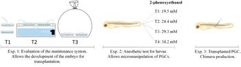
In this work, a series of experiments was conducted to achieve a germline chimera. Figure 1 shows the general view of the experiments, starting from incubation systems, anaesthetic for larvae, and subsequent transplantation of PGCs.
General procedure for embryo manipulation until the achievement of a Prochilodus lineatus germline chimera containing PGCs from Astyanax altiparanae. In the first series of experiments, incubation systems for dechorionated embryos of P. lineatus were evaluated. The best incubation system was used, hatched larvae were obtained, and then anaesthetic concentrations were evaluated to permit micromanipulation. The anaesthetised larvae received the PGCs from A. altiparanae and produced an interspecific germline chimera.

Induced spawning of streaked prochilod
All the reproductive procedures regarding broodstock selection, induced spawning, and incubation followed our previous procedure for the streaked prochilod (Yasui et al., Reference Yasui, Nakaghi, Monzani, Nascimento, Pereira dos Santos, Goes, Porto-Foresti and Senhorini2020). The broodstock were selected based on external secondary characteristics. Females present a rounded belly and a reddish in the urogenital papilla, and males have signs of reproduction and seminal release after gentle removal of the papilla area. After selection, reproduction was hormonally induced by using two doses of crude pituitary extract of common carp, Cyprinus carpio, which were applied at the base of the pectoral fin. Seven females received two doses: the first dose of 0.5 mg kg−1 and the second dose of 5 mg kg−1 8 h afterwards. Five males received a single dose of 5 mg kg−1 at the same time as the second dose for females. At 8 h after the second dose, the breeders were anaesthetised with clove oil (100 mg l−¹; Biodynamics, Brazil), and the gametes were collected by stripping. Oocytes were collected into a glass bowl without contaminants such as faeces, water, and urine. Similarly, the semen was collected in a 15 ml tube, and gamete activation was achieved with the immediate addition of water and manual homogenization.
Incubation of dechorionated embryos
After fertilization of P. lineatus, the embryos were divided into six aliquots (n = 3 intact with chorion and n = 3 dechorionated embryos). For chorion removal, a characin solution (12 mM NaCl, 1 mM KCl, 1.5 mM CaCl2 and 1.5 mM MgCl2) containing 0.05% pronase solution was used. Intact and dechorionated embryos were placed in 90-mm Petri dishes (∼50 embryos) and divided into three incubation systems: (1) control group with manual replacement of characin solution (every 1 h); (2) closed environment with high oxygen pressure and manual replacement of the solution; and (3) constant recirculation of the characin solution. The combinations resulted in six treatments. The recirculation of characin solution was performed with a pumping system in a 10-litre aquarium to the Petri dishes.
Embryo development was assessed using a stereomicroscope (Nikon SMZ 1500, Tokyo, Japan) connected to a charge-coupled device (CCD) camera (Ds-F1; Nikon, Tokyo, Japan) for observation at the main developmental stages: 2-cell, blastula, gastrula, somite, and hatching. After hatching, the percentages of normal and abnormal larvae were obtained based on the species morphological patterns.
Anaesthetic test for streaked prochilod larvae
Larvae of streaked prochilod at 1 dph were collected and then maintained individually in a 24-well plate containing 1000 µl of anaesthetic 2-phenoxyethanol at 19.5 mM, 24.4 mM, 29.3 mM, or 34.2 mM for 5 min. During this period, different parameters of anaesthesia were observed: induction of anaesthesia (numbness), heartbeat stopping, body curvature, and heartbeat frequency. Through this period, the larvae were checked for anaesthesia using a glass pipette to check the stages. The pipette was modified to avoid larva damage. After the anaesthesia, the larvae were collected and transferred to another plate containing water. Recovery was evaluated using the following criteria: recovery of movements, return of heartbeat, body recovery, heartbeat frequency, and survival. Heartbeat rate was measured using videos (1 min of observation).
Interspecific transplantation of PGCs
In this experiment, PGCs from Astyanax altiparanae were then transplanted into P. lineatus embryos using the incubation and anaesthesia procedure described above. First, the GFP-nos1 3′UTR mRNA was produced using the pCS2-GFP-nos1 3′UTR vector as a model, kindly provided by Professor Dr. Takafumi Fujimoto (Hokkaido University, Japan); this contained the Danio rerio nanos1 3′UTR mRNA (Köprunner et al., Reference Köprunner, Thisse, Thisse and Raz2001) fused with the coding region for green fluorescent protein (GFP). The vector was cleaved with the restriction enzyme NotI (IVGN0014, Anza™, Invitrogen™), and the mRNA was synthesized in vitro using a commercial mMESSAGE mMACHINE SPA transcription Kit® (AM 1340, Ambion®, Life Technologies™). The mRNA transcript was precipitated with LiCl and suspended in 200 mM KCl at the final concentration of 100 ng μl−1. The concentration and quality of the mRNA were measured on an QIAxpert spectrophotometer (Qiagen, Hilden, Germany).
Next, newly fertilized eggs of A. altiparanae were obtained using our previous protocol (Yasui et al., Reference Yasui, Senhorini, Shimoda, Pereira-Santos, Nakaghi, Fujimoto, Arias-Rodriguez and Silva2015). The embryos were dechorionated with 0.05% pronase dissolved in 20 ml of Holtfreter solution (NaCl 59.20 mM, KCL 0.67 mM, CaCl2 0.90 mM, NaHCO3 2.40 mM). After chorion digestion, the eggs were immediately transferred to 90-mm agar-plated Petri dishes. Dechorionated eggs were microinjected within the blastodisc at the 1–2-cell stage with GFP-nos1 3′UTR mRNA using a borosilicate micropipette (outer diameter 1 mm, and inner diameter of ∼2 µm) connected to a micromanipulator (Narishige M-152, Tokyo, Japan) and a microinjector (Eppendorf 5175. Cell Tram Vario, Hamburg, Germany). Injected eggs were maintained in Petri dishes at 28°C up to the blastula stage. At this stage, the blastoderm was detached from the yolk using a 25-gauge needle, therefore obtaining the blastosphere. The blastomeres were isolated by pipetting into 1.5 ml tubes containing 1 ml of Holtfreter’s solution with the addition of 0.25% sodium citrate (Sigma W302600, St. Louis, USA). The blastomere suspension was cultured in 100-mm Petri dishes containing Holtfreter’s solution for the later separation of GFP-positive cells.
At the 10-somite stage, GFP-positive cells (presumptive PGCs) were transplanted into P. lineatus larvae (1 dph) in the region of the gonadal ridges. A borosilicate micropipette of 20 μm inner diameter with a bevel of 30° was used.
After transplantation, the recipients were kept at 28°C in a biochemical oxygen demand (BOD) incubator; each PGC migration was observed under fluorescence stereomicroscopy (Nikon SMZ18, Tokyo, Japan) (excitation wavelength 470/40 nm) for such identification. Digital images were captured using a Nis-Ar Elements platform (Nikon, Tokyo, Japan). The hosts with transplanted GFP-positive cells were quantified. The percentages of survival and percentages of abnormal larvae were measured.
Results
Incubation of dechorionated embryos
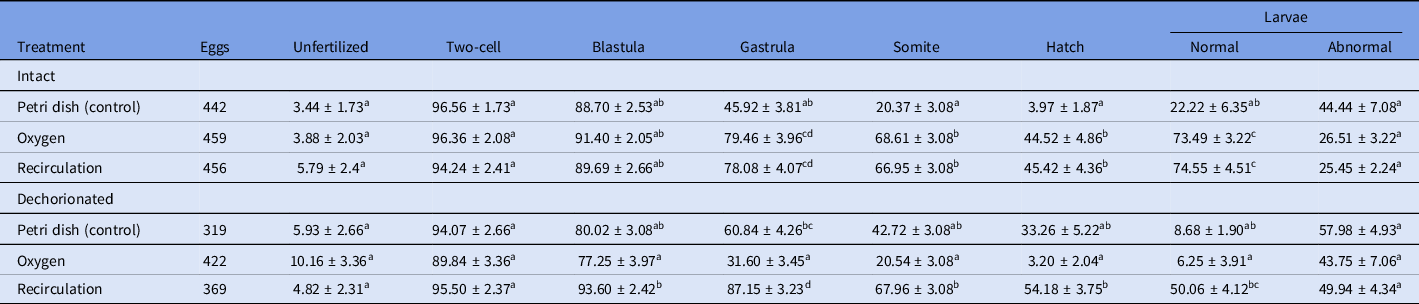
Different incubation systems influenced early development in both intact and dechorionated embryos (Table 1). The dechorionated embryos in the recirculation system had the highest hatching rate (54.18 ± 3.75%), although the intact embryos in the control group (static) had the lowest survival rates, with only 3.97 ± 1.87% at the hatching stage.
Early development of streaked prochilod Prochilodus lineatus embryos, maintained in different incubation systems. Intact (with chorion) and dechorionated embryos were maintained in Petri dishes and the main developmental stages evaluated. Data are shown as percentages ± standard error

a,b,cIdentical superscript letters within rows denote no significant differences by the Tukey’s multiple range tests (P > 0.05).
Survival rates decreased during the gastrula stage in all treatments. However, the lowest survival rates occurred with manual replacement of characin solution, in which there was no constant circulation of oxygen. Dechorionated embryos, maintained at high oxygen pressure, showed lower survival rates. Table 1 shows a higher percentage of abnormal larvae (43.75%) compared with other treatments. No significant difference was observed.
Higher percentages of normal larvae were found in intact and dechorionated embryos in the recirculation system with 74.55 ± 4.51% (P = 0.0101) and 50.06 ± 4.12% (P = 0.0642), respectively, when compared with the control group.
Anaesthetic test for streaked prochilod larvae
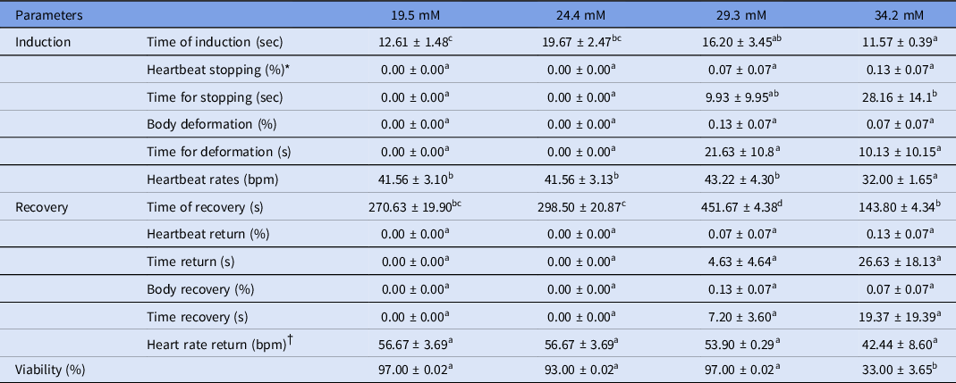
Table 2 shows the main parameters evaluated during anaesthesia of P. lineatus larvae. The time for anaesthesia induction in larvae ranged from 11.57 ± 0.39 s to 19.67 ± 2.47 s, and the larvae were anaesthetised within a similar period for all concentrations. However, a decrease in heartbeat rate was found at 34.2 mM 2-phenoxyethanol, an average of 32.00 ± 1.65 s (P = 0.0030) during anaesthetic induction.
Stages of induction and return of anaesthesia of streaked prochilod Prochilodus lineatus larvae submitted to different concentrations of anaesthetic 2-phenoxyethanol

a–dIdentical superscript letters within rows denote no significant differences by the Tukey’s multiple range test (P > 0.05).
* Heartbeat stopping indicates the percentage of larvae presenting heartbeat stopping.
† bpm, beating per minute.
As expected, the recovery period of the larvae submitted to the anaesthetic treatments increased gradually with the increase in the concentrations of 2-phenoxyethanol. However, the concentration of 34.2 mM produced the lowest survival of P. lineatus, presenting only 33% (Table 2). The concentration of 29.3 mM presented a return between 5 and 10 min with 451.67 ± 4.38 s (P = 0.8425). During the period of anaesthesia return, the results for the parameters of body curvature and heart rate were similar to those of induction. There was no significant difference within treatments.
Interspecific transplantation of PGCs
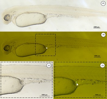
A single PGC of a A. altiparanae GFP-positive cell was transplanted in the region of the gonadal ridges of each P. lineatus larvae 1 day after hatching, totalling five transplants (Figure 2).
Transplantation of a single primordial germ cell from Astyanax altiparanae to Prochilodus lineatus larvae: (A, B) larva after transplant; (C, D) details of the highlighted region in (B).

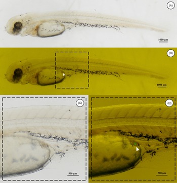
At 24 h after the procedure, GFP-positive cells were observed in two host embryos that remained in the peritoneal cavity in the transplanted area, but cells did not proliferate (Figure 3). On the following day, labelled cells were not identified. On the fourth day, the five transplanted P. lineatus larvae died.
Prochilodus lineatus larva transplanted with primordial germ cells from donors of Astyanax altiparanae: (A, B) larva at 1 day after the transplant; (C, D) detail of the highlighted region in (B).

Discussion
The procedure for micromanipulation of neotropical species was evaluated for the first time in dechorionated embryos. The results were optimized in the recirculation system for dechorionated embryos of P. lineatus regarding hatching and normal larvae, making it a positive result for germ cell transplantation.
The embryonic development duration was ∼15 h for the control treatments and 12 h for the recirculation and high oxygen pressure systems, measured from fertilization until larvae development. This information suggests that incubation systems may have influenced the early development at a constant temperature. In addition to being important in micromanipulation, temperature is an important parameter to determine the hatching period of intact and dechorinated embryos to synchronize the early development of donor and host species. The incubation temperature used in the recirculation system was 28°C, although Coelho et al. (Reference Coelho, Yo, Mira-López, Monzani, Arashiro, Fujimoto, Senhorini and Yasui2019) reported that P. lineatus embryos had a higher hatching rate at 26°C, with 67.3% and 58.8% of embryos intact and dechorionated, respectively. When evaluating the temperatures of 28°C and 24°C, Ninhaus-Silveira et al. (Reference Ninhaus-Silveira, Foresti and De Azevedo2006) achieved hatching after 14 h and 22 h, respectively.
In vitro culture of embryos from neotropical species is important for the development of laboratory studies, especially for the transplantation of PGCs. This emphasizes the importance of keeping the dechorionated embryos in manipulation laboratory systems in Petri dishes for microinjection, dissociation, and cell transplantation. In this study, our data strongly suggested that neotropical species present high oxygen demand during early development. The streaked prochilod spawns in high flow rate rivers during the peak of the rainy season. With a large perivitelline space, the demersal eggs permit mobility and incubation in open waters at a high oxygen availability. This suggests that incubation and maintenance in Petri dishes is difficult for embryos of such species in micromanipulation systems such as Petri dishes in static and limited oxygen waters. Indeed, other species whose micromanipulation was achieved in Petri dishes have been generally species with dense demersal eggs, in which natural spawning and incubation were achieved in static environments or for embryos that were incubated statically (Takeuchi et al., Reference Takeuchi, Yoshizaki and Takeuchi2001; Saito et al., Reference Saito, Pšenička, Goto, Adachi, Inoue, Arai and Yamaha2014).
Larval anaesthesia is also an important procedure that allows the performance of various procedures such as surgery (Murray, Reference Murray2002), transport (Parodi et al., Reference Parodi, Cunha, Becker, Zeppenfeld, Martins, Koakoski, Barcellos, Heinzmann and Baldisserotto2014), biometry (Hohlenwerger et al., Reference Hohlenwerger, Eduardo Copatti, Cedraz Sena, David Couto, Baldisserotto, Heinzmann, Caron and Schmidt2016), and transplantation in larvae (Bar et al., Reference Bar, Smith, Bubner, Yoshizaki, Takeuchi, Yazawa, Chen, Cummins and Elizur2016). This condition is characterized by a reduction in sensitivity that facilitates fish handling. In general, anaesthesia should be induced for a short period, preferably for less than 3 min and with minimal hyperactivity associated with low levels of stress and intoxication. In this work, the transplantation period between 5 and 10 min of anaesthesia was considered sufficient to accomplish approximately five transplants to larvae just exposed to the characin solution.
The induction and recovery times of the larvae were variable for the 2-phenoxyethanol doses as the larvae recovery period increased with the gradual increase in concentrations. Therefore, the concentration of 29.3 mM presented faster induction with 12 s and with recovery between 5 and 10 min. This result differs from previous studies with juveniles that reported that recovery time from anaesthesia was significantly shorter at higher concentrations of anaesthetic (Mylonas et al., Reference Mylonas, Cardinaletti, Sigelaki and Polzonetti-Magni2005; Toni et al., Reference Toni, Martos-Sitcha, Baldisserotto, Heinzmann, De Lima Silva, Martínez-Rodríguez and Mancera2015). This difference can be explained by the induction time of P. lineatus larvae, as they were exposed to the anaesthetic for 5 min, even with faster induction. However, immediately after induction, juveniles were removed from the anaesthetic and transferred to the water in other studies.
The survival rates observed in P. lineatus larvae after exposure to 34.2 mM anaesthetic suggest that the concentration of 29.3 mM of anaesthetic for this species cannot be exceeded. A dose of 29.3 mM presented a shorter induction period and recovery time of ∼10 min, which is considered sufficient for performing cell transplantation in larvae after exposure to the anaesthetic.
In the present study, PGCs from A. altiparanae were transplanted into the genital ridges of the anaesthetised P. lineatus. However, donor-derived germ cells were visualized 1 day after transplantation in only two individuals. In the following days, no cells were seen in the genital ridges of the host embryos. Although colonization of PGCs from GFP-labelled donors has not been observed in the genital ridges of hosts, Takeuchi et al. (Reference Takeuchi, Yoshizaki and Takeuchi2003) reported that PGCs transplanted into Oncorhynchus mykiss larvae may be evident on the 10th day after transplantation, and colonization efficiency was been traced for 30 days. However, Saito et al. (Reference Saito, Goto-Kazeto, Kawakami, Nomura, Tanaka, Adachi, Arai and Yamaha2011) revealed that the transplanted PGCs began to proliferate at the same age of development as the host PGCs, between 6 and 7 days in zebrafish. For the efficiency of PGC transplantation, correct cell migration and proliferation is necessary. However, in this study, success was not achieved in these procedures, suggesting that the recipient and/or stage used may be affecting such efficiency as they were not sterile. Furthermore, the time and conditions of PGC cultivation may be interfering with the viability and leading to changes in the expression level of genes related to the identity, migration, and proliferation of these cells, such as ddx4, dnd1, and nanos (Ciruna et al., Reference Ciruna, Weidinger, Knaut, Thisse, Thisse, Raz and Schier2002; Doitsidou et al., Reference Doitsidou, Reichman-Fried, Stebler, Köprunner, Dörries, Meyer and Raz2002; Nagasawa et al., Reference Nagasawa, Fernandes, Yoshizaki, Miwa and Babiak2013; Yön and Akbulut, Reference Yön and Akbulut2015; Rivers et al., Reference Rivers, Daly and Temple-Smith2020). Therefore, the production of germline chimeras still needs further study to improve the effectiveness of germ cell transplantation.
Conversely, Takeuchi et al. (Reference Takeuchi, Yoshizaki and Takeuchi2003) identified the larvae in salmonids as the best moment to perform the transplantation because the survival rate was higher (94%) when compared with embryos in the blastula and gastrula stages as hosts. In another study, the transplantation of PGC into larvae was performed in Oncorhynchus mykiss (Okutsu et al., Reference Okutsu, Suzuki, Takeuchi, Takeuchi and Yoshizaki2006), but only the transplantation of spermatogonial stem cells into larvae was carried out many times in different species (Morita et al., Reference Morita, Kumakura, Morishima, Mitsuboshi, Ishida, Hara, Kudo, Miwa, Ihara, Higuchi, Takeuchi and Yoshizaki2012; Farlora et al., Reference Farlora, Hattori-Ihara, Takeuchi, Hayashi, Octavera and Alimuddin2014; Bar et al., Reference Bar, Smith, Bubner, Yoshizaki, Takeuchi, Yazawa, Chen, Cummins and Elizur2016; Li et al., Reference Li, Fujii, Naito and Yoshizaki2017; Hattori et al., Reference Hattori, Yoshinaga, Katayama, Hattori-Ihara, Tsukamoto, Takahashi and Tabata2019).
In this study, a procedure for incubation, anaesthesia of larvae, and subsequent transplantation was established. This work is the first report regarding the transplantation of PGCs in Neotropical species. Several other parameters must be optimized to achieve a germline chimera. That information is an innovative, present application in basic and applied sciences for aquaculture and conservation of endangered genotypes.