Introduction
Cochlear implantation is a widely used surgical procedure for patients with sensorineural hearing loss.Reference Lima Júnior, Rodrigues Júnior Fde, Calhau, Calhau and Palhano1–Reference Sunkaraneni, Banerjee and Gray4 One of the major complications of cochlear implantation is intracranial haemorrhage, which may be life-threatening.Reference Sunkaraneni, Banerjee and Gray4–Reference Gosepath, Maurer and Mann9 If the surgeon is not aware of this complication, the diagnosis may be delayed, increasing the risk of death.Reference Benson6 Progressive accumulation of experience in cochlear implantation has enabled different methods of implant housing to be developed. Other authorsReference Venail, Sicard, Piron, Levi, Artieres and Uziel10–Reference Molony, Giles, Thompson and Motamedi13 have previously reported that they have abandoned bony tie-down sutures in preference to alternative implant housing techniques, in order to facilitate safer implantation.
Here, we report the fourth published case of subdural haematoma, and the eighth published case of intracranial haemorrhage, encountered following cochlear implantation. Possible mechanisms of injury are discussed and the standard methods of cochlear implantation are questioned, following further study of the current literature. The need for alternative implant housing techniques is suggested.
Case report
A three-year-old boy was admitted to the emergency department with loss of consciousness. He had a Glasgow coma score of 8 and dilatation of the right pupil. Systemic examination and laboratory findings were otherwise unremarkable.
One week previously, the child had undergone right-sided cochlear implantation for bilateral sensorineural hearing loss, at another hospital. We learned that, during this surgery, cerebrospinal fluid had been encountered and bleeding had occurred following drilling of the receiver bed. Bone wax had been applied for haemostasis.
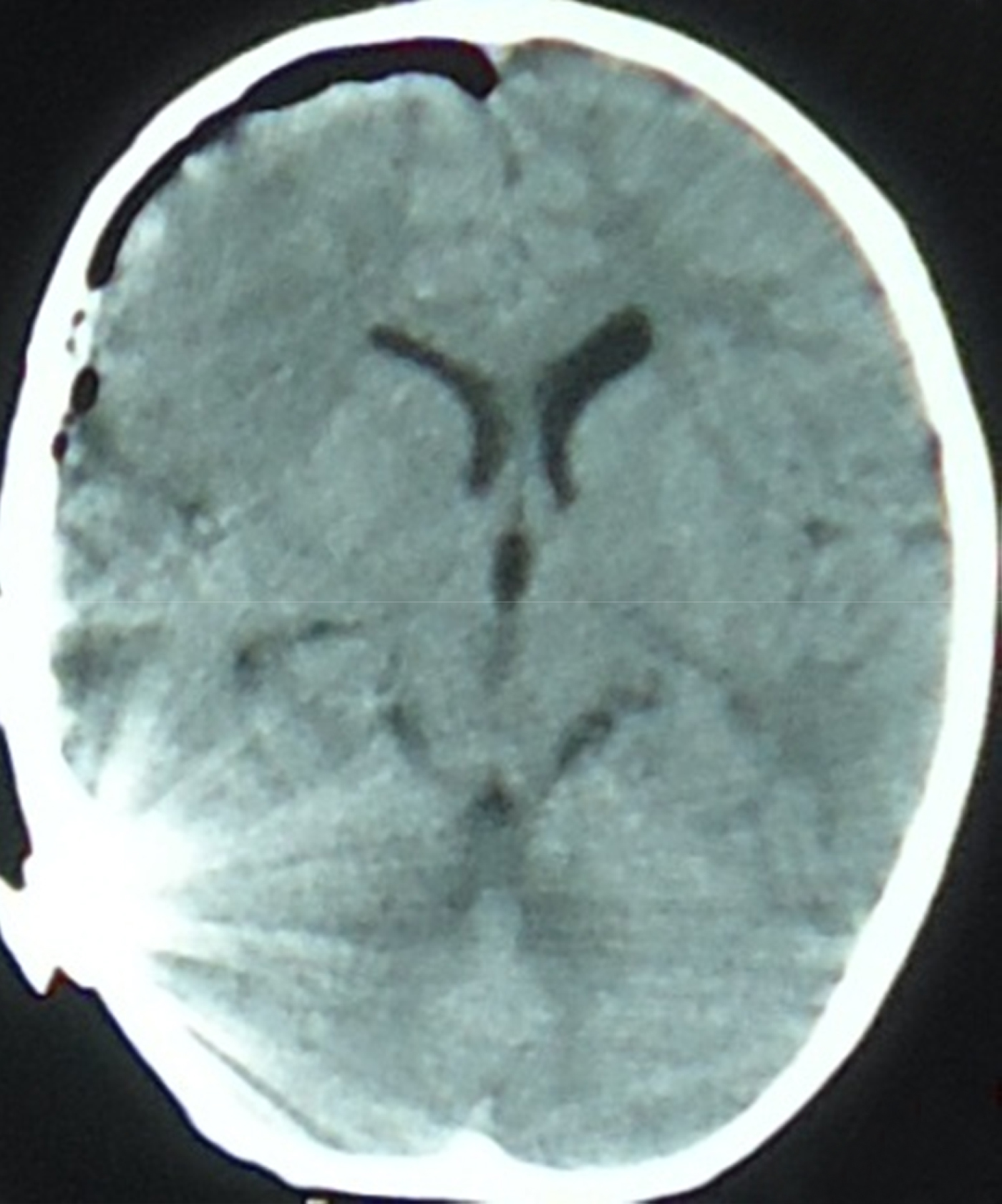
A cranial computed tomography (CT) scan revealed a large right frontoparietal subdural haematoma compressing the right cerebral hemisphere and resulting in a 20 mm midline shift (Figure 1).
Axial cranial computed tomography image taken on admission, showing a large right frontoparietal subdural haematoma compressing the right cerebral hemisphere and resulting in a 20 mm midline shift. Cochlear implant artefact is also seen.

The patient was immediately taken to the operating theatre and a subdural haematoma was evacuated via a right frontoparietal craniotomy. Craniotomy was performed in a manner which avoided harm to the cochlear implant device. The subdural clot, approximately 90 ml in volume, was located mainly posterior to the implant device over the right parietal region. After complete evacuation of the clot, we identified bleeding from an inferior anastomotic vein (vein of Labbé) beneath the device receiver. Appropriate haemostasis was achieved.
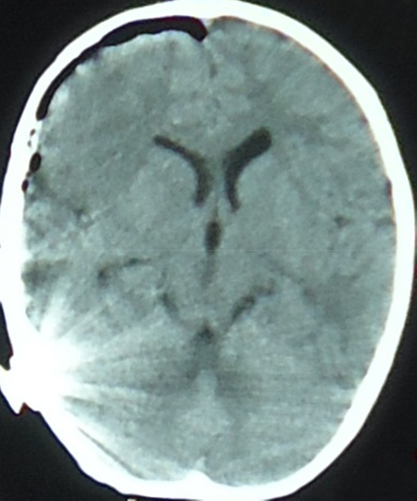
A post-operative cranial CT scan revealed that the haematoma had been evacuated successfully (Figure 2).
Axial cranial computed tomography image taken after right frontoparietal craniotomy, showing successful evacuation of the haematoma and disappearance of the midline shift. Cochlear implant artefact is also seen.
The post-operative course was entirely uneventful, and the patient was discharged from hospital without any neurological deficit.
Discussion
Cochlear implantation is a safe and effective surgical procedure with relatively low complication rates.Reference Lima Júnior, Rodrigues Júnior Fde, Calhau, Calhau and Palhano1, Reference Benson6, Reference Dodson, Maiberger and Sismanis7, Reference Gosepath, Maurer and Mann9, Reference Venail, Sicard, Piron, Levi, Artieres and Uziel10 Complications are divided into minor and major types. The prevalence of minor complications is 1–25.4 per cent and that of major complications is 2.1–11.7 per cent.Reference Stamatiou, Kyrodimos and Sismanis3, Reference Dodson, Maiberger and Sismanis7, Reference Venail, Sicard, Piron, Levi, Artieres and Uziel10 One of the major, life-threatening complications is intracranial haemorrhage. The prevalence of this extremely rare complication has been variously reported as 0 per cent,Reference Lima Júnior, Rodrigues Júnior Fde, Calhau, Calhau and Palhano1, Reference Loundon, Blanchard, Roger, Denoyelle and Garabedian2, Reference Venail, Sicard, Piron, Levi, Artieres and Uziel10 0.28 per centReference Dodson, Maiberger and Sismanis7 and 0.5 per centReference Stamatiou, Kyrodimos and Sismanis3 in different series.
Intracranial haemorrhage may occur in epidural, subdural or intracerebral locations. These complications may be fatal if not diagnosed and treated early. Seven cases of intracranial haemorrhage occurring after cochlear implantation have been reported so far.Reference Stamatiou, Kyrodimos and Sismanis3–Reference Gosepath, Maurer and Mann9 Two cases were epidural,Reference Barraclough, Pearman and Solanki5, Reference Gosepath, Maurer and Mann9 two were intracerebralReference Benson6, Reference Hagr and Bance8 and the remaining three were subdural haematomas.Reference Stamatiou, Kyrodimos and Sismanis3, Reference Sunkaraneni, Banerjee and Gray4, Reference Dodson, Maiberger and Sismanis7 Our patient represents the eighth case of intracranial haemorrhage, and the fourth case of subdural haematoma, reported after cochlear implantation.
Gosepath et al. Reference Gosepath, Maurer and Mann9 reported a case of epidural haematoma which became apparent 4 hours after cochlear implantation. They commented that this haematoma had possibly been caused by bleeding from bone or extradural veins, and emphasised that, if placement of the implant housing is associated with considerable bone work, epidural haematoma must be considered as a rare but life-threatening complication. Barraclough et al. Reference Barraclough, Pearman and Solanki5 reported another case of epidural haematoma noted within 12 hours of cochlear implantation, probably caused by trauma to the posterior parietal branch of the middle meningeal artery. In both of these particular cases, no dural injury was mentioned. Absence of dural exposure or injury should not rule out intracranial haemorrhage, as exemplified by these two cases.Reference Barraclough, Pearman and Solanki5, Reference Gosepath, Maurer and Mann9
Due to the potential risk of intracranial complications, Sunkaraneni et al. Reference Sunkaraneni, Banerjee and Gray4 reported that they had abandoned tie-down sutures in preference to an appropriately deep well with squared-off rims, and had subsequently witnessed no further complications of this nature. However, in patients with thin bones this technique may not be applicable, and thinness of the skull may necessitate dural exposure.Reference Barraclough, Pearman and Solanki5
Hagr and BanceReference Hagr and Bance8 reported a case of intracerebral haematoma seen immediately after cochlear implantation, and emphasised that the dura had been encountered and dural bleeding had occurred after drilling of the bone. They did not use anchoring sutures, but they believed that bleeding had occurred following drilling of the receiver bed.
BensonReference Benson6 reported another case of intracerebral haematoma diagnosed on the fourth day after cochlear implantation which had been completed using the standard method including tie-down sutures. During surgery, no dura had been exposed and no bleeding had occurred. However, this author commented that elevated blood pressure and straining with urination had probably caused previously sheared diploic vessels to open in a delayed fashion. It was suggested that new products be developed to eliminate the need for cortical fixation, in order to enable safer implantation.
Dodson et al. Reference Dodson, Maiberger and Sismanis7 reported a case of subdural haematoma detected immediately after cochlear implantation, in the recovery room. They believe that this haematoma had occurred secondary to opening of a diploic vein traversing the dura into the parietal cranium. Stamatiou et al. Reference Stamatiou, Kyrodimos and Sismanis3 reported another case of subdural haematoma seen within 24 hours of cochlear implantation, which they believed had occurred due to bipolar cauterisation of a prominent diploic vein. Sunkaraneni et al. Reference Sunkaraneni, Banerjee and Gray4 reported a case of subdural haematoma which had occurred one week after cochlear implantation, and suggested that it had been caused by bleeding from emissary veins opened by drilling. As in our case, these complications related to drilling of bone. Our patient's subdural haematoma was possibly due to direct injury to a superficial cortical vein which was anatomically located under the dura. In order for this type of injury to occur, the drill needs to penetrate the dura and pierce an underlying vein.
In cochlear implantation surgery, intracranial haemorrhage usually occurs after drilling the bone.Reference Stamatiou, Kyrodimos and Sismanis3, Reference Sunkaraneni, Banerjee and Gray4, Reference Benson6–Reference Gosepath, Maurer and Mann9 To avoid bone work related complications, different methods of device fixation have been developed which do not require drilling of bone. Molony et al. Reference Molony, Giles, Thompson and Motamedi13 compared the standard method and an alternative: periosteally placed tie-down sutures. They reported the prevalence of major complications as 4.1 per cent for the standard technique and 0 per cent for the alternative technique. No case of device migration was reported in their study, during a mean follow up of 2.4 years. Adunka et al. Reference Adunka and Buchman14 described another simple and effective implant fixation technique using periosteal sutures, in children, and reported no complications.Reference Adunka and Buchman14 Loh et al. Reference Loh, Jiang, Dezso and Fitzgerald O'Connor12 used a non-suture method of securing the cochlear implant by creating a bony groove, in children older than 18 months, and reported no implant migration. However, this technique may fail in some patients due to lack of thickness of cortical bone.Reference Barraclough, Pearman and Solanki5, Reference Loh, Jiang, Dezso and Fitzgerald O'Connor12 In another study,Reference Boscolo-Rizzo, Muzzi, Barillari and Trabalzini11 the receiver-stimulator of the cochlear implant was secured to the skull by a tailored flap of periosteum, without any bone work; no migration was reported during 48 months of follow up. Alexander et al. Reference Alexander, Caron and Wooley15 secured the implant using a tight periosteal pocket, placing the suture through the periosteum and soft tissue to collar the receiver in a modified well; they observed no complications with regard to device migration or extrusion.
Regarding the time course of intracranial haemorrhage development, there seems to be no association between haemorrhage and performance of cochlear implantation as a day-case procedure, since five cases were diagnosed within 24 hours, one on the fourth day and two on the seventh day after surgery.
• Intracranial haemorrhage is a rare but life-threatening complication of cochlear implantation
• A case is reported, and previous cases reviewed regarding causation
• Intracranial haemorrhage after cochlear implantation always occurs after drilling bone
• Non-drilling techniques may help to avoid this complication
Although there are no data available on the cosmetic results of these new surgical techniques, it seems that these procedures do not increase the risk of device migration or extrusion.
Conclusion
Cochlear implant surgeons must be aware of potential intracranial complications, and should question the current method of drilling. Drilling bone, in order to anchor sutures or to excavate the receiver bed for implant housing, increases the risk of intracranial haemorrhage, which has a high mortality rate. Alternative techniques may be considered to avoid this rare but life-threatening complication, always with due respect for the reliability and safety of such techniques.