INTRODUCTION
The Gastrotricha is a moderately sized taxon (~800 spp.) of microscopic (0.1–3.5 mm) marine and freshwater invertebrates characteristic of nearly all water bodies across the globe. In the oceans, gastrotrichs are well-known members of the benthic meiofauna and often recognized by their elaborate cuticle and the presence of numerous adhesive tubes. The Thaumastodematidae is the largest family of marine Gastrotricha in the order Macrodasyida, with more than 130 species in eight genera distributed worldwide (Todaro et al., Reference Todaro, Kånneby, Dal Zotto and Jondelius2011). The etymology of Thaumastodermatidae translates to ‘wonderful skin’ in reference to the elaborate cuticular scales and spines present in nearly every species of the family (Remane, Reference Remane1927). Tetranchyroderma is the most speciose genus and was originally named for the presence of tetrancres or 4-pronged spines (ancres) that characterized the earliest species descriptions (Remane, Reference Remane1926; but see Hummon & Todaro, Reference Hummon and Todaro2010). Since then, species with three-pronged spines (triancres), five-pronged spines (pentancres), and in some cases, a mixture of spine types (Lee & Chang, Reference Lee and Chang2014) have been described from across the genus. To date, five species in two genera, Tetranchryoderma and Pseudostomella, have been described with feathered triancres, which are flattened 3-pronged spines that are similar in outline to a bird's feathers (Ruppert, Reference Ruppert1970; Hochberg, Reference Hochberg2002, Reference Hochberg2008; Lee & Chang, Reference Lee and Chang2014). Herein, we describe the first species to bear two-pronged, feathered, spines (biancres) along with a unique combination of three-pronged feathered spines, and two other ordinary triancres types.
MATERIALS AND METHODS
Sediments were collected in June 2013 and July 2015 from an offshore sandy bottom located ~7 km east of the Fort Pierce Inlet, Florida. The Smithsonian Marine Station's RV ‘Sunburst’ was used to drag a small anchor dredge (29 × 12 cm opening) at ~12 m depth (Dredge in: 27°28.79′N 80°13.69′W; Dredge out: 27°29.08′N 80°13.76′W) for 15 min. Samples were placed into buckets and examined back at the Smithsonian Marine Station in Fort Pierce, Florida over the course of 5 days each time. Meiofauna was extracted from the sediments using the anaesthetization – decantation technique with isotonic MgCl2 (Pfannkuche & Thiel, Reference Pfannkuche, Thiel, Higgins and Thiel1988) and a 53 µm mesh sieve. Animals were sorted under a Leica EZ4 stereo microscope and subsequently mounted alive on glass slides in isotonic MgCl2. Individual specimens were examined with a Zeiss A1 compound microscope equipped with DIC and a Sony Handycam digital video camera. Measurements were taken with an ocular micrometer on relaxed specimens. Photographs and videos were recorded with a Sony Handycam digital camera. Lengths and positions of organ systems are described in terms of percentage body units, where total body length from anterior (U00) to posterior (U100) is 100 units.
Four specimens were fixed in 4% paraformaldehyde in 0.1 M phosphate buffer saline (pH 7.2) for at least 1 week. Specimens were then rinsed in PBS and stained with Alexa Fluor 488 phalloidin (Life Technologies) to document the musculature. Stained specimens were briefly rinsed in PBS before mounting in Fluoromount G (Southern Biotechnology Associates, Birmingham, AL) on glass slides. An Olympus FV 300 confocal laser-scanning microscope was used to visualize the specimens. An Argon laser (488 nm) was used to excite the samples, and Olympus software was used to capture the images. Confocal z-stacks were collected and processed as .TIF files and .MOV video files. Files were further processed with Volocity (Perkin Elmer) to generate z-projections. The specimen with the best preservation was selected as the holotype specimen and further processed to make a permanent slide.
Two specimens were fixed in 2.5% glutaraldehyde in 0.1 M sodium cacodylate buffer (pH 7.4) for several days prior to processing. Specimens were then rinsed in 0.1 M buffer for 1 h, postfixed in 1% OsO4 in 0.1 M buffer for 1 h, and then rinsed again in buffer prior to a standard ethanol dehydration series. Specimens were next critical point dried (Tousimis Samdri-795) and coated with gold using a sputter coater (Denton Vacuum Desk IV) prior to examination with an Amray 1400 SEM at the University of Massachusetts Lowell.
Sediments collected from the dive site were brought back to the University of Massachusetts Lowell and analysed for granulometry. Sediments were dried in an oven at 100°C for 24 h and then sorted on Gilson SS-15 sieve shaker with mesh sizes of 2, 1, and 500, 250, 125 and 63 µm. Sediment fractions were weighed and granulometric characters (mean, standard deviation (SD), skewness, kurtosis, median) were calculated using GRANPLOTS with line segments (Balsillie et al., Reference Balsillie, Donoghue, Butler and Koch2002).
Terminology for the description of the species follows that of Todaro (Reference Todaro2002).
SYSTEMATICS
Order Macrodasyida Remane, 1925 (Rao & Clausen, Reference Rao and Clausen1970)
Family THAUMASTODERMATIDAE Remane, 1926
Subfamily THAUMASTODERMATINAE Remane, 1927
Genus Tetranchyroderma Remane, 1926
Tetranchyroderma garraffonii sp. nov.
(Figures 1–5)
Zoobank lsid:zoobank.org:act:E5673944-347A-431B-B4BA-D49B4F0D9611
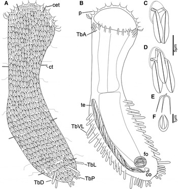
Tetranchyroderma garraffonii sp. nov.: (A) Schematic of dorsal view; (B) Schematic of ventral view; (C) Schematic of feathered biancre; (D) Schematic of feathered triancre; (E) Schematic of ‘pitchfork’ triancre; (F) Schematic of bowl-shape triancre. cet, cephalic tentacles; co, caudal organ; ct, cirrata tube; fo, frontal organ; p, papillae; TbA, anterior adhesive tubes; TbD, dorsal adhesive tubes; TbL, lateral adhesive tubes; TbP, posterior adhesive tubes; TbVL, ventrolateral adhesive tubes; te, testis.

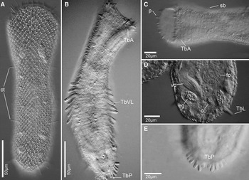
Tetranchyroderma garraffonii sp. nov.: (A) Dorsal view of a live adult; (B) Ventral view of a live adult; (C) Ventral view of the anterior part of the body; (D) Focus on reproductive organs at caudal end; (E) Focus on posterior adhesive tubes. co, caudal organ; ct, cirrata tube; eg, epidermal glands; fo, frontal organ; p, papillae; TbA, anterior adhesive tubes; TbL, lateral adhesive tubes; TbP, posterior adhesive tubes; TbVL, ventrolateral adhesive tubes; vd, vas deferens.

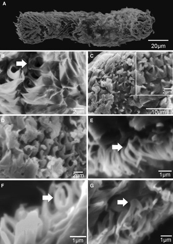
Tetranchyroderma garraffonii sp. nov.: SEM images of the cuticle. (A) Dorsal view of body; (B) Feathered ancres, arrow reveals two shafts connect by a ridge; (C) Focus on a feather scale base. Inset: Close-up of a biancre scale base. (D) Broken shafts of feathered scales; (E) ‘Pitchfork’ triancre; (F) Bowl-shape triancre; (G) Possible feathered biancre on lateral side of body.

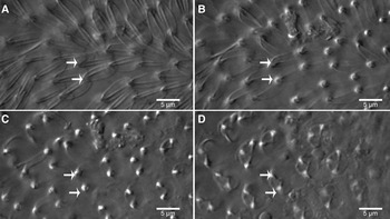
Tetranchyroderma garraffonii sp. nov.: DIC photomicrographs of feathered biancres (arrows) and triancres on the dorsal side of the body at different focal planes. (A, B) Focus on feathered scales; (C, D) stakes and bases.

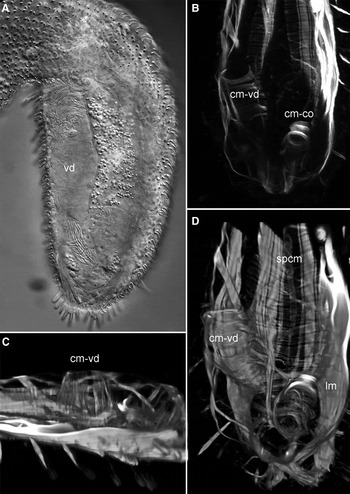
Tetranchyroderma garraffonii sp. nov.: (A) DIC photomicrographs of ventral view of caudal end, slightly squashed; (B–D) Confocal photomicrographs of phalloidin-stained specimen showing the muscular system of the posterior end: (B) Musculature as viewed from the ventral side; (C) Volocity-rendered view of the circular muscles from the lateral view (anterior to the left); (D) Volocity-rendered view of the circular muscles from the ventral side. cm-co, caudal organ circular muscles; cm-vd, vas deferens circular muscles; lm, longitudinal muscles; spcm, splanchnic circular muscles; vd, vas deferens.

TYPE MATERIAL
Holotype: 273 µm adult mounted in resin, USNM 1254654.
Paratypes: Two adults mounted on SEM stub, USNM 1254655; 1254656.
Zoobank access: http://zoobank.org/urn:lsid:zoobank.org:pub:E6C1976C-CCCB-41E5-AC5B-52B02AC0BA8C
Type locality: Bare sand, 12 m depth; sediment characteristics: mean, 1.3771 phi; SD, 1.1593; skewness, – 0.8047; kurtosis, 2.9128; median, 1.6924 phi.
Etymology: The species is named in honour of Dr André Garraffoni, the first author's (TQA) advisor, who has inspired TQA to work with this incredible taxon.
DIAGNOSIS
Specimens with body lengths of 212–287 µm long (mean 267.5 µm, n = 11). Maximum body width at mouth, neck, U50 and caudal end of body is 55/53/52/77 µm. Pharynx to 103 µm long (measured from the tip of the oral hood); oral hood to 31 µm long and fringed with papillae; mouth to 55 µm wide. Small cephalic tentacles present. Cuticle is covered with a mix of feathered biancres and feathered and ordinary triancres on dorsal and lateral surfaces. Epidermal glands present. Six TbA/side insert directly on the body surface posterior to the mouth rim. TbVL elongate, up to 19 per side. One TbL per side at U92 and one TbD inserted on posterior end at U96. Six-seven TbP per side, 2 of which form short caudal pedicles, 1 flanks the pedicle medially and 3–4 flank the pedicle laterally. Four cirrata tubes per side at U3/U37/U68/U95. Locomotory cilia form transverse rows on the ventral surface from the mouth margin to the caudum. Hermaphroditic, with paired posterior ovaries and a single testis on the right side as seen from the ventral side; egg dorsal at mid-body length; muscular caudal and sac-like frontal organs present.
DESCRIPTION
The type description is based on an adult specimen of 273 µm total body length (Holotype, Figures 1 and 2B), with ranges provided by additional adults. Oral hood to 31 µm long from mouth margin to tip of hood. Dorsal rim of hood (~2 µm) is smooth and without ornamentation. Sensory cilia to 6 µm long present at transition of scaled cuticle and smooth cuticle. Additional scattered sensory cilia, 6 µm long, emerge from small papillae at margin of hood. Numerous sensorial bristles to 12 µm are present along the lateral margins of the body. Cephalic tentacle present on either side of the hood and close to the base of the smooth cuticle; lateral head cilia up to 20 µm long surround the cephalic tentacle. Pharynx is 103 µm in length from tip of oral hood to PhIJ. Body width at mouth, neck and U50 are 55/40/52 µm, respectively.
Cuticle. Body covering of feathered biancres and feathered and ordinary triancres (Figures 1C and D, 3B–G, 4). All feather-like scales are flattened with a median rib. Biancres were not parallel but instead oriented slightly toward each other, often with the posterior edges touching or overlapping (Figures 1C, 3B, 4). Triancre scales appeared to be mostly parallel (Figures 1D and 4). The outline of most feathers was generally smooth when visualized with DIC microscopy (Figure 4), but with SEM, the individual ancres appeared to have a somewhat folded outline, which is probably an artefact of fixation or dehydration (Figure 3). Individual ancres are 3–8.5 µm long. Each ancre in a series (bi- or triancre) connects to a rounded scale base (1.8–5 µm) via a short shaft (Figure 3C and D); biancres have two shafts and triancres have three shafts. Each shaft arises from a thickened ridge of the scale base; biancre ridges form a V-shape (ends of the ‘V is where the shafts arise) (Figure 3B–D) and triancre-ridges form an upside-down T-shape (edges of the ‘T’ is where the shafts arise).
The ordinary triancres are present on the lateral surfaces of body and could only be observed under SEM, i.e. DIC microscopy was not sufficient to distinguish these ancre types, Two different triancre shapes were present (Figures 1D and E, 3E and F): The first ancre is bowl-shaped with a ~1.9 µm long (base 0.8 µm) (Figures 1F and 3G) and with curved tines almost touching each other at their tips. The second type of ordinary triancre has a ‘pitchfork’ shape with needle-like tines that project outward ~0.6 µm and then backward ~1.4 µm. The tines are connected directly to the body of scale by a small pedunculum ~0.5 µm long (Figures 1E and 3E).
Cirrata tubes. Four cirrata tubes to 10 µm long are present on the dorsolateral region of the body at U3/U37/U68/U95. Cirrata tubes have tiny spherical secretions on the inside and are distinct in appearance from the adhesive tubes.
Adhesive tubes. Adhesive tubes are present in five series: TbA, TbVL, TbL, TbDL and TbP. Anterior adhesive tubes (TbA) form a transverse row beneath the ventral mouth margin and arc slightly toward the lateral body margin. Six TbA per side, inserting directly on body surface, and somewhat staggered in placement. The most medial tubes are ~10 µm long (7.3–10 µm). The ventrolateral adhesive tubes (TbVL) are 6–23 µm long and extend from ~U48 to the caudal end U86. All adult animals show 19 tubes per side in the trunk region. A bilateral pair of dorsolateral adhesive tubes (TbDL), 23–25 µm long, was present at U94, and a bilateral pair of lateral adhesive tubes (TbL), 16 µm long, was located at U92. Posterior adhesive tubes (TbP) form two bilateral groups of 6–7 tubes on the caudum: 3–4 individual tubes insert directly on the lateral body wall next to a short pedicle that bears two tubes and one additional tube flank medially the pedicle. Most tubes are ~9 um long, with the medial tube of pedicles only reaching 6–7 µm.
Ciliature. The ventral ciliature forms a continuous series of transverse rows from the ventral mouth margin to the posterior end. Just beyond the opening to the caudal organ/anus region around U92 is another transverse row of active cilia.
Digestive tract. The digestive tract begins with a wide mouth (50–55 µm) with a mostly smooth margin and covered by an oral hood. The mouth leads to the pharynx that narrows to ~22 µm at the PhIJ (U37). Pharyngeal pores present at U32. The intestine is narrow to broad depending on its contents. Several specimens possessed diatom frustules in their guts. The anus opens ventrally at about U91.
Reproductive anatomy. Simultaneous hermaphrodites. A single testis is present on the right side (as seen from the ventral side) with the anteriormost portion close to the PhIJ; a vas deferens extends posteriorly to the base of the caudal organ (Figures 1B, 2D, 5A). The caudal organ (CO) is muscular and up to 14 µm long and 8 µm wide it is located at about U92 (Figure 2D). The frontal organ is sac-like and located anterior of the CO at about U89 (Figure 2D).
Musculature. The musculature consists of a series of strap-shaped longitudinal muscles that overlie splanchnic circular muscles, helicoidal muscles, and thin longitudinal bands. The general pattern of musculature is similar to what has been described in other species (Hochberg & Litvaitis, Reference Hochberg and Litvaitis2001), so here we focus on the musculature of the reproductive apparatus where most differences are noted. The posterior half of the vas deferens is enwrapped in thick bands of circular muscles that are distinct from the splanchnic component (Figure 5B). In lateral view (Figure 5C), they can be seen to project dorsal to the splanchnic circular muscles. The muscles are more abundant anteriorly and appear to become sparse posteriorly (Figure 5B and D). The caudal organ is also muscular. Circular muscles are present along ~75% of the length (beginning anteriorly) of the caudal organ; the posterior region of the caudal organ, where it appears to contact the vas deferens, does not have circular muscles (Figure 5D). There are no muscles associated with the frontal organ.
REMARKS
The genus Tetranchyroderma contains 85 described species (Lee & Chang, Reference Lee and Chang2014; Todaro, Reference Todaro2015), most of which bear ordinary triancres, tetrancres, pentancres, or a mixture of the two. Only two species are known to possess feathered triancres: T. adeleae Hochberg, Reference Hochberg2008 and T. pinnatum Lee & Chang, Reference Lee and Chang2014. The new species also possesses feathered triancres, but in addition, has feathered biancres as well, and ordinary triancres (two types described below), which is a novel discovery for the genus and family. The feathered biancres are similar in shape to the feathered triancres. DIC microscopy reveals the presence of these two feathered ancre types without the need for SEM, which is standard for describing the gastrotrich cuticle; however, we found that DIC alone cannot distinguish all ancre types. The biancres of T. garraffonii sp. nov. appear to be extremely delicate, and it would appear that a standard process of fixation and/or dehydration for SEM has caused them to curl and fold (Figure 5). A similar result appears to have happened to the feathered triancres of P. pinnatum (Lee & Chang, Reference Lee and Chang2014), and may also explain the curled ancre edges observed with TEM for T. adeleae (Hochberg, Reference Hochberg2002). In T. garraffonii sp. nov., the biancres and triancres possess a distinct midrib that extends down the length of each ancre; this midrib is absent in T. pinnatum but present in T. adeleae as well as all species of Pseudostomella that also possess feathered triancres: P. plumosa Ruppert, Reference Ruppert1970; P. klauserae Hochberg, Reference Hochberg2002; and P. megapalpator Hochberg, Reference Hochberg2002. The scale bases of the biancres and triances in T. garraffonii sp. nov. are round in shape and each appears to have a pattern of raised ridges that gives rise to each ancre type: a V-shape ridge gives rise to the biancres and an upside-down T-shaped ridge gives rise to the triancres. The shafts of the ancres do appear to project from the ridges (Figure 3B–D). The two different ridge patterns might have taxonomic value if other species are confirmed to possess similar ridges, since feathered scales are otherwise very similar to each other except in T. pinnatum where the scales are bifurcate. To date, ridge patterns have not been verified in all species (e.g. T. plumosa, T. adeleae).
The presence of two ancre types in a single species is relatively rare in the Thaumastodermatidae but still known from a few other species (see Thane-Fenchel, Reference Thane-Fenchel1970; Rao, Reference Rao1991; Hochberg, Reference Hochberg2008); however, this is the first description of a species that possesses two types of feathered ancres and two types of ordinary triancres. The ordinary triancres were extremely difficult to distinguish in this species because they are partially covered by the feathered ancres, especially with DIC optics, and are limited to the lateral sides of the body. Only with SEM could we visualize these normal ancres. The bowl-shape triancres are similar to those of T. aapton Dal Zotto et al. (Reference Dal Zotto, Ghiviriga and Todaro2010) but have different tine tips: T. garraffonii sp. nov. has spine-like tine tips and T. aapton has tine tips that are trifurcate. The ‘pitchfork’ triancre type of T. garraffonii sp. nov. has never been reported for any species of Tetranchyroderma, but Pseudostomella triancra Hummon, Reference Hummon2008 does have a similar ancre shape, though it is connected directly to the body whereas the ancres of T. garraffoni sp. nov. are connected to the body via a small pedunculum (Figure 3F). In addition to the feathered ancres and normal triancres, we may have also found a fifth scale type on the lateral side of the body (Figure 3G). This scale appears to be a feathered biancre composed of a shaft with two feathers rising from the same point (as opposed to separate shafts noted above for other biancres). The shaft is connected directly in the body and a scale base was not observed. Due the delicate structure of the cuticle armature, the SEM analysis did not provide a good image of this scale, and better analysis is required to confirm this finding.
In addition to the differences in scale types, other differences among the species include the presence of pestle organs (T. garraffonii sp. nov.), the number of adhesive tubes of the different series (T. adeleae: 4 TbA, 16 TbL (but may be considered TbVL), 0 TbDL, 3 TbP; T. pinnatum: 7 TbA, 17–30 TbVL; 0 TbL; 7–11 TbP), the structure of the caudal organ (pyriform in T. garraffonii and T. adeleae, tube-form in T. pinnatum), and the presence of elongate epidermal glands on the oral hood of T. adeleae.
FINANCIAL SUPPORT
This research is supported in part by Science without Borders/CNPq – Conselho Nacional de Desenvolvimento Cientifico e Tecnologico, Brazil. Collections of specimens for this research were supported by the National Science Foundation to RH (DEB 0918499). This is Smithsonian Contribution number 1024.