Introduction
Situated at the southernmost point of the Brazilian Amazon coast (western Atlantic Ocean), the Maranhão Gulf is shaped by the São Marcos and the São José bays, which are on either side of São Luís Island (Czizeweski et al. Reference Czizeweski, Pimenta and Saavedra2020). This region constitutes an estuarine complex extending for 5,414 km2 and is characterized by a diverse landscape that includes extensive mangroves, dunes, beaches, islands, estuaries, and one of the largest macrotidal ranges in Brazil, with an average tidal amplitude ranging from 3 to 7 m (Czizeweski et al. Reference Czizeweski, Pimenta and Saavedra2020; Lima et al. Reference Lima, Dias, Teixeira, Godoi, Torres and Araújo2021). Within this ecosystem, there is a rich diversity of fishes with over 300 species recorded (Instituto Maranhense de Estudos Socioeconômicos e Cartográficos – IMESC 2021), representing a wide range of potential hosts for parasites. Despite the high fish diversity in the Maranhão Gulf, studies focused on parasitic copepods on these hosts are scarce and only three species have been recorded so far, namely: Boylea inflata Paschoal, Couto, Nunes & Pereira, 2024 from Bagre bagre (Linnaeus, 1766), Notarius grandicassis (Valenciennes, 1840), and Sciades herzbergii (Bloch, 1794), Colobomatus conodoni Paschoal, Dias, Nunes & Luque 2025 from Conodon nobilis (Linnaeus, 1758), and Tuxophorus caligodes Wilson, 1908 from Echeneis naucrates Linnaeus, 1758 (Ferreira et al. Reference Ferreira, Menks, Nunes and Paschoal2022; Paschoal et al. Reference Paschoal, Couto, Nunes and Pereira2024, Reference Paschoal, Dias, Nunes and Luque2025).
The cyclopoid family Bomolochidae Claus, 1875 represents one of the most rich species groups of parasitic cyclopoid copepods worldwide, comprising about 167 species distributed in 25 genera (Paschoal et al. Reference Paschoal, Couto, Nunes and Pereira2024). These copepods are ectoparasites on marine fishes, mainly found in the branchial cavities, but occasionally in the nostrils, nasal sinuses, or orbits of their hosts (Boxshall and Halsey Reference Boxshall and Halsey2004; Casanova et al. Reference Casanova, Paschoal and Luque2023). Along the Brazilian coast, 21 species of bomolochids have been reported parasitizing bony fish, in which seven belong to the genus Bomolochus von Nordmann, 1832, three to the genera Acantholochus Cressey, 1984 and Ceratocolax Vervoort, 1965, two to the genera Hamaticolax Ho & Lin, 2006 and Orbitocolax Shen, 1957, and one to the genera Boylea Cressey, 1977, Neobomolochus Cressey, 1981, Nothobomolochus Vervoort, 1962 and Unicolax Cressey & Cressey, 1980 (Luque and Tavares Reference Luque and Tavares2007; Luque et al. Reference Luque, Vieira, Takemoto, Pavanelli and Eiras2013; Casanova et al. Reference Casanova, Paschoal and Luque2023; Paschoal et al. Reference Paschoal, Couto, Nunes and Pereira2024). However, the bomolochid fauna in Brazil remains poorly known, given the broad extent of the Brazilian coast and the high diversity of the local marine ichthyofauna, which surpasses 1,200 species (Casanova et al. Reference Casanova, Paschoal and Luque2023; Froese and Pauly Reference Froese and Pauly2025).
The littlescale threadfin Polydactylus oligodon (Günther, 1860) is a demersal polynemid fish, occurring from the Atlantic coast of USA (southern Florida) to southeastern Brazil (Coast of Santos), but not found in the Gulf of Mexico and in the western Caribbean Sea (Froese and Pauly Reference Froese and Pauly2025). This species commonly lives along sandy or muddy shores, feeding upon a variety of invertebrates (mainly crustaceans) and small fish (Marceniuk et al. Reference Marceniuk, Caires, Carvalho-Filho, Rotundo, Santos and Klautau2021; Froese and Pauly Reference Froese and Pauly2025). In Brazil, P. oligodon has low commercial value, commonly a bycatch in pink shrimp Farfantepenaeus brasiliensis (Latreille, 1817) fishing (Marceniuk et al. Reference Marceniuk, Caires, Carvalho-Filho, Rotundo, Santos and Klautau2021). Currently, only the nematode Procamallanus (Spirocamallanus) papillicaudatus (Bashirullah & Williams, 1980) is known to parasite this fish off Puerto Rico (Bashirullah and Williams Reference Bashirullah and Williams1980; Pereira and González-Solís Reference Pereira and González-Solís2022).
During parasitological studies on specimens of P. oligodon from the Brazilian coastal zone, some parasitic copepods were recovered from their nasal cavities. Detailed morphological study of these parasites revealed that they represented a new species of Naricolax, which is described herein. In addition, a dichotomous key for species of Naricolax is given.
Materials and methods
Fish sampling and processing
Fish were caught by artisanal fishermen, between March to April 2023, in the Maranhão Gulf (2°24′29″S, 44°05′52″W) (Figure 1), near the municipality of Raposa, State of Maranhão, Brazil. Hosts were measured and subsequently necropsied mostly fresh, but some specimens were kept frozen at −20°C, prior to parasitological examination. Their specific identification was based on Marceniuk et al. (Reference Marceniuk, Caires, Carvalho-Filho, Rotundo, Santos and Klautau2021), and the nomenclature and classification were updated according to Eschmeyer’s Catalog of Fishes (Fricke et al. Reference Fricke, Eschmeyer and Fong2025). Bomolochid copepods were collected from the host nasal cavities and were preserved in 80% ethanol for morphological analysis.
Sampling site of the present study: (A) Brazil, showing position of the state of Maranhão; (B) State of Maranhão, showing the Maranhão Gulf; (C) littoral of the municipality of Raposa, black circle showing sampling site.

Parasitological procedures
For light microscopical analysis, preserved copepod specimens were cleared in a drop of 85% lactic acid and the appendages were dissected and examined using the wooden slide technique established by Humes and Gooding (Reference Humes and Gooding1964). Drawings were made using a drawing tube attached to an Olympus BX51 microscope (Olympus Corporation, Tokyo, Japan). Measurements were made with a NIS-Elements software (v4.00.06; Nikon), using a Nikon Eclipse 80i microscope (Nikon, Tokyo, Japan) equipped with a DS-Ri1 camera (Nikon), and are expressed in micrometres as the range followed by the mean and standard deviation in parentheses. The descriptive terminology and classification follow Huys and Boxshall (Reference Huys and Boxshall1991) and Boxshall and Halsey (Reference Boxshall and Halsey2004), respectively. Prevalence and mean intensity are used according to Bush et al. (Reference Bush, Lafferty, Lotz and Shostak1997). Type specimens were deposited in the collection of the Museu de Zoologia da Universidade de São Paulo (acronym MZUSP), Brazil.
Results
Six specimens of P. oligodon (standard length 13.2–27.3 cm; mean ± standard deviation 18.7 ± 5.6 cm) were analysed, five of which were parasitized by at least one specimen of Naricolax. A total of eight female copepods were collected, showing prevalence of 83% and mean intensity of 1.6 copepods per infected fish (range 1–2).
Systematics
Class Copepoda Milne Edwards, 1840
Order Cyclopoida Burmeister, 1834
Family Bomolochidae Claus, 1875
Genus Naricolax Ho, Do & Kasahara, 1983
Type-species: Naricolax atypicus Ho, Do & Kasahara, 1983 by original designation
Naricolax zafirae sp. nov.
ZooBank registration: urn:lsid:zoobank.org:act:B08090CA-BC1C-4BF6-B80A-7C08ED4666D3
Material examined
Holotype (MZUSP-48257) and five paratype (MZUSP-48258) females collected in the nasal cavities of the littlescale threadfin Polydactylus oligodon (Günther, 1860) (Actinopterygii: Polynemidae) (type host) in the Maranhão Gulf, near the municipality of Raposa, State of Maranhão, Brazil (2°24′29″S, 44°05′52″W) (type locality). Two specimens were dissected and kept in the personal collection of the first author.
Etymology
The new species is named after Dr. Zafira da Silva de Almeida (1967–2021) from the Universidade Estadual do Maranhão, Brazil, for her contributions to the knowledge on functioning and conservation of the Amazonian fishery resources.
Description
Adult female [based on 08 specimens]. Body cyclopiform (Figure 2A) 728–844 (807 ± 40) long, measured on sagittal plane, from frontal margin of rostrum to posterior margin of caudal rami, excluding caudal setae; prosome length 404–508 (473 ± 32), maximum width 324–367 (344 ± 14) at posterior edge of dorsal cephalothoracic shield. Prosome comprising broad cephalothorax, incorporating first pedigerous somite, and free second to fourth pedigerous somites; third somite not overlapping fourth in dorsal view (Figure 2A). Urosome (Figure 2B) 270–350 (321 ± 26) long, comprising fifth pedigerous somite, genital double somite and three free abdominal somites. All urosomites wider than long (Figure 2B); genital double-somite 77–98 (88 ± 7) × 108–126 (118 ± 6), with strongly convex lateral margins; third to fifth urosomites progressively decreasing in size; anal somite weakly incised posteromedially. Surfaces of first and second urosomites smooth, lacking ornamentation; anal somite with sharp spinules on ventral surface (Figure 2C). Caudal rami (Figure 2C) about twice as long as wide, with sharp spinules on ventral surface; each ramus armed with two large medial setae plus two shorter terminal setae, one short subterminal dorsal seta, and one short lateral seta.
Naricolax zafirae sp. nov. adult female: (A) habitus, an = antennule, p2 = leg 2, p3 = leg 3, p4 = leg 4, p5 = leg 5, p6 = leg 6; (B) urosome, ventral; (C) anal somite and caudal rami, ventral; (D) rostral area, ventral; (E) antennule, ventral, white arrow showing modified seta, black arrowheads showing aesthetascs; (F) antenna, ventral. Scale bars: A, 200 µm; B–C, 40 µm; D, 20 µm; E–F, 30 µm.

Rostral area (Figure 2D) with T-shaped sclerite and armed with pair of small, acutely pointed tines. Antennule (Figure 2E) six-segmented, comprising broader proximal part and slender distal part; proximal part three-segmented with third segment divided by partial suture; distal part slender, comprising three segments. First segment with frontally-directed pedestal bearing three plumose setae proximally, modified 4th seta plus plumose 5th seta; modified 4th seta forming recurved spine-like element, with thickened basal part and three sensilla at tip. Second segment bearing five plumose setae, plus three naked setae dorsally, and three plumose setae plus four naked setae on ventral surface. Third segment bearing five plumose setae, distalmost long, extending beyond apex of antennule, two naked setae on dorsal surface plus one naked seta on ventral surface. segments four to six with setal formula: 3, 2 + 1ae, 7 + 2ae; fourth segment bearing one long plumose seta on ventral surface, extending beyond tip of antennule.
Antenna (Figure 2F) uniramous, three-segmented; comprising long proximal segment (coxobasis) bearing one naked seta, short middle segment (first endopodal) with one short naked seta and highly ornamented apical segment. Apical segment comprising partly fused second and third endopodal segments: proximal part (representing second endopodal segment) transformed into blunt distal process ornamented with ventral rows of spinules, continuous with multiple rows over ventral surface of segment, and armed with stout curved claw and distal pectinate process; distal part (third endopodal segment) bearing four curved claws and three unequal naked setae.
Labrum (Figure 3A) wider than long, ornamented with long, spiniform elements on ventral surface, and longitudinal depression medially. Mandible (Figure 3B) tipped with two unequal blades, each with single spinulate inner margin. Paragnath (Figure 3C) bipartite; basal part broad, distal process blunt, ornamented with shorter spines. Maxillule (Figure 3D) lobate, armed with one small, naked seta and three large, pilose setae. Maxilla (Figure 3E) two-segmented; proximal segment (syncoxa) larger, bearing 1 naked seta; second segment (basis) narrowing distally, bearing two spinulate apical elements plus one small naked seta. Maxilliped (Figure 3F) three-segmented; comprising syncoxa, armed with one seta in proximal half; basis armed with two unequal pilose setae; terminal (endopodal) segment forming sigmoid claw, and armed with one pilose proximal seta; claw simple, lacking accessory process.
Naricolax zafirae sp. nov. adult female: (A) labrum, ventral; (B) mandible, ventral; (C) paragnath, ventral; (D) maxillule, ventral; (E) maxilla, ventral; (F) maxilliped; (G) leg 5, ventral. Scale bars: A–C, 10 µm; D–E, 20 µm; F–G, 30 µm.

Legs 1 to 4 biramous, armature of legs (spines, Roman numerals; setae, Arabic numerals) as follows:

Leg 1 (Figure 4A) modified with flattened rami typical of bomolochids; members of leg pair joined by robust interpodal plate wider than long, with terminal row of spinules. Coxa with hirsute plumose seta, with blunt-apex drawn out into elongate tip; ventral surface with patch of setules and hair-like setules at outer margin. Basis with outer pilose seta; ventral surface with thumb-shaped seta and two rows of hair-like setules. Exopod three-segmented; outer spines on exopodal segments bear flagelliform tip and scale-like denticles on surface; last outer spine shortest, discernible in posterior aspect only; second and third segments with one and five plumose setae, respectively. Endopod three-segmented; all segments flattened and expanded transversely; first and second segments with inner plumose seta and ornamented with hair-like setules on outer margin; third segment armed with five plumose setae plus one reduced spine at outer margin.
Leg 2 (Figure 4B) with three-segmented rami; interpodal plate flattened and unornamented. Coxa with inner plumose seta and tiny spinules at outer margin. Basis with outer plumose seta. Exopodal spines bearing subterminal flagellum and bilateral denticulation, except for terminal spine of third segment with denticulation on outer margin only; second and third segments with one and five plumose setae, respectively. Endopodal segments broad and flattened, with hair-like setules on outer margin; first segment with one plumose inner seta; second segment with two plumose inner setae; third segment with two short spines and three plumose setae.
Naricolax zafirae sp. nov. adult female: (A) leg 1, ventral; (B) leg 2, ventral; (C) leg 3, ventral; (D) leg 4, ventral. Scale bars: A–D, 40 µm.

Leg 3 (Figure 4C) with three-segmented rami; interpodal plate flattened and ornamented with terminal row of spinules. Coxa with inner plumose seta and spine at outer margin. Basis with outer plumose seta. Exopodal spines bearing subterminal flagellum and bilateral denticulation, except for terminal spine of third segment with denticulation on outer margin only; inner margin of the first exopodal segment ornamented with row of hair-like setules, outer margin covered by tiny spines; second and third segments with one and five plumose setae, respectively. Endopodal segments elliptical and with hair-like setules on outer margin; first segment with one plumose inner seta; second segment with two plumose inner setae; third segment with two spines and two plumose setae.
Leg 4 (Figure 4D) with three-segmented rami; interpodal plate slender and ornamented posteriorly with row of thick spinules. Coxa lacking inner plumose seta. Basis with outer plumose seta. All exopodal spines bearing denticulation only on outer margin and subterminal flagellum; inner margin of first exopodal segment ornamented with row of hair-like setules; second and third segments with one and five plumose setae, respectively. Endopodal segments elongate and covered by tiny spines at distal margin; first and second segments with hair-like setules on outer margin; first segment bearing one plumose inner seta extending nearly to distal part of last segment; second segment with one inner plumose seta extending beyond tip of last segment; distal segment about 2.1 times as long as second segment, and bearing three elements distally; inner and outer distal spines with bilateral denticulation each, and with similar size. Apical seta plumo-spinulate, about 2.2 times as long as spines; shorter than last endopodal segment.
Leg 5 (Figure 3G) two-segmented; protopodal segment small, ornamented with marginal row of spinules and armed with one outer naked seta; free exopodal segment armed with subterminal naked spine plus three terminal elements, middle terminal seta naked, outer, and inner spines naked. Exopodal segment ornamented with spinules extending along outer and inner margins, plus distal rows.
Leg 6 (Figure 2A, 2B) represented by two setae on protuberance located at egg sac attachment area on genital double-somite.
Adult male. unknown.
Remarks
The instability of the genera within Bomolochidae has been a problem over decades of study, especially regarding their limits for differentiation (Vervoort Reference Vervoort1962, Reference Vervoort1969; Pérez-i-Garcia et al. Reference Pérez-i-García, Carrassón and Boxshall2017; Casanova et al. Reference Casanova, Paschoal and Luque2023; Paschoal et al. Reference Paschoal, Couto, Nunes and Pereira2024). Aiming at the best identification of the specimens collected from P. oligodon, the taxonomic analysis was performed considering the diagnoses of all genera of Bomolochidae. From the 25 genera currently assigned to this family, the new species clearly not belong to 21, since only the fourth seta on the antennule is modified into a curved spine-like element (Boxshall and Halsey Reference Boxshall and Halsey2004; Izawa Reference Izawa2021, Reference Izawa2023). In relation to the remaining four genera, i.e., Bomolochus, Megacolax Izawa, 2021, Naricolax and Unicolax, the present bomolochid cannot be placed in Bomolochus and Megacolax because it has the maxilliped claw lacking an accessory process (vs. accessory process present in these genera) (Boxshall and Halsey Reference Boxshall and Halsey2004; Ho and Lin Reference Ho and Lin2009; Izawa Reference Izawa2021). Furthermore, the new species was not allocated in Unicolax, based on its second endopodal segment of leg 3 that bears two setae (vs. one seta in the latter genus) (Boxshall and Halsey Reference Boxshall and Halsey2004). Therefore, the present bomolochid copepods from P. oligodon were assigned to Naricolax.
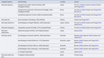
According to Walter and Boxshall (Reference Walter and Boxshall2025), the genus Naricolax currently comprises six valid species, namely, Naricolax atypicus Ho, Do & Kasahara, 1983, Naricolax hoi Hutson & Tang, 2007, Naricolax holinorum Izawa, 2021, Naricolax longispina Ho & Lin, 2005, Naricolax sphyraenae Izawa, 2020 and Naricolax stocki (Roubal, 1981). The new species differs from all the congeners by its pair of acutely pointed tines on the rostral area (vs. rostral area unarmed) (Ho et al. Reference Ho, Do and Kasahara1983; Ho and Lin Reference Ho and Lin2005; Hutson and Tang Reference Hutson and Tang2007; Izawa Reference Izawa2020, Reference Izawa2021, Reference Izawa2023). Moreover, N. zafirae sp. nov. has the last endopodal segment of leg 4 twice as long as the second segment of this ramus (vs. segments with equal length in the other congeners) (Ho et al. Reference Ho, Do and Kasahara1983; Ho and Lin Reference Ho and Lin2005; Hutson and Tang Reference Hutson and Tang2007; Izawa Reference Izawa2020, Reference Izawa2021, Reference Izawa2023). In conclusion, these features represent clear autapomorphies of the new species within Naricolax, consequently supporting its establishment. Details on host and geographic occurrence of Naricolax spp. can be found in Table 1.
Species of Naricolax associated with their hosts, localities, and references

In addition, the new species can be also differentiated from N. atypicus, N. holinorum, N. sphyraenae and N. stocki by having a T-shaped rostral area (vs. protruded in N. atypicus and concave in N. holinorum, N. sphyraenae, and N. stocki) (Ho et al. Reference Ho, Do and Kasahara1983; Izawa Reference Izawa2020, Reference Izawa2021, Reference Izawa2023), as well as from N. hoi by the apical seta on the last endopodal segment of leg 4 shorter than ramus (vs. longer than ramus in the latter species) (Hutson and Tang Reference Hutson and Tang2007). Finally, N. zafirae sp. nov. differs from N. longispina by the outer spine on the second endopodal segment of leg 3 shorter than this segment (vs. longer than the segment in the latter species) (Ho and Lin Reference Ho and Lin2005).
Discussion
In the present study, the affinities of the new species with Naricolax were confirmed based on the modified fourth seta of antennule, the morphology of the maxilliped claw, the number of setae on the second endopodal segment of leg 3, and the caudal rami with two major setae (Ho et al. Reference Ho, Do and Kasahara1983; Boxshall and Halsey Reference Boxshall and Halsey2004). Moreover, the presence of tines on the rostral area was used to differentiate the new species from the congeners, for the first time. An analogous situation can be observed for Bomolochus multiceros Pillai & Natarajan, 1977, a parasite of Ariomma indica (Day, 1871) from India, which is the unique member of Bomolochus lacking rostral tines (see Ho and Lin Reference Ho and Lin2009). In a similar sense, the shape of the rostral area was also used by Hutson and Tang (Reference Hutson and Tang2007) to differentiate N. hoi from the remaining congeners. Therefore, the evaluation of the shape of the rostral area and the presence of tines on it, would be useful for identifying species of Naricolax in further taxonomic studies.
The genus Naricolax was established by Ho et al. (Reference Ho, Do and Kasahara1983), to accommodate N. atypicus from the hexagrammid fish Hexagrammos otakii Jordan & Starks, 1895 off Kojima Bay, Japan. After the erection of this genus, five other species have been described infesting fishes from six actinopterygian families (Ariidae Bleeker, 1858, Hexagrammidae Jordan, 1888, Lateolabracidae Ghedotti, Davis & Smith, 2018, Leiognathidae Gill, 1893, Sparidae Rafinesque, 1818 and Sphyraenidae Rafinesque, 1815), of six orders (Acanthuriformes, Acropomatiformes, Carangaria, Eupercaria, Perciformes, and Siluriformes) off Australia, Japan, Korea, and Taiwan (see Table 1). Thus, the new species represents the first Naricolax described in Atlantic waters, and the seventh valid species in the genus.
The family Polynemidae Rafinesque, 1815 includes marine and brackish (sometimes riverine) fish, with global distribution along tropical and subtropical seas, and is represented by 42 species from eight genera (Froese and Pauly Reference Froese and Pauly2025). This family has great economic importance for artisanal fishermen, in which more than 100,000 t was harvested in the recent years (Girard et al. Reference Girard, Davis, Baldwin, Dettaï, Martin and Smith2022). However, only a few species of polynemid fishes have been targeted for parasitological studies, such as Eleutheronema tetradactylum (Shaw, 1804) (Zischke et al. Reference Zischke, Cribb, Welch, Sawynok and Lester2009; Moore et al. Reference Moore, Stapley, Allsop, Newman, Ballagh, Welch and Lester2011; Bharadhirajan et al., Reference Bharadhirajan, Gopalakrishnan, Raja, Murugan, Vijayakumar and Rahman2013), Filimanus heptadactyla (Cuvier, 1829) (West Reference West1992), Galeoides decadactylus (Bloch, 1795) (Diebakate and Raibaut Reference Diebakate and Raibaut1996), Polydactylus sextarius (Bloch & Schneider, 1801) (Gudivada et al. Reference Gudivada, Vankara, Hemalatha and Vijayalakshmi2014), and Polydactylus quadrifilis (Cuvier, 1829) (Capart Reference Capart1959). Regarding parasitic copepods, polynemid fishes have been parasitized by members of the families Caligidae Burmeister, 1835, Lernanthropidae Kabata, 1979, Lernaeopodidae Milne Edwards, 1840, Pennellidae Burmeister, 1835, and Philichthyidae Vogt, 1877 (Boxshall and Halsey Reference Boxshall and Halsey2004; Moore et al. Reference Moore, Stapley, Allsop, Newman, Ballagh, Welch and Lester2011; Bharadhirajan et al., Reference Bharadhirajan, Gopalakrishnan, Raja, Murugan, Vijayakumar and Rahman2013). Therefore, the present study provides the first data on parasitic copepods from P. oligodon, as well as indicates that members of the Bomolochidae can be parasites of Polynemidae. These findings also suggest a higher biodiversity of parasites from polynemid fish, than that is currently known.
With the new species described here, a dichotomous key for species of Naricolax (based on adult females) is presented as follows:
1. Rostral area with one pair of acutely pointed tines; last endopodal segment of leg 4 twice as long as previous segment.…………………………………………. N. zafirae sp. nov.
– Rostral area without such ornamentation; endopodal segments of leg 4 with similar length…………………………………………………………………………………………………….... 2
2. Outer spine on second exopodal segment of leg 3 longer than two distal exopodal segments combined……………………………………………………………………………. N. longispina
– Outer spine on second exopodal segment of leg 3 shorter than two distal exopodal segments combined……………………………………………………………………………………….... 3
3. Rostrum T-shaped; last endopodal segment of leg 4 with outermost element longer than innermost element.…………………………………………………………………………. N. hoi
– Rostrum not as above; last endopodal segment of leg 4 with outermost element shorter than innermost element……………………………………………………………………….... 4
4. Modified 4th seta on first antennulary segment expressed as one stout process……………………………………………………………………………………………………….... N. sphyraenae
– Modified 4th seta on first antennulary segment forming recurved spine-like element…………………………………………………………………………………………………….... 5
5. Middle seta within the 3 distal elements on the free exopodal segment of leg 5 slightly longer than the others……………………………………………………………. N. holinorum
– Middle seta within the three distal elements on the free exopodal segment of leg 5 twice as long as the others………………………………………………………………………….... 6
6. Spines on last endopodal segment of legs 3 and 4 coarsely serrate………………………………………………. N. atypicus
– Spines on last endopodal segment of legs 3 and 4 finely spinulate……………………………………………………... N. stocki
Data availability
The authors confirm that the data supporting the findings of this study are available within the article.
Acknowledgements
We wish to thank Nivea Fernanda Maria Ferreira Costa, Saturno de Sousa Dias and Filipe Ribeiro Menks from the Universidade Federal do Maranhão in São Luis for helping with parasitological examination of fish. Thanks are also due to Dr Kunihiko Izawa from Izawa Marine Biological Laboratory, Japan, for providing literature.
Author contributions
F.P., J.V.C., and J.L.S.N. performed field collections and parasitological survey. F.P., E.J.L.P., and J.V.C. analysed the copepods and prepared the illustrations. F.P. and F.B.P. wrote the first draft of the manuscript. All reviewed the manuscript and approved the final version.
Funding
F.P. was supported by the UERJ-PROTEC program, Brazil. J.V.C. was supported by a student fellowship from Coordenação de Aperfeiçoamento de Pessoal do Ensino Superior (CAPES), Brazil. J.L.S.N. was supported by Fundação de Amparo à Pesquisa e ao Desenvolvimento Científico e Tecnológico do Maranhão (FAPEMA/BEPP02588/2023). F.P.B. was supported by a FAPEMIG/CNPq researcher fellowship. This work partially funded by Conselho Nacional de Desenvolvimento Científico e Tecnológico (CNPq Universal, process number 404083/2021-8 A/C).
Competing interests
The authors declare that they have no conflict of interest.
Ethical standards
All applicable institutional, national and international guidelines for the care and use of animals were followed.