Introduction
Cricopharyngeal dysfunction is a common cause of dysphagia,Reference Halvorson and Kuhn1 consisting of a failure of appropriate relaxation of the cricopharyngeus muscle during the pharyngeal phase of swallowing. Typical symptoms include dysphagia, globus sensation, coughing or choking episodes while eating, and regurgitation. Cricopharyngeal dysfunction is responsible for a spectrum of disease, ranging from asymptomatic cricopharyngeal bar to the formation of a Zenker's diverticulum.Reference Tieu and Hunter2 In this paper, we focus on the management of isolated cricopharyngeal dysfunction, in the absence of a diverticulum.
Materials and methods
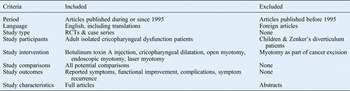
A comprehensive search was performed using Ovid (Medline, Embase), the Cochrane Library, PubMed and Google Scholar. The term ‘cricopharyngeal’ was used in conjunction with the terms ‘management’ and ‘treatment’, and with the terms ‘dysphagia’, ‘dysfunction’, ‘spasm’ and ‘hypertrophy’. The last date of the final search was 19 May 2015. Results were limited to articles published in English from 1995 to the present day. The inclusion and exclusion criteria are shown in Table I.
Inclusion and exclusion criteria

RCT = randomised, controlled trial
The data collected from the articles reviewed included information regarding the participants, interventions, comparisons, outcomes, study design and study size, as per the Preferred Reporting Items for Systematic Reviews and Meta-Analyses (‘PRISMA’) statement.Reference Moher, Liberati, Tetzlaff and Altman3
Results
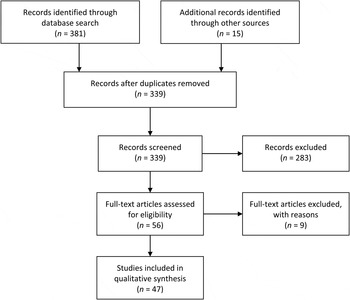
The initial search identified 339 unique titles (Figure 1). The titles and abstracts of these articles were reviewed to check for relevance. The full text was obtained for 56 relevant articles. The reference sections of these articles were examined to identify additional articles of relevance.
Preferred Reporting Items for Systematic Reviews and Meta-Analyses (‘PRISMA’) flow diagram.

No relevant randomised trials were identified. A number of case series were used to perform a qualitative analysis. There was a high degree of heterogeneity with regard to the interventions, comparisons and outcomes measured in these studies, and this precluded any quantitative analysis. Most case series reported positive outcomes, suggesting a risk of publication bias.
Pathophysiology
The upper oesophageal sphincter consists of the cricopharyngeus muscle, along with the lower fibres of the inferior constrictor muscle and the upper part of the cervical oesophagus.Reference Halvorson and Kuhn1 At rest, this sphincter is in a state of contraction; relaxation occurs just prior to the arrival of a food bolus as part of the pharyngeal phase of swallowing. Failure of this relaxation may relate to: disruption of the central or peripheral nervous systems, changes in the muscle itself, or operative procedures to the head and neck.Reference Manison4
Assessment
A thorough patient history and examination is mandated; this should indicate the level of dysphagia and aid patient selection for further intervention. A barium study of the upper oesophagus may show a cricopharyngeal bar (Figure 2), although this can be present in normal subjects.Reference Halvorson5 Videofluoroscopy can provide additional, dynamic information about the swallow.Reference Lawson and Remacle6 Oesophageal manometry may also be used, though opinion varies as to its value.Reference Halvorson5–Reference Ozgursoy and Salassa9 Gastroscopy is indicated in cases where there are clinical or radiological features suggestive of malignancy.
Barium swallow study image, showing a prominent cricopharyngeal bar, causing narrowing of hypopharyngeal lumen and obstruction to the flow of contrast.

Treatment
Following a thorough assessment, treatment should be tailored to the particular patient, taking into account the severity of symptoms and other co-morbidities.Reference Solt, Bajor, Moizs, Grexa and Horváth10–Reference Schneider, Thumfart, Pototschnig and Eckel20 Many would advocate empirical treatment with proton pump inhibitors for a period of three months before surgical intervention is considered, on the basis that reflux disease may trigger cricopharyngeal dysfunction.Reference Brady, Stevenson, Somers, Hough and Di Giandomenico21 Swallowing therapy may also be performed by speech therapists as a first-line measure, prior to consideration of surgical intervention.Reference Jones, Knigge and McCulloch22
Botulinum toxin A
Injection of the cricopharyngeus muscle with botulinum toxin A under direct vision was described in 1994,Reference Schneider, Thumfart, Pototschnig and Eckel20 and a series using a percutaneous, computed tomography guided technique in awake patients was published shortly thereafter.Reference Atkinson and Rees23
A review performed by Kelly et al. in 2013 identified 20 studies in which the cricopharyngeus muscle was injected with botulinum toxin.Reference Kelly, Koszewski, Jaradeh, Merati, Blumin and Bock24 The majority of these were case series with short-term follow up, with success rates ranging from 43 to 100 per cent (as measured using validated scoring tools such as the Eating Assessment Tool (‘EAT-10’)Reference Belafsky, Mouadeb, Rees, Pryor, Postma and Allen25). Techniques included rigid oesophagoscopy under general anaesthetic, flexible endoscopy with sedation, and percutaneous injection using local anaesthetic.
Kelly et al. also reported their case series of 49 patients including both operating theatre and clinic procedures; they found a 65 per cent success rate with no severe complications or deaths.Reference Kelly, Koszewski, Jaradeh, Merati, Blumin and Bock24 However, their report made it clear that repeated injections with botulinum toxin are often necessary to achieve or maintain a good effect, and it is not yet possible to predict (through pre-operative investigations) which patients will benefit from the procedure.Reference Kelly, Koszewski, Jaradeh, Merati, Blumin and Bock24
Dilatation
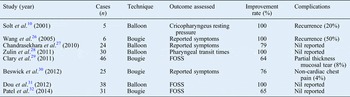
Dilatation of the cricopharyngeus muscle may be performed using either a balloon dilatorReference Ozgursoy and Salassa9 or bougie dilator (Savary–Gilliard dilator).Reference Wang, Kadkade, Kahrilas and Hirano26 A number of case series published in recent years (Table II) indicate that both techniques are safe and effective, although some patients experienced short-term recurrence and required repeat procedures in order to maintain symptomatic improvement.Reference Solt, Bajor, Moizs, Grexa and Horváth10, Reference Wang, Kadkade, Kahrilas and Hirano26–Reference Salassa33 It has thus been suggested that dilatation might be used as a first-line intervention, prior to more definitive management.Reference Solt, Bajor, Moizs, Grexa and Horváth10
Case series involving cricopharyngeus muscle dilatation

FOSS = Functional Outcome Swallowing ScoreReference Salassa33
Cricopharyngeal myotomy
Open cricopharyngeus myotomy was first described by Kaplan in 1951.Reference Kaplan11 This usually consists of a left lateral cervical incision, which is used to access and divide the cricopharyngeus muscle externally down to mucosa.Reference Tieu and Hunter2, Reference Lawson and Remacle6 Given this approach, there is a risk of damage to the recurrent laryngeal nerve or to the contents of the carotid sheath. Other reported complications include localised infection, haematoma, fistula formation, aspiration pneumonia and tracheostomy.Reference Mason, Bremner, DeMeester, Crookes, Peters and Hagen8, Reference Brigand, Ferraro, Martin and Duranceau34
Endoscopic approaches classically make use of a rigid endoscope under general anaesthesia, to identify and divide the cricopharyngeus muscle, from mucosa through to prevertebral fatty tissue.Reference Lawson and Remacle6 In the absence of a Zenker's diverticulum, the use of endoscopic stapling devices is not practicable, as the staplers cannot gain purchase on the cricopharyngeus muscle posteriorly.Reference Tieu and Hunter2 Instead, the incised mucosa may be left to heal by secondary intent, or endoscopically clippedReference Dale, Mackeith, Burgess, Rourke, Winter and Corbridge16 or sutured.Reference Ho, Morzaria and Damrose35, Reference Mortensen, Schabery, Genden and Woo36
Techniques described have included the use of a steel scalpel,Reference Mosher12 diathermy knife,Reference Dohlman13 carbon dioxide (CO2) laser,Reference Ozgursoy and Salassa9, Reference van Overbeek, Hoeksema and Edens14, Reference Takes, Van Den Hoogen, Henri and Marres37–Reference Bergeron and Chhetri40 potassium titanyl phosphate (KTP) laserReference Halvorson and Kuhn1, Reference Halvorson5, Reference Kuhn and Bent19 and harmonic scalpel.Reference Fama, Moore and Kasperbauer15 There is no evidence of any significant variation in outcomes between these different techniques. The risks associated with endoscopic myotomy include dental trauma, pharyngeal wall tear, fistula formation, and complete hypopharyngeal or oesophageal perforation.Reference Pollei, Hinni, Hayden, Lott and Mors18 A recurrence rate of around 10 per cent should be expected, irrespective of the technique or repair used.Reference Dale, Mackeith, Burgess, Rourke, Winter and Corbridge16, Reference Pollei, Hinni, Hayden, Lott and Mors18 A range of studies have assessed the safety of CO2 and KTP laser techniques; both were found to have low rates of morbidity, with outcomes comparable to non-laser and open techniques.Reference Lawson and Remacle6, Reference Dale, Mackeith, Burgess, Rourke, Winter and Corbridge16, Reference Pollei, Hinni, Hayden, Lott and Mors18, Reference Ho, Morzaria and Damrose35–Reference Pitman and Weissbrod42
In individuals with limited neck movement, cricopharyngeus myotomy may be performed using a flexible scope.Reference Altman, Genden and Moche17 However, this remains a relatively novel procedure, and further evidence is required to establish its safety and efficacy.Reference Halland and Baron43
There exist only a small number of (non-randomised) studies comparing different forms of cricopharyngeus myotomy in patients without Zenker's diverticulum. Dauer et al. compared open myotomy (8 cases) with endoscopic CO2 laser myotomy (14 cases).Reference Dauer, Salassa, Iuga and Kasperbauer41 They found that endoscopic treatment had a similar success rate to open cases, but was associated with shorter operative times and in-patient stays. While complication rates were similar between the two groups, major complications occurred in the open myotomy group: prolonged intubation led to tracheostomy in one patient with medical co-morbidities, while another developed a pharyngocutaneous fistula. In a recent pilot study, Arenaz Bua et al. compared endoscopic CO2 laser myotomy (four cases) with balloon dilatation (four cases), and found both to be effective over a six-month period.Reference Arenaz Bua, Olsson, Westin, Rydell and Ekberg44 However, limited case numbers and a short follow-up period make it difficult to draw conclusions.
Conclusion
There exists a range of therapeutic options for the management of isolated cricopharyngeal dysfunction. It may be useful to perform botulinum toxin A injection or dilatation as a first-line procedure given their potential diagnostic value, particularly in patients who are elderly or have multiple co-morbidities. Patients who fail to benefit from these techniques or experience recurrence may then proceed to surgical intervention. Endoscopic myotomy provides an effective alternative to the classical open technique, with potentially reduced morbidity. However, further studies are required to compare the efficacy of the various treatment modalities.