Introduction
Aneurysm of the petrous portion of the internal carotid artery (ICA) is uncommon. The petrous and carotid segments of the ICA remain subject to pathology, the most important being aneurysm formation. Such aneurysms often present as an expansile mass with cranial nerve dysfunction and epistaxis. Imaging scans should be analysed carefully in order to gain a definitive diagnosis. Some aneurysms are too large to be treated with endovascular occlusion techniques; in such cases, ligation of the parent vessel is indicated. In the case reported below, the patient continued to experience persistent, mild epistaxis despite ICA ligation. This resulted from continued filling due to reconstitution of the left carotid siphon via the left external carotid artery arising from the internal maxillary artery; this reperfusion was clearly demonstrated on post-operative magnetic resonance angiography (MRA).
Case report
A 30-year-old man presented with multiple episodes of epistaxis and blood-stained sputum. On further questioning, he also reported several episodes of left visual field blurring; however, his visual field was normal on examination. The patient had no previous medical illnesses and denied any history of trauma or head injury.
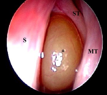
Endoscopic examination revealed a smooth mucosal polyp prolapsing through the left sphenoid ostium into the sphenoethmoidal recess (Figure 1).
Endoscopic examination demonstrating a left sphenoethmoidal recess polyp (*). S = septum; ST = superior turbinate; MT = middle turbinate

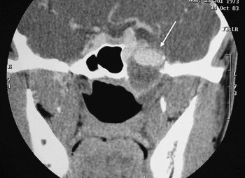
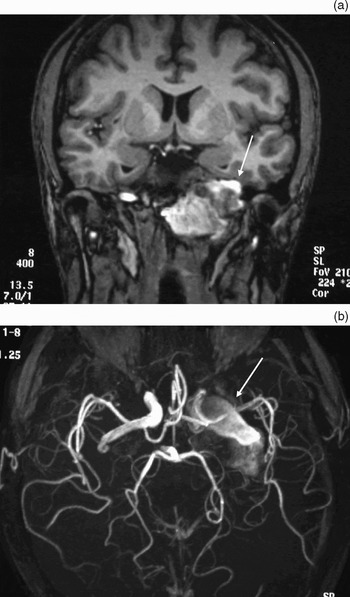
Computed tomography (CT) scanning showed an expansile, 4.6 × 3.5 cm lesion within the left temporal bone, which was expanding and effacing into the carotid canal, petrous apex, middle cranial fossa floor and clivus. This lesion also extended medially to produce a bulge on the lateral sphenoid wall (Figure 2). Subsequent magnetic resonance imaging (MRI) and MRA scans confirmed a fusiform aneurysm of the left internal carotid artery (ICA) in the horizontal portion of the petrous temporal bone, hence the diagnosis of a giant left petrous carotid aneurysm (Figure 3).
Coronal computed tomography scan showing an expansile lesion (arrow) effacing into the carotid canal, petrous apex, middle cranial fossa floor and clivus.

Coronal (a) magnetic resonance imaging scan and (b) magnetic resonance angiography scan, showing a left fusiform aneurysm (arrow) of the left internal carotid artery in the horizontal portion of the petrous temporal bone.

The patient was admitted for a baseline transcranial Doppler monitoring study and carotid angiography with balloon occlusion test.
Results for transcranial Doppler monitoring of the anterior circulation were within normal limits. Cerebrovascular reactivity using 8 per cent carbon dioxide challenge was normal for both middle cerebral arteries. The mean velocity of left middle cerebral artery flow dropped from 60 cm/second to 48–50 cm/second during balloon occlusion test of the left ICA. However, the left ICA balloon occlusion test indicated good flow from the anterior communicating artery and posterior communicating artery via the left vertebral artery.
Endovascular intervention options were discussed with the interventional radiologists. They opined that the aneurysm was too large for endovascular occlusion. Therefore, the option of ICA ligation, and its inherent complications, was discussed with the patient. The patient subsequently consented and underwent this procedure. Post-operatively, recovery was uneventful and the patient was discharged home well.
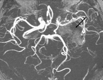
A repeat MRA performed 10 days post-operatively revealed an abrupt termination to contrast flow into the aneurysm, which was compatible with post-surgical changes secondary to ICA ligation (Figure 4).
Magnetic resonance angiogram performed 10 days post-operatively, showing abrupt termination of contrast flow to the aneurysm (arrow).

However, three months post-operatively, the patient still complained of mild epistaxis and occasionally coughing up blood. A repeat MRA and angiogram confirmed stable occlusion of the left ICA, with no visible filling of the previous aneurysm.
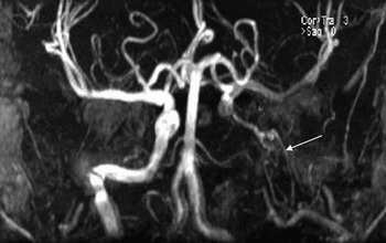
Nine months post-operatively, the patient began to complain of left temporal headaches and mild epistaxis. Cranial nerve examination was unremarkable. A repeat MRA scan showed continued occlusion of the left ICA and no obvious filling of the residual aneurysm. However, there was reconstitution of the left carotid siphon via the left inferolateral trunk arising from collaterals from the third segment of the internal maxillary artery. Therefore, a small amount of blood may still have been entering the aneurysm and causing periodic symptoms (Figure 5).
Magnetic resonance angiography scan performed nine months post-operatively, showing continued occlusion of the left internal carotid artery. However, there is reconstitution of the left carotid siphon via the left inferolateral trunk arising from the collaterals from the third segment of the internal maxillary artery (arrow), which may indicate that a small amount of blood is still entering the aneurysm and causing the periodic symptoms.

The patient continued to experience mild epistaxis periodically. Two years after the ICA ligation, repeated MRA and angiography confirmed stable occlusion of the left ICA, with no filling of the previous aneurysm. At the time of writing, the patient was in his fourth year of follow up, with no major complications.
Discussion
Aneurysms of the internal carotid artery within the petrous temporal bone are extremely rare. They are characteristically giant, fusiform in morphology and congenital.Reference Halbach, Higashida and Hieshima1 Most cases have no obvious aetiology and are presumed to be congenital or developmental in origin. The aetiology of congenital aneurysms is thought to involve a developmental weakness of the arterial wall where the embryonic arteries have regressed, in particular the caroticotympanic artery.Reference Kudo and Colley2 Other causes involving trauma, mycotic and other middle-ear infections, neoplasm and mastoid surgery have been described.Reference Constantino, Russell and Reisch3 Most cases present in the third and fourth decades, with equal sex preponderance.Reference Reece, Higgins, Hardy and Moffat4
The clinical presentation of internal carotid artery (ICA) aneurysms depends on the direction of expansion. Our patient presented with epistaxis, headaches and episodes of left visual field blurring. This corresponds with a medially expanding aneurysm, which typically presents with epistaxis, headaches and cranial nerve palsies from the IIIrd to the VIIIth cranial nerves.Reference Halbach, Higashida and Hieshima1 The above symptoms, coupled with an endoscopic appearance of a smooth mucosal polyp in the sphenoethmoidal recess area,Reference Sethi, Lau, Chee and Chong5 should prompt suspicion of an aneurysm. Diagnostic imaging should be performed first, before taking a biopsy, in order to rule out an aneurysm. A biopsy in these instances may lead to catastrophic consequences.
Aneurysms can easily be demonstrated on cross-sectional imaging. A combination of CT, MRI and MRA is usually diagnostic.Reference Reece, Higgins, Hardy and Moffat4 On CT scanning, petrous aneurysms usually appear as destructive lesions of the petrous bone orientated along the carotid canal. There is usually well corticated expansion of the carotid canal.Reference Moonis, Hwang, Ahmed, Weigele and Hurst6 On MRI, the lesion demonstrates complex signal intensity owing to turbulent flow within the aneurysm. There are regions of flow void and intense enhancement compatible with a vascular lesion.Reference Moonis, Hwang, Ahmed, Weigele and Hurst6 A differential diagnosis of glomus jugulare, glomus tympanicum, high jugular bulb, aberrant carotid artery and even persistent stapedial artery should be borne in mind.Reference Moonis, Hwang, Ahmed, Weigele and Hurst6, Reference Moffat and O'Connor7 In our patient, conventional angiography was performed in order to confirm the diagnosis, and was also used post-operatively as the patient still experienced minor symptoms despite surgical intervention.
Intervention for giant aneurysms aims for symptomatic relief in the hope of reducing episodes of haemorrhage and thromboembolism. Headaches can often be cured after several months but cranial nerve palsies are not always reversible.Reference Coley, Clifton and Britton8 Intervention may comprise either endovascular occlusion or ICA ligation. Giant aneurysms are now increasingly being treated by endovascular occlusion, using metallic coils or detachable balloons, with preservation of the parent artery.Reference Halbach, Higashida and Hieshima1, Reference Kudo and Colley2, Reference Higashida, Halbach, Dowd, Barnwell, Dormandy and Bell9 The petrous temporal bone is a surgically inaccessible area; therefore, detachable balloon occlusion of the ICA below and above the aneurysm and/or coil embolisation are good alternatives.Reference Higashida, Halbach, Dowd, Barnwell, Dormandy and Bell9 The other added advantage of endovascular carotid occlusion is that a balloon occlusion test can be performed prior to definitive occlusion, in order to assess tolerance to vessel sacrifice and also to test the ability of the collateral circulation to compensate cerebral blood flow to the affected side. In our patient, a baseline transcranial Doppler monitoring study and a carotid angiogram with balloon occlusion test were performed as routine procedures prior to any planned surgical intervention.
Endovascular occlusion of aneurysms with preservation of the parent vessel can only be performed in a limited number of cases, because the majority of reported petrous aneurysms are fusiform in shape.Reference Halbach, Higashida and Hieshima1 Thromboembolic events may occur during placement of the embolic device, or the device itself may migrate. Many such events manifest as transient ischaemic attacks which do not result in permanent deficits.Reference Higashida, Halbach, Dowd, Barnwell, Dormandy and Bell9 Treatment by volume expansion, antiplatelet agents and heparin provides effective prophylaxis and/or reversal in most cases. Placement of an embolic device is also hazardous due to the fragile nature of the aneurysm wall, which may not support the device, potentially leading to disastrous consequences.Reference Crow, Scott, Guinto, Chaljub, Wright and Rabassa10
There seems to be a lack of documented evidence on the long term follow up of patients treated with such endovascular techniques. However, after short term follow up one study found a 3 per cent incidence of continued filling of unclippable carotid aneurysms following placement of balloons across or proximal to the aneurysm neck.Reference Larson, Tew, Tomsick and van Loveren11 Placement of balloons distal to the aneurysm becomes necessary when there is evidence of continued filling into the aneurysm. Such patients are candidates for placement of distal balloons and/or a coil.Reference Coley, Clifton and Britton8
Parent vessel occlusion, such as common carotid or internal carotid ligation or direct surgery, has long been used in the treatment of aneurysms.Reference Gelber and Sundt12 In our case, the aneurysm was too large to be treated with endovascular occlusion techniques, so the parent vessel was ligated. However, this procedure carries a risk of reperfusion phenomenon due to anastamosis from the ipsilateral and contralateral carotid systems.Reference Cohen, Anastassov and Chuang13 This was evident in our case initially – even after ICA ligation, there was evidence of continued filling from reconstitution of the left carotid siphon via the left external carotid artery arising from the internal maxillary artery, which may indicate that a small amount of blood was still flowing into the aneurysm and causing the periodic symptoms.
Ipsilateral cerebral infarction as a result of hypoperfusion secondary to balloon occlusion has been reported, even when patients have tolerated test occlusion.Reference Coley, Clifton and Britton8 The incidence of subsequent infarction is about 4 per cent for cavernous carotid aneurysms treated by endovascular occlusion.Reference Higashida, Halbach, Dowd, Barnwell, Dormandy and Bell9 On the other hand, ICA ligation carries a 17 per cent long term incidence of stroke.Reference Roski, Spettzler and Nulsen14
Conclusion
We present a rare case of a giant petrous carotid aneurysm that presented with epistaxis and a sphenoethmoidal polyp. The clinical symptoms corresponded to the direction of expansion of the aneurysm. Imaging was crucial in establishing a definitive diagnosis. Intervention techniques should be individualised in order to achieve safe outcomes. Internal carotid artery (ICA) ligation was performed, without any remarkable post-operative complication. Persistent epistaxis following ICA ligation was a result of the reperfusion phenomenon.