Introduction
The retinal pigmented epithelium (RPE) resides adjacent to the outer neural retina and plays a critical role in retinal development and also in the maintenance of photoreceptor function in the mature organism. Throughout life, RPE cell production is maintained in the peripheral retina at a low level, while there is evidence for RPE cell loss centrally (Del Priore et al., Reference Del Priore, Kuo and Tezel2002; Al-Hussaini et al., Reference Al-Hussaini, Vugler, Semo and Jeffery2008). Some newly generated RPE cells migrate centrally to compensate for this age-related loss (Al-Hussaini et al., Reference Al-Hussaini, Vugler, Semo and Jeffery2008). Maintenance of the balance between age-related cell loss and cell production is probably an important homeostatic mechanism in the RPE.
The production of RPE cells is partially regulated by melanin because in albino rats, excess RPE cell numbers enter the cell cycle and many become polyploid without completing cell division (Adams et al., Reference Adams, Shahabi, Hoh-Kam and Jeffery2010). This leads to progressive ocular pathology (Shahabi et al., Reference Shahabi, Lenassi and Jeffery2011). While the regulators of cell generation in the mature central nervous system are largely unknown, it has been demonstrated that glatiramer acetate induces elevated neuronal production in the adult hippocampus (Butovsky et al., Reference Butovsky, Koronyo-Hamaoui, Kunis, Ophir, Landa, Cohen and Schwartz2006). Glatiramer acetate is a random polymer of four amino acids; glutamic acid, lysine, alanine, and tyrosine that appears to act as an immunomodulator. It has been used in the treatment of multiple sclerosis, although its exact mode of action is unknown (Carpintero et al., Reference Carpintero, Brandt, Gruaz, Molnarfi, Lalive and Burger2010; Van Kaer, Reference Van Kaer2011). There is a link between this drug and the eye as it has been claimed that glatiramer acetate also reduces age-related deposits behind the human retina (Landa et al., Reference Landa, Butovsky, Shoshani, Schwartz and Pollack2008; Landa et al., Reference Landa, Rosen, Patel, Lima, Tai, Perez, Aizman and Garcia2011). These focal deposits known as drusen are a risk factor for age-related macular degeneration (Sarks, Reference Sarks1976; Green, Reference Green1999; Hollyfield, Reference Hollyfield2010). As glatiramer acetate elevates cell production in the brain and has been linked to a potential ocular therapy for age-related macular degeneration, we ask here whether it impacts on RPE cell production in mature rats. If so, this may represent a therapeutic route for disease states that target the RPE.
Materials and methods
Ten Lister hooded female rats were used at approximately 8 months old. They were housed with a 12-h light/dark cycle in a temperature-controlled environment and fed ad libitum. All experimental procedures were carried out under local ethical guidance and the U.K. (Scientific Procedures) Act 1986. Animals were either injected with 0.2 ml phosphate-buffered saline (PBS) (n = 5) or 0.2 ml glatiramer acetate (n = 5500 μg/rat) followed by 0.8 ml Bromodeoxyuridine (5-bromo-2-deoxyuridine, BrdU; Sigma-Aldrich, Gillingham, UK, 50 mg/kg). Animals were injected everyday for 14 days and were then sacrificed using CO2. Their eyes were removed and were fixed in 4% paraformaldehyde in PBS for 1 h.
The RPE and attached choroid were isolated and radial incisions made to produce RPE sheet whole mounts (Al-Hussaini et al., Reference Al-Hussaini, Schneiders, Lundh and Jeffery2008). One eye from each animal was processed for the cell cycle marker Ki67 and one for BrdU, to reveal patterns of cell division. For Ki67, whole mounts were blocked for 2 h in 5% normal donkey serum in PBS with 3% Triton X-100 and incubated overnight in rabbit anti-Ki67 (1:1000; Vector Laboratories, Peterborough, UK), diluted in 1% normal donkey serum in PBS with 3% Triton X-100. After PBS washing, eyecups were incubated in the secondary antibody, donkey antirabbit Alexa Flour 488, for 2 h in 1% normal donkey serum in PBS with 0.3% Triton X-100. Nuclei were subsequently stained with 4,6-diamidino-2-phenylindole (DAPI; Sigma-Aldrich, Gillingham, UK). The tissue was then washed in PBS followed by washing in Tris buffer saline (pH 7.4). They were mounted with Vectashield (Vector Laboratories, Peterborough, UK).
For BrdU, the processing was identical to that of Al-Hussaini et al. (Reference Al-Hussaini, Vugler, Semo and Jeffery2008). Briefly, whole mounts were stained with a mouse anti-BrdU primary antibody and a donkey antimouse Alexa Flour 568 as the secondary. Before the secondary antibody could be used, antigen retrieval was undertaken by applying 6 m hydrochloric acid diluted in 1% Triton X-100 in PBS for 30 min onto the whole mounts. The tissue was washed and mounted as above. Individual RPE sheets were not double stained for both Ki67 and BrdU because in previous experiments where this was undertaken, some Ki67-labeled cells were ghost like in appearance. This raised the concern that the antigen retrieval process necessary for BrdU label was in some way reducing the quality of the Ki67, undermining the value of double labeling the whole mounts. All cell counts were made at a magnification of 200×. Once a cell was identified, it was examined at 400× on channels for both the cell label and DAPI. Cell counts were made over the entire RPE surface.
To control for potential differences in the area of the RPE whole mounts, they were measured from low power images. Further, the number of cells counted in each whole mount has been normalized for area by dividing the total number of labeled cells present by the area of the RPE surface. Statistical comparisons were made using the nonparametric Mann–Whitney U test, which makes no assumptions regarding the normality of the data distribution. Data are plotted as means with standard deviations.
Results and discussion
The RPE whole mounts were approximately 65 mm2 in area. There was little variation in this area between animals and nonsignificant differences between the animals injected with PBS or glatiramer acetate (Mean experimental group area = 66.85 mm2, s.d. = 3.93; Mean control group area = 66.75 mm2, s.d. = 3.93, P = 1.00).
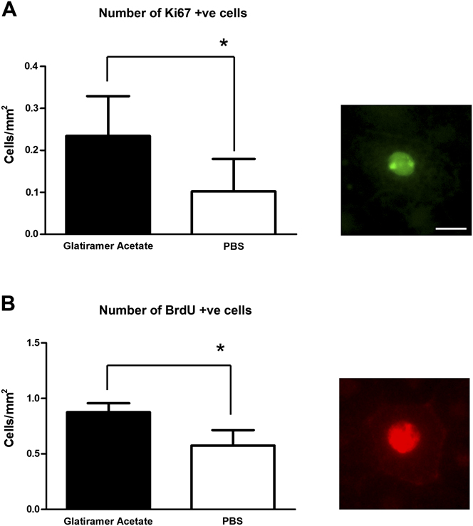
Ki67-positive RPE cells were present in allthe eyes examined. There were on average seven positive cells present in the RPE sheets of animals injected with PBS. However, in those injected with glatiramer acetate, the number increased to an average of 16 positive cells. Hence, the number of Ki67-positive cells doubled in the drug-treated animals. This difference was significant (Mean experimental group = 15.4, s.d. = 5.41; Mean control group = 6.8, s.d. = 4.96, P = 0.0297). In every case, Ki67-positive cells were confined to the peripheral retina as revealed previously (Al-Hussaini et al., Reference Al-Hussaini, Vugler, Semo and Jeffery2008). The difference between the experimental and control groups corrected for area remained statistically significant (Fig. 1A; Mean experimental group = 0.234, s.d. = 0.095; Mean control group = 0.012, s.d. = 0.008, P = 0.0297). These data reveal that significantly more RPE cells were in the cell cycle in the peripheral retina in those animals injected with glatiramer acetate than those injected with PBS.
Ki67 (A) and BrdU (B) labeling of the RPE corrected for the number of cells (mm2). Presence of a cell within the cell cycle was assessed with Ki67 at a single time point. BrdU was injected over 14 consecutive days. Significantly elevated levels of Ki67 (P = 0.0297) and BrdU (P = 0.0040) were found following administration of glatiramer acetate demonstrating that it increases cell proliferation. The photomicrographs show a positively labeled RPE cell for ki67 (Green, A) and BrdU (Red, B). However, while Ki67-positive cell numbers doubled, BrdU-positive cell numbers only increased by approximately 50% following 14 consecutive daily pulses, which represents 3% per day. It is possible that many cells entering the cell cycle, as shown with the Ki67 label, fail to complete appropriate cell division, as revealed by BrdU numbers. These cells may either die or may be arrested in the cell cycle. Graphs for the absolute number of cells in each group were almost identical to those shown corrected for area. Scale bar = 25 μm for both photomicrographs.

Ki67 labeling of the RPE does not reveal that cells are actually dividing, only that they are in the cell cycle when the animal was killed. Not all cells that enter the cell cycle complete cell division. Cell cycle entry can be a prelude to cell death or to a polyploidy state where the cell is arrested in the cell cycle (Cernak et al., Reference Cernak, Stoica, Byrnes, Di Giovanni and Faden2005; Yang & Herrup, Reference Yang and Herrup2007; Shahabi et al., Reference Shahabi, Lenassi and Jeffery2011). To confirm that cell division was taking place, the number of BrdU-labeled RPE cells were counted. The average number of BrdU-positive cells found in PBS-treated animals was 40 per RPE sheet, while in the experimental group, the average increased to 60 per sheet, which represents a 50% increase over the 14-day period. This difference was statistically significant (Mean experimental group = 58.8, s.d. = 8.671; Mean control group = 38.8, s.d. = 10.99, P = 0.0159). The number of BrdU cells was again corrected for area, and again, this difference was statistically significant (Fig. 1B; Mean experimental group = 0.876, s.d. = 0.080; Mean control group = 0.576, s.d. = 0.138, P = 0.0040). However, Ki67 labeling represents cells in the cell cycle at the time of death, while the BrdU label was cumulative over 14 days. If the percentage increase in BrdU is averaged per day, in glatiramer acetate-injected animals, it is only 3%. While the vast majority of BrdU-labeled cells in both groups were located in the periphery, a small number were present outside this region, consistent with the findings of Al-Hussaini et al. (Reference Al-Hussaini, Vugler, Semo and Jeffery2008), and the hypothesis that some cells that divide in the periphery may migrate centrally.
This study shows that relatively brief exposure to glatiramer acetate elevates cell production in peripheral RPE cells. The mechanism by which this occurs is unclear; however, as argued by Butovsky et al. (Reference Butovsky, Koronyo-Hamaoui, Kunis, Ophir, Landa, Cohen and Schwartz2006), who demonstrated elevated hippocampal neurogenesis following glatiramer acetate administration, the process is likely to depend in part on local immune activity that is associated with microglia production of growth factors such as insulin-like growth factor 1 (IGF1). This may rest on the finding that glatiramer acetate induces a phenotypic switch in microglia in the brain to dendritic cells that produce IGF1 (Butovsky et al., Reference Butovsky, Koronyo-Hamaoui, Kunis, Ophir, Landa, Cohen and Schwartz2006).
There is likely to be a balance between cell production and death over extended periods in the ageing RPE (Del Priore et al., Reference Del Priore, Kuo and Tezel2002). The data on aging RPE are very mixed with morphological studies providing evidence for both age-related cell loss, age-related cell increase, and for a relatively static state (Gao & Hollyfield, Reference Gao and Hollyfield1992; Panda-Jonas et al., Reference Panda-Jonas, Jonas and Jackobczyk-Zmija1996; Harman et al., Reference Harman, Fleming, Hoskins and Moore1997; Del Priore et al., Reference Del Priore, Kuo and Tezel2002). However, the spread of data is large in all these studies. It is likely that there are numerous variables impacting on the regulation of RPE cell numbers with age, and that we are only aware of some of these. In spite of such differences, it is clear that RPE cells are generated in the mature peripheral retina in a life-long manner (Al-Hussaini et al., Reference Al-Hussaini, Vugler, Semo and Jeffery2008). Further, the only study to mark dying cells in the mature RPE has only been able to demonstrate them at central locations (Del Priore et al., Reference Del Priore, Kuo and Tezel2002).
The results of this study clearly reveal with two separate markers that glatiramer acetate increases levels of proliferation in the mature RPE. While the numbers we reveal are not large, it is important to note that, particularly for the Ki67 label, this is a snap shot of a dynamic process, in which there is a constant low level of proliferation over life (Al-Hussaini et al., Reference Al-Hussaini, Vugler, Semo and Jeffery2008). However, one clear facet of this study is the disparity between the data generated using the cell cycle marker Ki67 and that using the marker of cell division, BrdU. The Ki67 data revealed an approximate 100% increase in the number of cells in the cell cycle at a single time point following drug administration, while 14 sequential daily injections of BrdU elevated cell division by only approximately 50%, which is 3% per day. There are many variables that can lead to discrepancies between positively labeled cell numbers with the two markers, in particular the fact that Ki67 labels all phases of the cell cycle, while BrdU is only incorporated in S phase, which accounts for 10–30% of the cell cycle depending on total DNA load. Given that there was a 100% increases in Ki67 on a single day, it might be expected that the elevation in BrdU would be much greater. However, the large difference between the two markers may indicate that many cells in the cell cycle do not progress to full cell division.
The unexpectedly low number of BrdU cells is unlikely to be due to it being diluted out through multiple cell divisions over the 14 days. Separately, we have carried out multiple injections of BrdU and traced the progressive accumulation of labeled RPE cells. This showed that labeled RPE cells increase in number in a roughly linear manner within the time window used here with no indication of numbers declining or leveling off.
There are two alternative explanations for the discrepancy. First, cell cycle proteins can be up regulated in mature cells when they initiate caspase-related apoptosis (Cernak et al., Reference Cernak, Stoica, Byrnes, Di Giovanni and Faden2005; Yang & Herrup, Reference Yang and Herrup2007). It is possible that encouraging cells into the cell cycle at inappropriate stages may result in their failure to complete cell division and they fail to survive. Second, RPE cells can enter the cell cycle but then may be unable to progress through to full cell division and to become polyploid (Adams et al., 2009: Shahabi et al., Reference Shahabi, Lenassi and Jeffery2011). There is a tendency towards this in the rodent RPE where the majority of central RPE cells are binucleated (Ts’o & Friedman, Reference Ts’o and Friedman1967). In human, binucleation is uncommon and associated with pathology, specifically proximity to drusen (Al-Hussaini et al., Reference Al-Hussaini, Schneiders, Lundh and Jeffery2009). However, when pigment is absent, albino RPE cells enter the cell cycle in large numbers as melanin precursors are known to regulate the cell cycle (Illia & Jeffery, Reference Illia and Jeffery1999; Shahabi et al., Reference Shahabi, Lenassi and Jeffery2011). These RPE cells become trapped going through inappropriate nuclear division becoming large and distorted but failing to complete cell division. These cells eventually become dysfunctional, with elevated cell death that impacts on the adjacent neural retina (Shahabi et al., Reference Shahabi, Lenassi and Jeffery2011). Whether this is the case with glatiramer acetate is unclear, but the discrepancy between the two markers implies that either cell death or polyploidy may be taking place, although the impact of this may not be apparent until animals age further.
We did not notice any progression towards polyploidy in the tissue examined, although the treatment period may not have been long enough for this to be obvious. The alternative may be cell death. However, repeated attempts to mark dying RPE cells have been unsuccessful, as they are difficult to identify, probably due to their rapid rate of clearance and possibly due to their partial masking by melanin. They can be revealed locally following laser lesions (von Leithner et al., Reference von Leithner, Ciurtin and Jeffery2010) and have been rarely revealed in aged human RPE whole mounts (Del Priore et al., Reference Del Priore, Kuo and Tezel2002), but here, the number of RPE cells is vast in comparison with the number present in rodents.
There are likely to be homeostatic mechanisms regulating the size of the RPE population over life. While there may be situations in which it is beneficial to elevate RPE production, particularly in response to specific damage, its elevation generally may not be advantageous. However, any potential detrimental impact may not be apparent until the system has been exposed to glatiramer acetate for a period and the animal progressed through to later maturity.
Acknowledgments
This research was supported by The Rosetrees Trust.