Introduction
The order Rhabditida Chitwood, Reference Chitwood1933 represents a variety of niches and constitutes largely bacterivores, with the exception of some diplogastrid predators which often demonstrate facultative bacteriovory. Andrássy (Reference Andrássy2005) considered Bunonematoidea (Paramonov, 1956) Andrássy, Reference Andrássy1971 as one of the superfamilies of suborder Rhabditina Chitwood, Reference Chitwood1933 and considered Pterygorhabditidae Goodey 1963 and Bunonematidae Micoletzky, 1922 as its families. Bunonema Jägerskiöld, Reference Jägerskiöld1905; Serronema (Paesler, 1957) Goodey, 1963; Rhodolaimus Fuchs, 1930; Rhodonema Andrássy, Reference Andrássy1983; Craspedonema Richters, 1908; Sachsium Andrássy, Reference Andrássy1971and Aspidonema (Sachs, Reference Sachs1949) Andrássy, Reference Andrássy1958are the representative genera of the family Bunonematidae. Andrássy (Reference Andrássy2005) considered Pterygorhabditis and Pterygorhitis as the representative genera of the family Pterygorhabditidae. Earlier (Andrássy, Reference Andrássy1971) he split the genus Pterygorhabditis to raise the genus Pterygorhitis and transferred the species P. panopla Bernard, Reference Bernard1979 and P. hungarica Andrássy, Reference Andrássy1982 to the latter, while the status of other species, namely P. pakistanensis Timm, Reference Timm1957 (type species), P. superbus Huseni, Ahmad and Firdausi, Reference Huseni, Ahmad and Firdausi1997 and P. saprophila Khan & Tahseen, Reference Khan and Tahseen2005, remained unchanged. The genus Aspidonema (Sachs, Reference Sachs1949) Andrássy, Reference Andrássy1958 has five reported species, namely A. stammeri (Sachs, Reference Sachs1949) Andrássy, Reference Andrássy1958; A. scheucherae (Sachs, Reference Sachs1949) Goodey, 1963; A. ruehmi (Sachs, Reference Sachs1949) Goodey, 1963, A. weingartnerae (Sachs, Reference Sachs1949) Andrássy, Reference Andrássy1958 and A. sachsi (Meyl, Reference Meyl1961) Andrássy, Reference Andrássy1971. However, A. sachsi is not considered dependable as it has been raised on a single male by Meyl (Reference Meyl1961).
The present paper deals with the description of two new species, Pterygorhabditis punctata n. sp. and Aspidonema formosa n. sp. collected from aquatic locations, unusual habitats for these genera.
Materials and methods
Nematodes were obtained from fixed samples stored in 4% formaldehyde from previous surveys. They had been extracted from soil samples by Cobb's (Reference Cobb1918) sieving, decanting and modified Baermann funnel technique (Cobb, Reference Cobb1917). Fixed specimens were processed to anhydrous glycerin (Seinhorst, Reference Seinhorst1959) and mounted on slides with the wax ring technique (De Maeseneer & D'Herde, Reference De Maeseneer and D'Herde1963). Nematodes were measured with an ocular micrometer and illustrated with the aid of a drawing tube. Light microscopy images were taken with a Jenoptik digital camera ‘ProgRes’ (Jenoptik, Jena, Germany) attached to an Olympus BX–51 DIC microscope (Olympus, Japan).
Results
Description of Pterygorhabditis punctata n. sp.
General
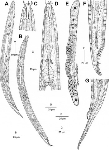
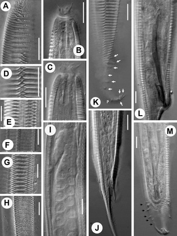
Females (figs 1, 2, 3 and table 1): body medium-sized, stout, tapering at both extremities, more at posterior end. Cuticular framework provided with heavily sclerotized, transverse bars having branched ends, forming hexagonal blocks (fig. 2D–G) arranged in eight longitudinal rows. Ends of transverse bars of cuticle ramifying into 2, 3 or 4 finer processes that interlock with processes of adjacent bars to form rhomboids with a zigzag pattern, longitudinally (figs 2D, 3L). Dot-like punctations interspersed with transverse bars (fig. 2H). Left side without discernible ridges. Right and left sides of body annulated; annuli 2.0–2.5 μm wide in different body regions. Lip region offset, 10–11 μm in diameter; three distinct lobe-like lips on right side (fig. 2B), three lips with frilled margins on left side (fig. 2C); lips provided with minute inner labial sensilla, raised outer labial sensilla, four long setose cephalic sensilla (fig. 2B, C). Amphid apertures not seen. Stoma prismatic, rhabditoid type, about one-fifth to one-sixth of pharyngeal length or 2.3–2.6 times labial diameter. Cheilostom tubular, weakly cuticularized (figs 1C and 2B, C); gymnostom cuticularized, with parallel walls, stegostom surrounded by pharyngeal collar for about 25–35% of stoma length (fig. 1C); metastegostom with a fine denticle on each plate (fig. 1C). Telostegostom obscure. Glottoid apparatus absent (fig. 2B, C). Pharynx with weakly swollen or cylindroid corpus, 85–107 μm long, 4.0–5.5 times isthmus length or 59.7–65.3% of pharyngeal length. Isthmus 25–32 μm long. Basal bulb strong, oblong to square-shaped, 30–42 μm × 22–27 μm, with grinder and single-chambered haustrulum. Nerve ring surrounding pharynx at 70–78% of pharyngeal length from anterior end. Secretory–excretory pore not visible. Body width at pharyngeal end 3.8–4.1 times labial diameter. Cardia 4–7 μm long with conical flaps. Intestinal cells large with prominent nuclei, boundary of intestinal lumen thin. Rectum length 3.0–3.5 times anal body diameter, terminating as a tube formed by external cuticular sheath. Reproductive system didelphic, amphidelphic. Ovaries dorsally reflexed with 2–3 tiers of oocytes in germinal zone. Anterior genital branch 2.6–2.8 times longer than posterior one. Posterior genital branch greatly reduced with small ovary having oocytes. Eggs not observed in utero. Vagina 15–23 μm long, with cuticularized lumen, anteriorly directed occupying a third to a half of corresponding body diameter. Vulva slit-like, post-equatorial. Distance between vulva and anus 3.2–4.4 times tail length. Tail 3.0–4.5 times anal body diameter long, uniformly tapering into a fine, usually dorsally curved, tip.
Pterygorhabditis punctata n. sp.: (A) entire female; (B) entire male; (C) anterior end; (D) pharyngeal region; (E) female reproductive system; (F) male posterior region; (G) female posterior region.

Pterygorhabditis punctata n. sp.: (A) anterior body region showing transverse cuticular bars (smooth and ramified ones); (B) anterior end with labial sensilla and stoma (right side); (C) anterior end with labial sensilla and stoma (left side); (D–G) body region (right side) showing cuticular pattern; (H) body region showing punctations; (I) male reflexed testis and a part of vas deferens; (J) female posterior region with anal cuticular sleeve; (K, L) male posterior region (lateral); (M) male posterior region (ventral) (scale bars = 10 μm).

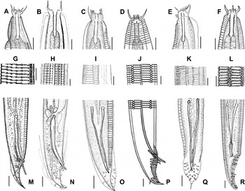
Comparison of anterior body region, cuticular ornamentation and male posterior region in species of Pterygorhabditis Timm, Reference Timm1957. (A, G, M) P. hungarica (after Andrássy, Reference Andrássy1982); (B, H, N) P. panopla (after Bernard, Reference Bernard1979); (C, I, O) P. superbus (after Huseni, Ahmad & Firdausi, Reference Huseni, Ahmad and Firdausi1997); (D, J, P) P. pakistanensis (after Timm, Reference Timm1957); (E, K, Q) P. saprophila (after Khan & Tahseen, Reference Khan and Tahseen2005); (F, L, R) P. punctata n. sp. (scale bars = 10 μm).

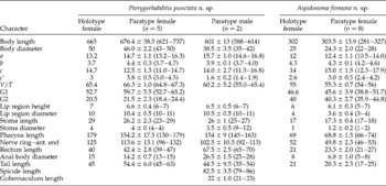
Morphometric characteristics (measurements in μm) of Pterygorhabditis punctata n. sp. and Aspidonema formosa n. sp.; mean ± standard deviation (range).

a= body length/body diameter; b= body length/pharynx length; c= body length/tail length; c′ = tail length/anal body diameter; V= (distance from anterior region to vulva/body length) × 100; T= (length of testis/body length) × 100; G1 = (length of anterior gonad/body length) × 100; G2 = (length of posterior gonad/body length) × 100.
Males (figs 1, 2, 3, table 1): body slender, almost straight upon fixation. Similar to female in general morphological characters except smaller body size. Testis single, dorsally reflexed; reflexed part 38–67 μm long. Spicules long, slender, 3.0–3.2 times longer than cloacal body diameter with slightly differentiated capitula, fused distally (figs 2L, M, 3R). Gubernaculum small, spatula–shaped, about 20–27% of spicule length. Bursa reduced, open, pseudopeloderan type, leaving small tail spike free. Bursal papillae setose, nine pairs, pre-cloacal pair not seen; post-cloacal papillae in 3+3+P+1+2 configuration (figs 1F, 2K, M). Papillae GP1, GP2 and GP3 less spaced, robust; GP4 smaller and thinner, separated from robust GP5 and GP6; phasmids anterior to dorsally directed GP7; GP8 and GP9 closely placed near tail tip.
Type habitat and locality
Samples containing P. punctata n. sp. were collected from the shore of a canal at Bulandshahr (geographical coordinates: 28.40 296°N, 77.85 824°E), Uttar Pradesh, India.
Type specimens
Holotype female, five paratype females and two paratype males on slide, Pterygorhabditis punctata n. sp. no. NOSR/1–4 deposited in Nematode Collection, Department of Zoology, Aligarh Muslim University, Aligarh, Uttar Pradesh, India.
Diagnosis and relationships
Pterygorhabditis punctata n. sp. is characterized by a medium-sized body; presence of flattened hexagonal blocks arranged in eight longitudinal rows in both sexes, three lobe-like lips on the right side and three reduced lips with frilled margins on the left side; a weakly cuticularized cheilostom; each metastegostomal plate with a minute denticle, pharyngeal corpus uniformly wide; female reproductive system with a greatly reduced posterior branch and males with long, slender, distally fused spicules, a spatula-shaped gubernaculum, a reduced, pseudopeloderan bursa and nine pairs of prominently setose, post-cloacal genital papillae.
Pterygorhabditis punctata n. sp. most closely resembles P. pakistanensis Timm, Reference Timm1957 in morphometric and morphological details, but differs in having greater a (body length/body diameter: 13.5–16.3 vs. 11.0–13.1) and c (body length/tail length: 11.0–14.7 vs. 8.3–9.4) values; a lesser number (8 vs. 10–12) of flattened hexagonal cuticular blocks in the female; lips on left side frilled (vs. indistinct); each metastegostomal plate with one denticle (vs. denticle not reported); males with conspicuous spatula-shaped (vs. blunt) gubernaculum; GP1 and GP2 post-cloacal (vs. pre-cloacal) and only the fourth pair (vs. sixth and seventh pairs thin and smaller) and the bursa reduced (vs. moderately developed).
Pterygorhabditis punctata n. sp. resembles P. superbus Huseni, Ahmad & Firdausi, Reference Huseni, Ahmad and Firdausi1997 in most morphometric characteristics but differs in having a longer body (0.6–0.7 mm vs. 0.3–0.5 mm); greater V value ((distance from anterior region to vulva/body length) × 100: 65–67 vs. 57–60); left side with indiscernible ridges (vs. six longitudinal ridges); cheilostom simple (vs. cuticularized); posterior genital branch greatly reduced (vs. equal to anterior genital branch); vagina anteriorly directed (vs. at right angle to main body axis); and males with longer (79–86 μm vs. 51–60 μm) spicules; larger (21–23 μm vs. 13–15 μm), proximally spatula-shaped (vs. attenuated) gubernaculum and pre-cloacal papillae absent (vs. four pre-cloacal pairs).
The new species also resembles P. saprophila Khan & Tahseen, Reference Khan and Tahseen2005 in most morphometric details but differs in having a longer body (0.6–0.7 mm vs. 0.41–0.51 mm) with cuticle tightly applied (vs. forming wing) in anterior region; greater V value (65–68 vs. 53–54); left side with indiscernible ridges (vs. two faint longitudinal lines); faint (vs. conspicuous) cheilostom; lips on right side conspicuous, lobe–like (vs. indistinct); pharyngeal corpus weakly swollen or cylindrical (vs. strongly swollen); posterior genital branch greatly reduced (vs. equal to anterior genital branch) and males with longer (79–86 μm vs. 55–57 μm) spicules; larger (21–23 μm vs. 14–15 μm), proximally spatula-shaped (vs. attenuated) gubernaculum and nine pairs of genital papillae with pre-cloacal papillae absent (vs. four pre-cloacal pairs).
Etymology
The species epithet ‘punctata’ denotes the presence of fine yet conspicuous scattered punctations besides the ridges and a rhomboid system of markings.
Remarks
The morphological feature, e.g. metacorpus, could have been taken as a character of comparison with P. superbus Huseni, Ahmad & Firdausi, 1997; however, it has been stated to be cylindrical in the original description (p. 46 para. 1) but illustrated to be prominently swollen (fig. 3C) apud Huseni et al. (Reference Huseni, Ahmad and Firdausi1997). The new species seems to be a close relative of P. pakistanensis Timm, Reference Timm1957 in many aspects; however, there are few noticeable differences: besides the lesser number of cuticular blocks in females, the interlocking of transverse cuticular bars seems to be more complex (fig. 3L vs. 3J) in the present species. Even if the differences of metastegostomal tooth and allometric ratios (a and c) are overlooked, the other characters, namely a conspicuous spatula-shaped gubernaculum and a fairly reduced bursa with dissimilar configuration of papillae, make P. punctata distinct from P. pakistanensis. It further appears that in the original illustrations of P. pakistanensis Timm, Reference Timm1957, Figures 1B and 1D are not indicated with appropriate scale bars and Figures B, C, D and H seem to have been drawn at the same scale.
Andrássy (Reference Andrássy2005) raised the genus Pterygorhitis for those species of Pterygorhabditis that have 1–3 head shields, a markedly swollen corpus and relatively thicker spicules, and transferred Pterygorhabditis panopla and Pterygorhabditis hungarica to the former genus. The presence of the shield is a character that poses ambiguity. Pterygorhitis panopla has a single head shield as also confirmed by Holovachov et al. (Reference Holovachov, Esquivel and Bongers2003). The shield, in our opinion, can also be due to an artefact that may occasionally appear during the fixation or dehydration process. Therefore, it can be a supplementary character but not the differentiating one. Another feature, the swollen corpus, is too flimsy a character to have a diagnostic value on its own. Spicules which are equally long in Pterygorhitis only differ on account of thickness but that, too, is a comparative feature. Spicules as illustrated in the Costa Rica population of P. panopla (Holovachov et al., Reference Holovachov, Esquivel and Bongers2003, Figure 2F) look slightly different, with a ventral triangular process similar to that of some species of Rhabditidae. Therefore, instead of grouping these species into two genera separated merely on very minor characters, we prefer placing the two species P. panopla Bernard, Reference Bernard1979 (fig. 3B, H, N) and P. hungarica Andrássy, Reference Andrássy1982 (fig. 3A, G, M) under the ‘panopla’ species group, while the others are placed under the ‘pakistanensis’ species group (fig. 3D, J, P).
Amended diagnosis
Pterygorhabditis is characterized by small to medium-sized individuals with a relatively plump body; a cuticle with conspicuous transverse annulations, with the right side adorned with 6–12 longitudinal blocks having a rhomboidal pattern, demarcated by ridges; the left side bearing 2–6 longitudinal lines or ridges; the lip region bearing setose appendages; small amphids; stoma rhabdtitoid type usually with inconspicuous metastegostomal denticles; glottoid apparatus absent; pharyngeal collar absent or very small; pharyngeal corpus weakly developed to strongly swollen; female reproductive system didelphic, amphidelphic with vulva equatorial to slightly post-equatorial; males with very long spicules with variable thickness, usually fused at the tip, bursa generally reduced, asymmetrical, with 7–9 pairs of genital papillae.
The species of genus Pterygorhabditis can be differentiated on the basis of the type of longitudinal cuticular patterns on the right side, the number of the lines/ridges on the left side, the presence or absence of an oval shield in the anterior region, the shape/thickness of spicules and also the number and arrangement of male genital papillae. The other features, namely labial sensilla, metastegostomal structure, nature of the pharyngeal corpus, length of female gonad, position of vulva, etc. are the features that show considerable variation, hence they cannot be relied upon exclusively for species identification.
Key to species of Pterygorhabditis Timm, Reference Timm1957
-
1 Larger species (L > 0.6 mm)2
–Smaller species (L < 0.6 mm)3
-
2 Ornamented cuticle divided into 10–12 longitudinal blocks with rhomboidal pattern in female, male with two pairs of pre-cloacal genital papillaepakistanensis
–Ornamented cuticle divided into eight longitudinal blocks with rhomboidal pattern in female, male without pre-cloacal genital papillaepunctata n. sp.
-
3 Neck region with flattened annules forming oval shield, spicules robust, c. 2–3 times thicker than spicule head4
–Neck region with simple coarse annulations, spicules attenuated, about the same thickness as spicule head5
-
4 Neck region with a large oval shield, right side with prominent longitudinal ridgespanopla
–Neck region with three oval shields, longitudinal ridges indiscerniblehungarica
-
5 Left side with two longitudinal lines, vulva with cuticular flap and males with nine pairs of genital papillaesaprophila
–Left side with four longitudinal lines, vulva without cuticular flap and males with eight pairs of genital papillaesuperbus
Description of Aspidonema formosa n. sp
General
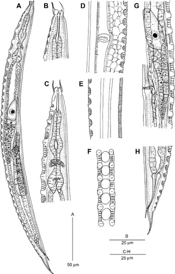
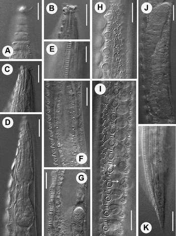
Female (figs 4, 5, 6 and table 1): body 0.28–0.32 mm long, plump, ventrally curved upon fixation, tapering at both extremities. Right side of body bearing two striated ridges, two rows of reticulate network, two rows of warts and a single mid-lateral row of round to ovoid shields (fig. 5I). Warts forming 35–48 pairs with smooth (fig. 5F) or bifid apices (fig. 5G); 2.5–5.0 μm high having faint, weakly developed skeletal rods within a few specimens (fig. 5H, I). Each two consecutive warts placed 5–8 μm apart with 4–6 fine ribs in between. Anterior warts up to level of gymnostom, and posterior ones slightly beyond rectum, are smaller. Striated ridges (figs 4E, 5E) form the outermost border of reticulate networks of hexagonal or polygonal pattern that surround the rows of warts and shields. Shields represent rounded, smooth membranous structures loosely fitting into large hexagonal/polygonal sockets/depressions alternate to wart pairs (fig. 5I). Shields are displaced from their position (fig. 5I) with a slight pressure or friction in a few specimens. Five longitudinal ridges present on left side of body (fig. 5K). Lip region set-off from adjoining body; inner and outer labial sensilla not distinct; cephalic sensilla with leaf-like bases, 6–8 μm long. Amphid apertures not seen. Cuticular fold at neck base not observed. Stoma rhabditoid-type, length about one-fourth of pharyngeal length or 2.5–3.0 times labial width. Cheilostom conspicuous, rod-like; gymnostom occupying more than 50% of stomal length; metastegostom anisomorphic (figs 4B, C, 5C), without armature. Telostegostom distinct. Pharynx with strongly swollen corpus having thickened lumen, narrow isthmus and ovoid, basal bulb 12–14 × 10–12 μm, with grinder and single-chambered haustrulum. Pharyngeal corpus 1.4–1.9 times longer than isthmus and basal bulb together. Body width at pharyngeal end 2.7–3.8 times labial width. Nerve ring encircling isthmus at 67–75% of pharyngeal length. Secretory–excretory pore and hemizonid not seen. Cardia conoid, 2–3 μm long. Intestine with wide lumen. Rectum 2.6–4.5 times anal body diameter long, extending into tube formed by external cuticle. Reproductive system didelphic, amphidelphic. Both genital branches equally developed. Ovaries large, dorso-laterally reflexed, with two rows of oocytes in germinal zone. Anterior ovary on right and posterior ovary on left side of intestine, both their apices crossing each other. Spermatheca and crustaformeria not distinguishable. Neither eggs nor spermatozoa observed in genital tract. Vagina length one-third of corresponding body diameter; vulva a transverse slit with an ovoid rim, slightly post-equatorial. Vulva–anus distance 4.9–6.6 times tail length. Tail conoid, uniformly tapering to a fine terminus.
Female Aspidonema formosa n. sp.: (A) entire body; (B) anterior end; (C) pharyngeal region; (D) vulval region (ventral); (E) body region (left side) showing longitudinal ridges; (F) body region (right side) showing shields surrounded by warts; (G) reproductive system (anterior branch); (H) posterior region.

Female Aspidonema formosa n. sp.: (A) anterior end showing rectangular cuticular shields; (B) anterior region showing the level of first wart pair with respect to stoma; (C) anterior end showing labial sensilla; (D) pharyngeal region; (E) body region (left side) showing striated ridge; (F, G) body region (right side) showing warts; (H, I) body region showing dot-like pattern and shields; (J) reproductive system; (K) posterior region (scale bars = 10 μm).

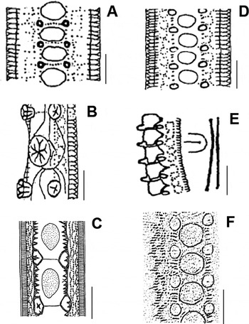
Cuticular ornamentation in species of Aspidonema (Sachs, Reference Sachs1949) Andrássy, Reference Andrássy1958: (A) A. stammeri (after Meyl, Reference Meyl1961); (B, C) A. scheucherae (after Meyl, Reference Meyl1961; Holovachov & Bongers, Reference Holovachov and Bongers2001); (D) A. ruehmi (after Meyl, Reference Meyl1961); (E) A. weingartnerae (after Meyl, Reference Meyl1961); (F) A. formosa n. sp. (scale bars = 10 μm).

Male: unknown.
Type habitat and locality
Aspidonema formosa n. sp. was obtained from a small ditch (alluvium soil rich in organic matter) at Sikandra Rao, Aligarh, Uttar Pradesh, India.
Type specimens
Holotype female and eight paratype females on slide Aspidonema formosa n. sp. NOA/1–6, deposited in the Nematode Collection, Department of Zoology, Aligarh Muslim University (geographical coordinates: 27.9108°N, 78.0797°E), Aligarh, Uttar Pradesh, India.
Diagnosis and relationships
Aspidonema formosa n. sp. is characterized by a small body with the right side having two rows of 35–48 conical to bifid warts, circular in outline, flanking a median row of oval to round, smooth membranous shields and flanked by a well-developed network on the sides; warts with faint or indiscernible skeletal rods; anisomorphic metastegostom without discernible armature, swollen pharyngeal corpus, female genital branches equal-sized, reflexed parts of ovaries crossing each other, vulva slightly post-equatorial and males unknown.
Aspidonema formosa n. sp. most closely resembles A. ruehmi (Sachs, Reference Sachs1949) Goodey, 1963 in most morphometric and morphological characteristics, but differs in having a relatively greater c value (12–18 vs. 11–13); warts circular (vs. transversely oval) in outline, greater number of wart pairs (35–48 vs. 30–34) supported by no, or a weakly developed, skeletal rod (vs. two distinct internal rods) and in the absence of males (vs. males present in A. ruehmi apud Andrássy, Reference Andrássy1983).
The new species differs from closely related A. stammeri (Sachs, Reference Sachs1949) Andrássy, Reference Andrássy1958 in having greater c (12–18 vs. 9–12) and smaller V values (54–56 vs. 59–64); fewer pairs of warts (35–48 vs. 50–59); middle row of weak and membranous (vs. cuticularized) shields with those at anterior and posterior extremities large and rectangular (vs. small, rounded) in shape; and in the absence of males (vs. males present).
Etymology
The species name is derived from the Latin word (formosus = beautiful) denoting its beautiful cuticular ornamentation.
Remarks
The genus Aspidonema is recorded for the first time from India. All five previously described species were reported from Germany, Hungary and Turkey, and were extracted from compost (Sachs, Reference Sachs1949; Andrássy Reference Andrássy1983), horse and cow dung (Andrássy, Reference Andrássy2005) or rotting material (Holovachov & Bongers, Reference Holovachov and Bongers2001).
Aspidonema formosa n. sp. was collected from a ditch; however, it was in close proximity to a farmyard manure storage pit. The species is represented by a population of females only. Amixis could be the probable mode of reproduction, although this is subject to verification. It is a fact that the morphometrics of all the congeners do not vary significantly, except for very few attributes. Hence, the differentiation has to be based entirely on the morphological characters that are relatively consistent and reliable. The shape of the labial sensilla, metastegostomal structure, nature of pharyngeal corpus, length of female gonad, position of vulva, etc., all have considerable intra-population variation and hence are not reliable for differentiation of species. The types of cuticular ornamentation, i.e. the number and structure of warts, shields and networks, besides the number of longitudinal ridges on the left side, form important characters for differentiation. Figure 6 compares the cuticular ornamentation in different species so as to draw the line of distinction. The species A. stammeri, A. ruehmi and A. formosa n. sp. show a more or less similar nature of ornamentations. However, besides their number and pattern, the proportionate sizes of the warts and shields also have distinctive value. The other congeners, namely A. sheucharae and A. weingartnerae, differ significantly on account of dissimilar warts and shields. Holovachov & Bongers (Reference Holovachov and Bongers2001) gave a comprehensive description of A. sheucharae with detailed scanning electron microscopy to reveal finer details of the cuticle. Aspidonema formosa markedly differs from A. sheucherae in the shape of warts (rounded vs. triangular), shape of shields (rounded to oval vs. ellipsoidal) and the nature of the network (polygonal vs. elongate/rectangular type).
Amended diagnosis
Aspidonema is characterized by small-sized nematodes with the right side ornamented with paired warts flanking a median row of larger, usually round to oval, membranous shields and surrounded by a punctated network latero-dorsally and latero-ventrally; the left side bearing lines or thin ridges; the lip region expanded with setose cephalic sensilla; stoma rhabditoid, usually with sclerotized cheilostom, metastegostom usually without denticles, occasionally anisomorphic; pharyngeal collar short; pharyngeal corpus usually swollen; female reproductive system didelphic, amphidelphic with reflexed ovaries; vulva equatorial or post-equatorial in position; rectum long; males with long, slender spicules; bursa asymmetrical and tail forked distally in males.
The genus has four known species besides A. formosa n. sp. The species A. sachsi has not been considered among the valid species of the genus as it is based on a single male specimen. A species needs to sustain itself and, in order to produce its progeny, the female is required, which is unknown in this case, hence the status of the species is uncertain. Also, a number of morphological characters are not shown by males or have disparity with female characters; therefore, A. sachsi is not considered to be a valid species.
Key to species of Aspidonema (Sachs, Reference Sachs1949) Andrássy, Reference Andrássy1958
-
1 Warts fewer, up to 35 pairs2
–Warts more than 35 pairs3
-
2 Warts 20–26 pairs, provided with 3–6 skeletal rods, shields roundedscheucherae
–Warts 30–35 pairs, provided with 2 skeletal rods, shields ovalruehmi
-
3 Warts oval in cross-section, shields closely packed with angular edgesweingartnerae
–Warts rounded in cross-section, shields with curved edges, spaced4
-
4 Warts 50–59 pairs, c= 9–12, V= 59–64stammeri
–Warts 35–49 pairs, c= 12–18, V= 54–56formosa n. sp.
Discussion
Bunonematids, like rhabditids, are saprobionts that prefer organic matter-enriched habitats harbouring a rich bacterial flora. They have been reported from a variety of habitats, namely soil, moss, forest duff, decaying plant residue, moist soil at the bank of a water body or from the dung and frass of bark beetles. To survive and flourish in these decomposing types of habitat these nematodes seem to be facultative anaerobes and are also able to tolerate changing osmotic pressures, like other saprobiotic nematodes (Sudhaus, Reference Sudhaus, Makarov and Dimitrijević2008). Their presence in remote locations, including the crevices and tunnels of trees, away from the soil environment, may suggest phoresy, a mechanism involving the use of an organism (usually an insect) as a carrier for transportation to a new and more suitable habitat away from the unfavourable one. The phoretic relationship is, therefore, considered a type of commensalism in which these nematodes associate themselves with insects of different species, to obtain the benefit of transportation. Many bark beetles, scarab beetles, etc. serve as transport hosts. The species of Pterygorhabditis have been largely reported from arboreal sites, namely from under the bark of trees or from the frass of the bark beetles, indicating the possibility of such associations. The species of Aspidonema have mostly been reported from cow dung and compost, which harbour a variety of insects having the potential to be phoretic associates. Habitats of P. punctata n. sp. and A. formosa n. sp. may appear different, but in both cases the association with insects cannot be ruled out due to their closeness to farmyard manure reservoirs or other rotting substances. It has to be further established whether the commensal nematodes complete their life cycles in hollow spaces of the host without obtaining nutrients from the host – a condition called entoecy – or whether they are simply necromenic, thus invading and feeding on the transport host after its death.
Bunonematids seem to be equipped with a series of cuticular adornments, presumably to survive in the adverse environments. Several studies have been conducted in the past to ascertain the cuticular/extracuticular origin of their ornamentations. Raski & Jones (Reference Raski and Jones1973), while elucidating the transmission electron microscopic (TEM) details of the bunonematid cuticle, reported that the striations of ridges, skeletal rods of warts and the palisade rods originate from an inner cortical layer, visible under light microscopy but not under scanning electron microscopy (SEM). Another ultrastructural study on the cuticle of three species of Bunonema Jägerskiöld, Reference Jägerskiöld1905 revealed that the ridges and tubercles are formed due to evagination and thickening of the median zone of the cuticle (Van Gansbeke et al., Reference Van Gansbeke, Bert, Govaerts, Claeys, Van Driessche, Manhout and Borgonie2004), thus confirming their cuticular origin. However, the polygonal network was stated to lie above the epicuticle, hence being extracuticular in origin. That was the reason the network was rubbed free, because of its superficial placement in B. richtersi Jagerskiöld, 1905 and B. hysseyi Bernard, Reference Bernard1979, but not the tubercles, which remained firmly attached (Raski & Jones, Reference Raski and Jones1973; Bernard, Reference Bernard1979). In Rhodolaimus dimorphicus the tubercles were also detached but in a broken state (Bernard, Reference Bernard1979) confirming their deeper, cuticular origin. The removal of shields in A. formosa by slight friction is also indicative of its superficial extracuticular status.
Financial support
The present work was supported financially by the Department of Science and Technology, New Delhi and Ministry of Environment and Forests, New Delhi, India.
Conflict of interest
None.