I. INTRODUCTION
The extensive applications of high-speed-cutting machines and materials difficult to process could raise extremely strict requirements for excellent red hardness and wear resistance of cutting tools.Reference Zhu, Zhang and Ding1 Normally, commercial Fe-based tool steels could not be suitable for the meets of high-speed cutting or dry cutting. TiN, TiC hard ceramic films with ultra-high hardness synthesized on tool steels could prolong service life and improve cutting efficiency.Reference Wang, Xue, Cao and Zhao2 However, the disadvantages of hard ceramic film including high residual stress, thin thickness, and weak bonding with substrate could seriously limit the wide industrial application in high-speed cutting.Reference Huang, Ma and Chen3
High-entropy alloys (HEAs), first proposed in 2004, have been drew great attention in metallic materials science and engineering because of their excellent mechanical performances.Reference Wu, Jiang, Jiang, Pan, Cao, Deng, Wang and Li4 It is remarkably considered that HEAs could be one of the three research directions in metallic field.Reference Zhang, Zuo, Tang, Gao, Dahmen, Liaw and Lu5 Owing to the high-entropy effect in thermodynamics, the explored HEAs could be classified by main phase structures into three groups: face-centered cubic (FCC),Reference Pickering, Muñoz-Moreno, Stone and Jones6 body-centered cubic (BCC),Reference Gao, Gao, Hawk, Ouyang, Alman and Widom7 and hexagonal close-packedReference Zhao, Qiao, Ma, Gao, Yang, Chen and Zhang8 structures plus a few intermetallic compounds.Reference Gwalani, Choudhuri, Soni, Ren, Styles, Hwang, Nam, Ryu, Hong and Banerjee9 Multiple alloy elements with atomic radius difference and strong interatomic interaction contained in the HEA systems could enhance severe lattice distortion and sluggish diffusion.Reference Juan, Tseng, Hsu, Tsai, Tsai, Lin, Chen, Lin and Yeh10 Therefore, BCC structural HEAs possess outstanding hardness and prominent thermal stability, which have been regarded as a potential for high temperature materials.Reference Senkov, Senkova and Woodward11 Generally, the bulk HEAs fabricated by repetitive vacuum arc melting technique could obtain uniform microstructures with much difficulty.Reference Han, Chen, Zhao, Fan, Yang, Shao and Yao12 Besides, many precious metals and complex preparation process could raise huge costs, which may restrict the development and application.Reference Guo, Shang and Liu13
Laser cladding, an advanced additive manufacturing technique, with its characteristics of high-temperature heating and rapid cooling could acquire uniform coating with desirable mechanical performances, whose thickness may reach millimeter scale.Reference Shang, Axinte, Sun, Li, Li, Du, Qiao and Wang14 Moreover, the rapid solidification could result in the nonequilibrium solute retention effect, further improving solid solubility of alloy elements and reduce component segregation.Reference Wu, Zhang, Zhang, Zhang and Dong15 Therefore, laser cladding might be a unique method for preparing HEA coatings on the tool steel substrate. Up to now, many novel HEA coatings fabricated by laser cladding have been developed.Reference Zhang, Zuo, Tang, Gao, Dahmen, Liaw and Lu5
In previous studies, laser cladding Fe5Cr5SiTiCoNbMoW HEA coating has been successfully fabricated on the Q235 steel substrate. The phase structure and wear resistance of as-cladding coating were investigated.Reference Guo, Liu and Zhou16 However, Q235 steel is a kind of structural material, not belonging to cutting tools, which cannot fully show HEA coating’s outstanding performances. In this paper, Fe5Cr5Co5SiTiNbMoW HEA coating was designed as follows: (i) Fe, Co, and Cr could easily form solid solution because of similar atomic sizes and electronegativities. (ii) Si, Ti, Nb, Mo, and W are common alloying elements in tradition alloys. Co-adding these elements into the HEA could obtain more excellent strengthening effect. And the coating was fabricated by laser cladding on W6Mo5Cr4V2 tool steel. The microstructure and hardness transition after various annealing temperatures were researched in detail. Besides, the annealing strengthening mechanism has been also discussed.
II. MATERIALS AND EXPERIMENTAL METHODS
A. Coating preparation
The Fe, Cr, Co, Si, Ti, Nb, Mo, and W metal powders with the purity more than 99.6% and the size about 150–250 mesh were used for raw materials. The weighed powders in specific proportion of 5:5:5:1:1:1:1:1 were dry blended for 4 h in the stainless steel jar using planetary ball mill (QM-3SP2, Nanda Instrument Co., Ltd., Nanjing, China). After being sifted, the mixed alloy powder was stored in a vacuum drying oven. The W6Mo5Cr4V2 tool steels with a size of Ø 50 × 10 mm were selected as substrates for laser cladding. The surface was polished by using a grinding machine, and then cleaned by alcohol to get rid of dirt and oil. Afterward, the substrates were placed in a vacuum drying oven for backup.
The fiber laser machine (YLS-6000, IPG Photonics Corporation, Oxford, Massachusetts) was used for laser cladding. The 1.5 mm mixed alloy powder in thickness was preplaced on the substrate. The optimal process parameters were as follows: spot diameter D = 3.5 mm, laser power P = 4.0 kw, and scanning rate v = 6 mm/s. During the laser cladding process, the pure argon gas was used to prevent the coating from oxidation. After laser cladding, the HEA coatings were annealed at 800, 850, 950, and 1050 °C for 4 h, using the SYX-6-13 heat treatment furnace.
B. Coating characterization
The phase structures of HEA coating were examined by X’Pert Powder X-ray diffraction (XRD) with copper target (λ = 1.5406 Å). The XRD was operated at 45 kV and 200 mA, a scanning range of 20°–80°, and a scanning length of 0.02°. The cross-sectional microstructure of HEA coating before and after various annealing temperatures was observed by using an OLYMPUS GX51 metallurgical microscope (OM; OLYMPUS, Tokyo, Japan), a Supra-40 scanning electron microscope (SEM), and an energy dispersion spectrum (EDS). The microhardness of cross-sectional coatings was measured by using a JMHV-1000AT microhardness tester (DONGHUA, Shanghai, China) under a load of 200 g for 10 s. Each point was measured more than three times to narrow error.
III. EXPERIMENTAL RESULTS
A. Phase structure and microstructure of as-cladding HEA coating
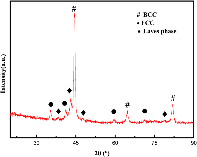
Figure 1 shows the XRD pattern of as-cladding Fe5Cr5Co5SiTiNbMoW HEA coating. The HEA coating is mainly composed of BCC and FCC plus a few Laves phases. The corresponding angles of BCC phase peaks are 44.44°, 64.66°, and 81.84°. The lattice constant is thus calculated to be 0.288 nm, similar with α-(Fe, Cr) phase (PDF # 41-1466). Additionally, the angle of (111) peaks in FCC phase is 35.38°, and its lattice constant is calculated to be 0.440 nm, well matched to (Nb, Ti)C (PDF # 47-1418).
XRD pattern of as-cladding HEA coating.
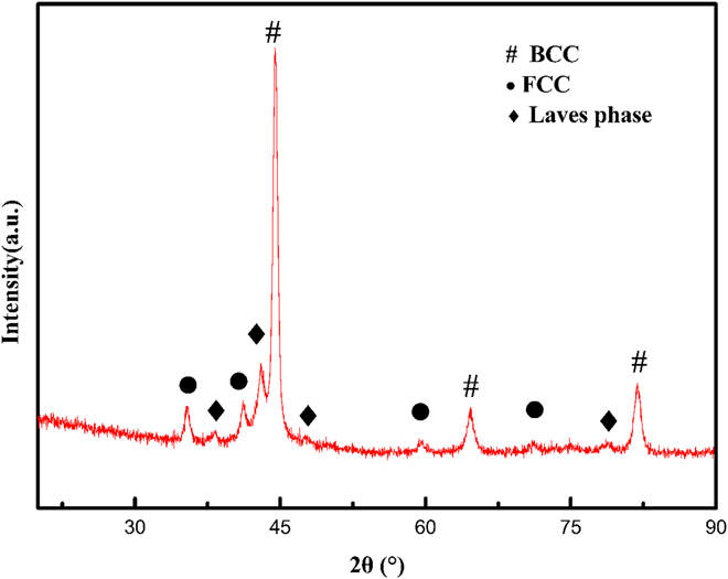
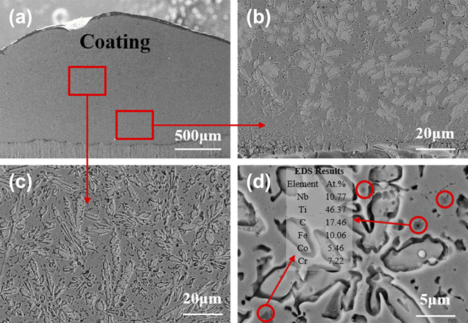
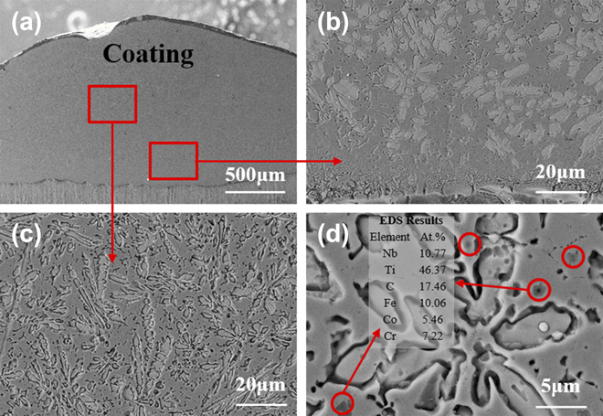
Figure 2 shows the cross-sectional microstructure of as-cladding HEA coating. Figure 2(a) shows the whole cross section morphology. The coating is about 1.2 mm in thickness. There are no microcracks or holes existing in HEA coating. Figure 2(b) shows the bottom microstructure of HEA coating. The coating is of metallurgical bonding with the substrate. Figure 2(c) shows the microstructure of the center area. Amounts of fine dendrites could be widespread in the coating with no specific growth direction. Meanwhile, there exist some dendrites with large sizes because Laves and FCC phases might be an excellent heterogenous nuclear center.Reference Yang, Zhang, Zhou, Guo, Ren, Yang and Yang17 Figure 2(d) shows the enlarged drawing of Fig. 2(c). The nanosized particles disperse and distribute in the coating. The EDS results indicate that the particles could be FCC structural (Ti, Nb)C with a small amount of Fe, Co, Cr elements contained.
Cross-sectional microstructure of as-cladding HEA coating: (a) the entire morphology, (b) microstructure of bottom region, (c) microstructure of center region, and (d) analysis of particle phase.
B. Phase structure of as-annealing HEA coating
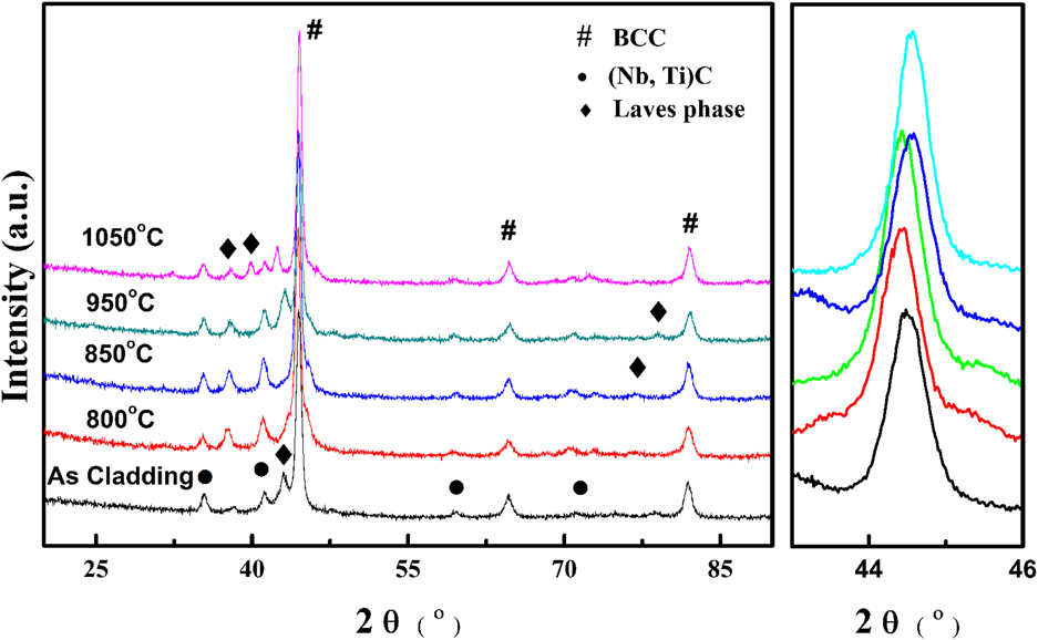
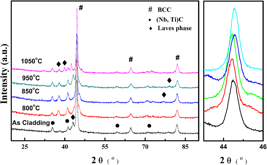
Figure 3 shows the XRD patterns of Fe5Cr5Co5SiTiNbMoW HEA coating before and after annealing. There after annealing below 950 °C, no other intermetallic phases appeared in the coating. However, while annealing at 1050 °C for 4 h, amounts of Laves phases produce because solute atoms possessing strong vitalities in high temperature could easily diffuse to form intermetallic compounds. From the detailed analysis of 43.5°–46°, it can be suggested that annealing below 850 °C, (110) peak of BCC phase shifts to the left. And then with the annealing temperature increasing, the (110) peak shifts to the right, lattice distortion effect decreasing, which could be consistent with that the high-temperature diffusion of solute atoms produces intermetallic compounds, reducing lattice expansion.
XRD patterns of HEA coating before and after annealing.
C. Microstructure transition of HEA coating after annealing
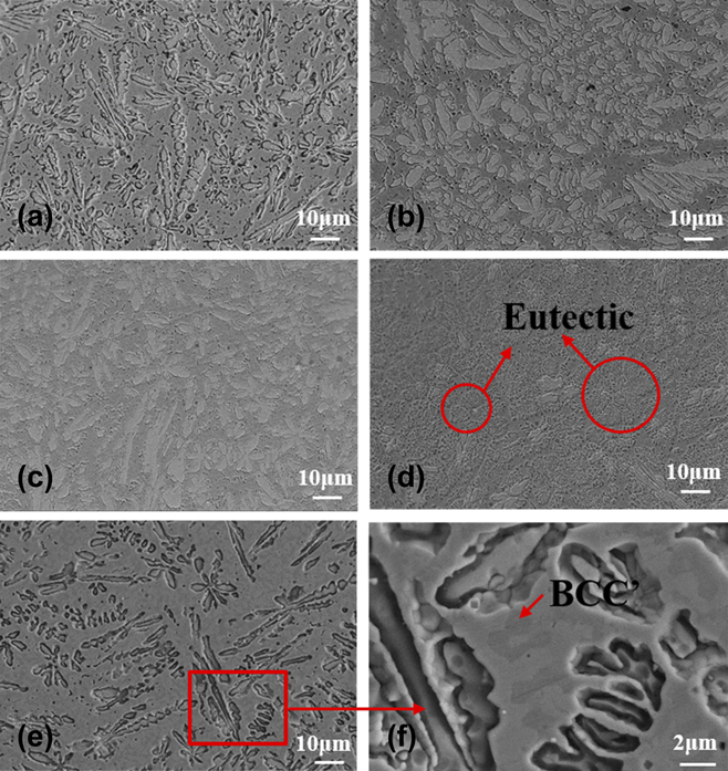
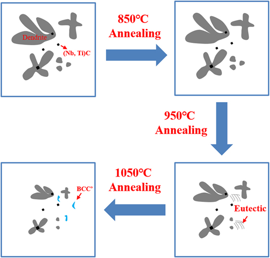
Figure 4 shows the microstructures of HEA coating after various annealing temperatures. Figure 4(a) shows the microstructure of as-cladding HEA coating. Figures 4(b) and 4(c) show the microstructures of HEA coating after 800 °C and 850 °C, respectively. The morphologies of as-annealed HEA coatings below 850 °C remained little changed, compared with as-cladding HEA coating. Figure 4(d) shows the microstructure of coating after annealing at 950 °C. Amounts of crystal whiskers are appeared in the coating, which is explained that high temperature could improve diffusion coefficient of solute atoms.Reference Schuh, Mendez-Martin, Völker, George, Clemens, Pippan and Hohenwarter18 The dendrites are decomposed into eutectic structure. The microstructures after 1050 °C annealing are shown in Figs. 4(e) and 4(f). The sizes of dendrites become smaller. And the eutectic structure disappears. The BCC′ phases could be observed in the matrix.
Microstructure of HEA coating with various annealing temperatures (a) as-cladding, (b) annealing at 800 °C, (c) annealing at 850 °C, (d) annealing at 900 °C, (e) annealing at 1050 °C, and (f) amplification analysis of coating annealing at 1050 °C.
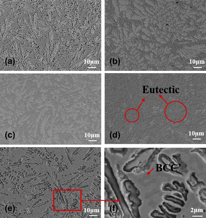
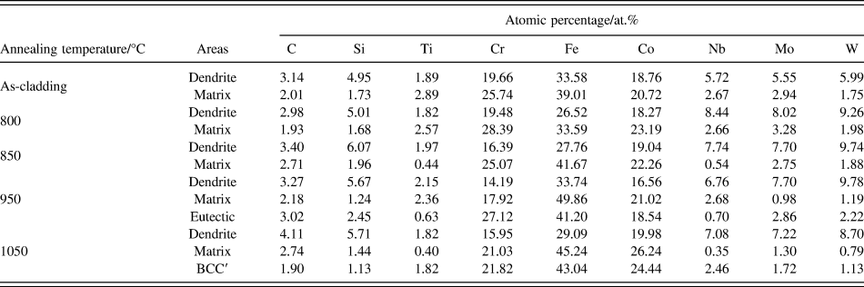
Table I shows the EDS analysis results of HEA coating before and after annealing. Each value is averaged of multiple measurements. Here in as-cladding coating, the main elements could be Fe, Co, and Cr with the content almost 70%. Furthermore, the Fe content is higher than Co and Cr because a few molten substrates mixed with alloy powders to form HEA coating in the laser cladding process. The Si and high-melting-point elements of Nb, Mo, W are enriched in the dendrite. After annealing at 950 °C, the Si, Mo, and W elements were segregated in the crystal whiskers. While annealing at 1050 °C, the amount of Nb, Mo, and W elements contained in BCC′ phase is higher than that of the BCC matrix.
The EDS results of Fe5Cr5Co5SiTiNbMoW HEA coating before and after annealing.

D. Diffusion layer of as-annealing HEA coating
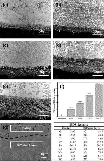
Figure 5 shows the OM and SEM images of the interface between HEA coating and substrate before and after various annealing temperatures. Figure 5(a) shows the interface of as-cladding HEA coating. Before annealing process, there exists an obvious white band in the bonding area. Figures 5(b)–5(e) show the interface morphologies after annealing at 800 °C, 850 °C, 950 °C, and 1050 °C, respectively. The black area shown in the figures could be named as diffusion layers. It is seen that with the annealing temperatures increasing, the thickness of the diffusion layer increases, and the result is shown in Fig. 5(f). After annealing at 1050 °C, the diffusion layer reaches about 90 μm. Moreover, the dendrites in the diffusion layer are coarser than that of away from the interface. Figure 5(g) shows the SEM image of the diffusion layer after annealing at 1050 °C. The dendrites in the diffusion layer are decomposed. Many second phases are large-scale separated in the grain boundaries. The elemental composition of the diffusion layer and coating are shown in the EDS results. The Cr and C contents contained diffusion layer are higher than that of coating. And high-melting-point elements of Nb, Mo, and W were also enriched in the diffusion layer.
OM and SEM images of the interface between HEA coating and substrate before and after various annealing temperatures. (a) OM image of as-cladding coating, (b) OM image of coating annealing at 800 °C, (c) OM image of coating annealing at 850 °C, (d) OM image of coating annealing at 950 °C, (e) OM image of coating annealing at 1050 °C, (f) thickness of diffusion layers, (g) SEM image of diffusion layer after annealing at 1050 °C, and EDS results of the coating and diffusion layer.

E. Microhardness evolution
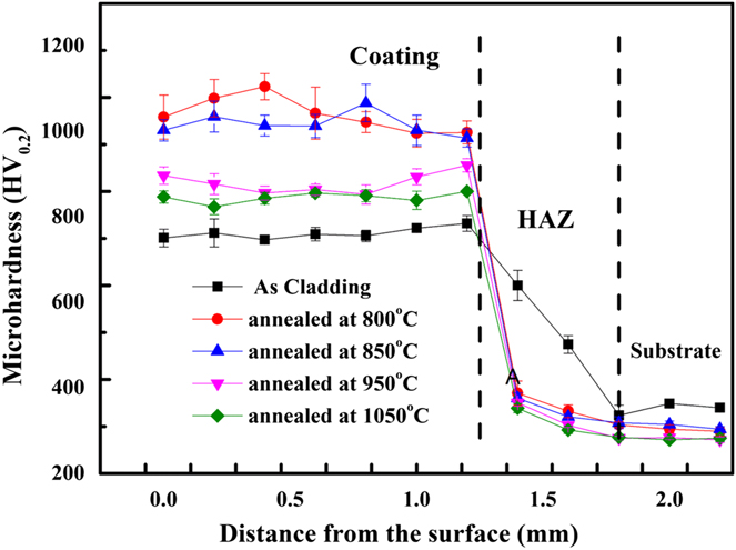
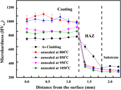
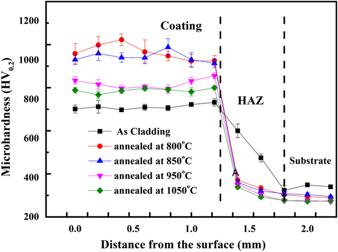
Figure 6 shows the microhardness evolution of HEA coating before and after annealing. The as-cladding HEA coating exhibits relatively low hardness value, about 780 HV0.2. The heat-affected zone of the substrate near cladding coating possesses high hardness because laser heating and rapid cooling results in laser quenching strengthening effect. But after high-temperature annealing, the hardness increases. In the classical theory, annealing treatment could reduce hardness of the work pieces, improving cutting performance.Reference Kotan, Saber, Koch and Scattergood19,Reference Miracle and Senkov20 Surprisingly, the coating possesses high hardness, about 1050 HV0.2, after 800 °C annealing, which is of huge improvement compared with that of as-cladding coating. Also, while annealing temperature reached 1050 °C, the hardness is higher than that of as-cladding coating. Normally, the W18Cr4V and W6Mo5Cr4V2 tool steels after several complex heat treatments could be soft in the working condition above 600 °C. Hence, the HEA coating exhibits excellent softening resistance ability. This kind of HEA could be a desirable coating material for high-speed cutting.
Microhardness evolution of HEA coating before and after annealing.
IV. DISCUSSION
A. Phase formation in HEA coating
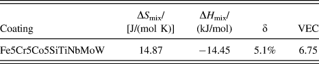
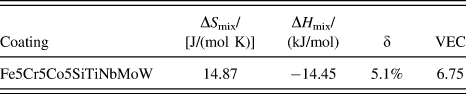
In the XRD analysis, although a small amount of Laves phases and (Nb, Ti)C exist in the HEA coating, the amount of BCC solid solution could be of overall majority. Up to now, many literature studies have reported the BCC and FCC phase formation rules of HEAs, according to the physicochemical empirical formulae proposed by Guo and Zhang.Reference Miracle and Senkov20,Reference Guo, Ng and Liu21 Hence, the relevant thermodynamic parameters, including enthalpy of mix (ΔH mix), configuration entropy of mix (ΔS mix), average atomic size difference (δ), and average valence electron concentration (VEC) as well as average melting point (ΔT m), are shown in Table II. Here, the ΔS mix is 1.78R J/(mol K) (where R is the gas constant), far higher than that of equiatomic HEAs composed of five elements (1.61R).Reference Yeh, Chen, Lin, Gan, Chin, Shun, Tsau and Chang22 Thus, the high entropy of HEA coating could promote the production of single solid solution, inhibiting brittle intermetallic phases. The ΔH mix and δ are calculated as −14.41 kJ/mol and 5.1%, respectively, fully complying with Zhang’s BCC solid solution formation criterion. Furthermore, the value of VEC calculated as 6.75 is below 6.87, further confirming the Guo’s BCC formation rule.Reference Guo, Ng, Lu and Liu23 Another reason for the formation of single solid solution is that laser rapid solidification would restrict the atomic migration and nucleation of second phase, improving the stability of nonequilibrium BCC solid solution. Additionally, some nanoparticle (Nb, Ti)C phases were widely dispersed in the coating, as shown in Fig. 2. The enthalpy of Nb–C and Ti–C (ΔH Ti–C = −109 kJ/mol, ΔH Nb–C = −102 kJ/mol) is extremely “negative”. Therefore, the Nb and Ti atoms prior to combine with C atoms contained in the molten substrate, forming strong carbides.
Related thermodynamic parameters of the Fe5Cr5Co5SiTiNbMoW HEA.
B. Microstructure evolution
In the microstructure of as-cladding coating, it is clearly observed that fine dendrite and tiny (Nb, Ti)C carbides. The grain refining is mainly related to two factors. One is based on sluggish atomic diffusion effect. The grain nucleation and growth could be controlled by partitioning of multiple alloy elements and substitution diffusion. In the HEA system, high atomic interaction and size difference could enhance viscosity of liquid metal. According to Stokes–Einstein Eq. (1),Reference Mason24
 $${D_{{\rm{eff}}}} = {{kT} \over {3\pi \eta \alpha }}\quad ,$$
where D eff is the effective diffusion coefficient, k is the Boltzmann constant, α is the interatomic spacing, T is the Calvin temperature, and η is the liquid viscosity. Hence, high viscosity could limit the solute diffusion, restricting phase nucleation and growth in the solidification process. Another one could be that laser rapid solidification shortened solute diffusion time, inhibiting grain growth.
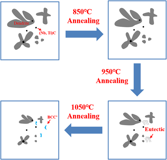
The diagram of the microstructure evolution before and after annealing is displayed in Fig. 7. The microstructure evolution may be concluded that with annealing temperature increased, the BCC matrix is decomposed to form the BCC′ phase. The size and number of dendrites and Laves phases are diminished. However, nanosized (Nb, Ti)C particles stably exist in the coating. The coating exhibits excellent microstructural stability above 1050 °C, which might be closely related to the sluggish diffusion effect. Whereas after annealing, the diffusion layer occurred in the bonding interface between substrate and coating because of diffusion of C, Nb, Mo, and W atoms into the diffusion layer. Besides, the increase of the thickness could be caused by that high temperature would improve atomic diffusion activation energy, accelerating diffusion length.
Diagram of microstructure evolution before and after annealing.
C. Strengthening mechanism
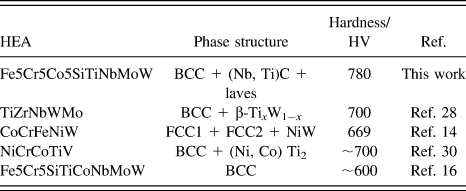
The hardness results indicate that as-cladding Fe5Cr5Co5SiTiNbMoW HEA coating exhibits significantly high hardness about 780 HV0.2. Notably, Table III shows the phase structure and hardness of some HEA systems fabricated by laser cladding. The mean hardness in our work could be higher than that of these recently reported HEAs. Similarly, the following reason could result in high hardness shown in the Fe5Cr5Co5SiTiNbMoW HEA coating. First, amounts of different alloy elements, especially big-sized Ti, Nb, Mo, and W atoms contained in the HEA, result in severe lattice distortion effect, enhancing the solid solution strengthening effect of the BCC phase.Reference Han, Chen, Zhao, Fan, Yang, Shao and Yao12 Also, nanosized (Nb, Ti)C and Laves phase were observed in the coating, which could improve the precipitation strengthening effect. Likewise, Cheng found that TiC–TiB2 phases were in situ formed in laser cladding CoCrCuFeNi HEA coating, resulting in hardness promotion.Reference Cheng, Liu, Liang and Chen25 Additionally, laser rapid solidification could restrict the grain growth, causing remarkable grain refinement strengthening.Reference Shuai, Yang, Wu, Lin, Liu, Zhou, Feng, Liu and Peng26
The phase structure and hardness of some reported HEA coating fabricated by laser cladding.

Besides, our HEA coating exhibits excellent softening resistance ability. Even after annealing at 1050 °C for 4 h, this HEA coating shows high hardness about 850 HV0.2, in which corresponding Rockwell hardness is nearly 66 HRC. It might be a new and surprising exploration in the HEA research field. The Fe5Cr5Co5SiTiNbMoW HEA coating fabricated by laser cladding displayed annealing strengthening ability, opposite to the traditional engineering metal materials.Reference Cavaleiro, Marques, Fernandes, Caevalho and Hosson27,Reference Zhang, Zhou, Yu and Li28 Especially, the coating shows high average hardness about 1050 HV0.2 annealed at 850 °C for 4 h. This phenomenon could be explained by XRD patterns of HEA coating after 850 °C annealing. The (110) peak of the BCC phase shifts to lower degree, indicating lattice expansion intensification. Therefore, BCC solid solution after annealing at 850 °C possesses a higher solid solution strengthening effect, resulting in increasing microhardness of the coating. Similar research results have been obtained that laser cladded refractory TiZrNbWMo HEA coating had ultra-high hardness about 1300 HV after annealing at 800 °C in Zhang’s publication.Reference Zhang, Zhou, Yu and Li28 Differently, the thickness of our HEA coating is about twice wider than that of Wang’s. Likewise, Huang also found high-temperature annealing could not decrease the microhardness of laser cladded TiVCrAlSi HEA coating.Reference Huang, Zhang, Vilar and Shen29
V. CONCLUSION
(1) A crack-free and microstructure-dense Fe5Cr5Co5SiTiNbMoW HEA coating has been successfully fabricated by laser cladding on W6Mo5Cr4V2 tool steel.
(2) The as-cladding HEA coating is mainly composed of BCC solid solution phase and (Nb, Ti)C plus few Laves phase. The microstructure mainly consists of dendrites, matrix, and nanosized (Nb, Ti)C particles. The high melting point elements of Nb, Mo, W were segregated in the dendrites.
(3) The microstructure has little changed below 850 °C annealing. Above 850 °C, the dimensions of dendrites become smaller, the BCC matrix decomposes to from eutectic, and then transforms into the BCC′ phase.
(4) The average microhardness of as-cladding HEA coating is about 780HV0.2. After annealing, the coating possesses prominent hardness improvement. Especially, the average hardness after annealing at 850 °C for 4 h could reach 1050 HV0.2. The annealing strengthening mechanism is attributed to enhancing the solid solution strengthening effect of the BCC phase. Therefore, the laser cladding Fe5Cr5Co5SiTiNbMoW HEA coating might be a potential application on dry and high-speed cutting.
ACKNOWLEDGMENTS
The author is grateful for financially support by the National Natural Science Foundation of China (No. 51671061), High-level Innovative Talents Plan of Guizhou province [No. (2015)4009], and Science and Technology Planning Project of Guizhou province [No. (2017)5788].