Introduction
In 1906, Robert Bárány published his original research on the caloric test and his convection theory to outline the mechanisms behind the generation and direction of caloric nystagmus. Today, how calorics are induced, what they mean and what structures of the inner ear produce them is still debated.Reference Longridge, Mallinson and Pothier1, Reference Longridge, Mallinson and Pothier2 The one certain fact is that the tested side is the side from which the response arises; irrigation of the left ear produces a nystagmus generated by the left ear. The fact that a caloric test is side-specific will always be crucial from a clinical point of view, because unilateral caloric abnormalities can dictate the side on which surgical or medical management of a disease is undertaken.
In 1964, Jongkees and Phillipszoon described the ‘sum over difference’ calculation for slow phase velocity of caloric responses to determine which side was pathological.Reference Jongkees and Philipszoon3 They utilised a ‘two standard deviations from normal’ distribution curve for delineating their normal population.
Caloric testing stood as the only effective method of quantifying vestibular function until computerised dynamic posturography (Neurocom, Clackamas, Oregon, USA) was introduced. The latter is an internationally used standardised sway and balance assessment technique, which also defines ‘normal performance’ as falling within two standard deviations of the mean for an age-matched population. Lipp and Longridge showed that although posturography is a less specific detector of vestibular disease (it does not lateralise pathology), it is significantly more sensitive than calorics at detecting vestibular disease.Reference Lipp and Longridge4
Vestibular-evoked myogenic potentials were initially recognised as being a vestibular response to sound in the 1960s.Reference Nguyen, Welgampola and Carey5 Cervical vestibular-evoked myogenic potential testing was developed as a clinical test in the 1990s and is now used as a routine diagnostic assessment.Reference Curthoys6 Ocular vestibular-evoked myogenic potentials were first described in 2005.Reference Rosengren, Todd and Colebatch7 The responses can be induced by a tendon hammer on the head, a mini-vibrator or a sound stimulus (click or tone). To date, the focus has been on how reliable the measured response is and which are the best parameters to evaluate. Bone conducted cervical and ocular vestibular-evoked myogenic potentials create vibrational waves that conduct across the skull; these are not focused on one side, but directly stimulate the otoliths in both ears simultaneously.Reference Nguyen, Welgampola and Carey5 The interaural asymmetry ratio reported in a vestibular-evoked myogenic potential assessment is the ‘otolithic’ equivalent of the standard canal paresis score (i.e. the traditional measurement technique of caloric evaluation during video-oculography). This ratio is a powerful calculation for identifying the affected side.Reference Curthoys6
Despite the fact that the caloric testing protocol is well recognised throughout the world, the degree of caloric response asymmetry considered abnormal varies (25 per cent, 20 per cent and 17 per cent are the values quoted by various sources).Reference Mallinson, Longridge, Pace-Asciak and Ngo8 Each laboratory has its own accepted norms. The same is true for vestibular-evoked myogenic potential testing. Each centre needs to establish local standards by testing normal people with the centre's own equipment, to confirm that their technique produces results similar to other centres, in order to provide normative data.Reference Nguyen, Welgampola and Carey5
Both cervical and ocular vestibular-evoked myogenic potentials offer several parameters for detecting abnormalities. In addition to the interaural amplitude ratio (the parameter traditionally used), wave latency can also be compared with normative data (similar to auditory brainstem response testing).
Because of the caveats outlined by Nguyen et al.,Reference Nguyen, Welgampola and Carey5 we utilise sound stimuli. The present paper describes a sensitive, accurate and reliable technique for recording sound-induced cervical and ocular vestibular-evoked myogenic potentials to delineate unilateral and bilateral vestibular pathology. The reason for emphasising tones is that the interaural attenuation of an insert tone stimulus is 90 dB.Reference Katz, Medwetsky, Burkard and Hood9 This means that, as is the case with calorics, the stimulated ear is the one from which the stimulus response is arising. This cannot be said with either form of vibration used in vestibular-evoked myogenic potentials (tendon hammer or mini-vibrator). A click is a more generalised sound stimulus, where the attenuation is less specific to the frequency so specific side localisation is less conclusive.
Many of the patients we assess have vestibular dysfunction due to trauma. Concomitantly, they often also incur neck injury. This precludes the ‘head lift’ and ‘head turn’ techniques, which are frequently used cervical vestibular-evoked myogenic potential assessment techniques.
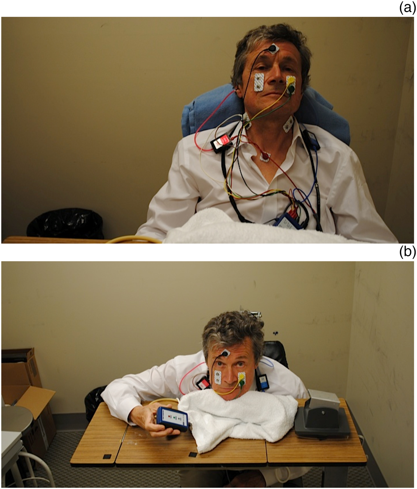
We devised a technique of carrying out cervical and ocular vestibular-evoked myogenic potential testing with the patient in a sitting position looking forward. Using this technique (illustrated by one of the authors in Figures 1a and 1b), both sides are tested without the patient having to move. Tonic sternocleidomastoid muscle tension is approximately equal using this technique. This precludes limitations caused by the head turn and head lift techniques due to neck pain.
(a) One author (NSL) demonstrates the electrode array for cervical and ocular vestibular-evoked myogenic potential assessment that enables cervical vestibular-evoked myogenic potential testing to be followed immediately by ocular vestibular-evoked myogenic potential testing without the patient having to move from the initial test position. (b) The same author (NSL) shows the cervical vestibular-evoked myogenic potential testing position with the head pushing down on a towel-covered sphygmomanometer cuff. In order to undertake ocular vestibular-evoked myogenic potential testing, the identical head position is maintained and the subject is instructed to elevate their gaze to a fixed target on the opposite wall at 30–35 degrees above centre gaze.

Materials and methods
Disposable wet gel electrodes (GN Otometrics, Chicago, Illinois, USA) are placed using differential electrodes on the sternocleidomastoid muscle, with a ground electrode in the centre of the forehead and a reference electrode below the sternal notch. The differential electrodes are centred over the palpated belly of the sternocleidomastoid muscle; the top electrode records the flexion of the sternocleidomastoid muscle and the bottom electrode of the pair records the cervical vestibular-evoked myogenic potentials. Impedances are kept below 5 KΩ.
Cervical vestibular-evoked myogenic potential testing is performed with the patient in a seated position behind a bedside tray. The patient is adjusted so that their chin rests comfortably on a blood pressure cuff; this cushions the chin, distributes the pressure and prevents point tenderness.
We use a commercially available evoked potential system (ICS Chartr EP 200; GN Otometrics). This system has a built-in electromyography (EMG) monitor with visual feedback, which assesses the level of tonic EMG and indicates if the level is inaccurate. The EMG is monitored and a response window created. The tonic contraction window is set between 50 μV and 100 μV; this is a level that is easily maintained by the patient. Data are only collected if a sternocleidomastoid muscle contraction is maintained within this window. Figure 1a shows our electrode montage.
The patient is instructed to push down with the chin and hold the indicator light at the acceptable level (Figure 1b). Tests on both ears are completed without the patient having to move their head or body from the initial test position. This setup results in a ‘symmetrical’ flexion of the sternocleidomastoid muscle when recording from both sides, and allows measurement of each side with the patient in an identical position.
It has been shown by Vanspauwen et al.Reference Vanspauwen, Wuyts and Van de Heyning10 and others that the threshold for cervical vestibular-evoked myogenic potential response is close to 80 dB. A screening recording is first carried out at 70 dB to detect a subthreshold response. We then use a cervical vestibular-evoked myogenic potential stimulus of a 95 dB, 500 Hz air conduction rarefaction tone burst, delivered at a 5.4 Hz stimulation rate, utilising a Blackman window (the window recommended by the manufacturer) with 2-0-2 ms rise-plateau-fall pattern.
Sound is delivered using 300 Ω Otometrics™ Otoinsert earphones to each ear sequentially. Measurements are taken from the ipsilateral electrode pair. Two runs are completed to confirm a reproducible wave, with a maximum of 125 sweeps each, and averaged. A third run is completed if the responses are inconsistent or not reproducible. After three runs, if there is no reproducible response, the response is recorded as absent.
The ocular vestibular-evoked myogenic potential technique also uses disposable wet gel electrodes positioned below the centre of the eye; the top (active) electrode is situated directly underneath the eye and the bottom (reference) electrode is situated 1 cm below. A ground electrode is placed in the centre of the forehead. Impedances are again kept below 5 KΩ. The patient is positioned leaning forward, and is instructed to place their chin on the meal table in a manner identical to the cervical vestibular-evoked myogenic potential technique, and to look straight ahead at a target. Using this technique, we are able to prevent any related head movement during ocular vestibular-evoked myogenic potential testing. The table is set at a predetermined height, and the patient's chair is situated a fixed distance from the adjacent wall. The patient is then instructed to elevate their eyes and fixate on a target that is elevated 30 degrees from their fixated centre gaze. The examiner sits behind the patient and places a hand firmly on the back of the head, to confirm that no head movement takes place during the ocular vestibular-evoked myogenic potential assessment that might affect gaze angle.
The ocular vestibular-evoked myogenic potential stimulus is a 97 dB, 500 Hz air conduction rarefaction tone burst, delivered at 5.1 Hz stimulation rate, utilising a Blackman window (the window recommended by the manufacturer) with a 2-0-2 ms rise-plateau-fall pattern. Sound is delivered using 300 Ω Otoinserts to each ear sequentially. Measurements are taken from the contralateral electrode pair. Two runs are completed, with a maximum of 100 sweeps each, and averaged. Again a third (and sometimes even a fourth) run is completed if the responses are inconsistent or not reproducible, before a response is reported as ‘absent’.
Figure 1a indicates the electrode placement and Figure 1b shows how cervical and ocular vestibular-evoked myogenic potentials can be carried out simultaneously using our technique; both figures show one of the authors demonstrating the configuration.
Results
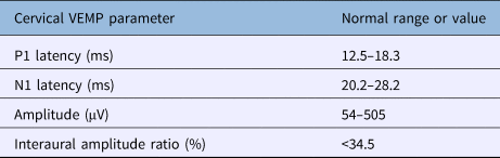
As discussed by Nguyen et al.,Reference Nguyen, Welgampola and Carey5 it is necessary to develop and validate an appropriate normative database for cervical vestibular-evoked myogenic potentials. We compared normative data obtained at several other centres with results from 80 normal ears collected in our laboratory (Table 1).
Our cervical vestibular-evoked myogenic potential norms

VEMP = vestibular-evoked myogenic potential
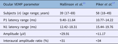
In order to develop our ocular vestibular-evoked myogenic potential database, we again incorporated normative data from a number of different centres. We then collected data from a control group of 20 normal people aged 20–70 years in order to validate these data on our own system. (This exercise did not require ethics approval, as it was deemed to be a quality assurance review by our hospital and university ethics board.) These data were very close to the normative database utilised by other centres (Table 2).Reference Mallinson, Kuijpers and Longridge11–Reference Nguyen, Welgampola and Carey16
Ocular vestibular-evoked myogenic potential normative data from different centres

VEMP = vestibular-evoked myogenic potential; NR = not reported
In collecting our normative data, ocular vestibular-evoked myogenic potential responses were obtained in 39 of 40 normal ears. One 28-year-old individual had unilaterally absent ocular vestibular-evoked myogenic potentials and ipsilateral high tone sensorineural hearing loss. A pre-existing ear disorder was assumed, and the measurement from this ear was not included in our normative data. Data on the remaining 39 ears comprised our normative database.
Discussion
Our ocular vestibular-evoked myogenic potential normative data are comparable to normative data utilised in different centres, with the exception of one outlier study by Piker et al.Reference Piker, Jacobson, McCaslin and Hood17 (Table 3). Their data showed a relative offset compared to all other reported normative data. Their study utilised older equipment, the Nicolet Viking™ evoked potential system, which allows latency adjustments to compensate for stimulus artifact removal, transit times through transducer tubing and so on. On this piece of equipment, the latency is adjustable and needs to be set correctly. The ER3A insert phones with tubing (as used in Piker and colleagues’ studyReference Piker, Jacobson, McCaslin and Hood17) have a longer latency than ER5A inserts with no tubing, and we speculate that compensation for this may not have been taken into account. This correction factor, if pre-programmed, may have been overridden, which could explain the outlying results.
Comparison of our ocular vestibular-evoked myogenic potential norms with those of Piker et al.

VEMP = vestibular-evoked myogenic potential
The present paper describes a simple technique for undertaking cervical and ocular vestibular-evoked myogenic potential testing in a single position. This can be done simultaneously if sufficient channels are available (simultaneous recording was first described in 2009 by Chou et al.Reference Chou, Wang and Young18). This has the benefit of minimising the sound stimulus to the cochlea. When assessing the otoliths using cervical and ocular vestibular-evoked myogenic potentials, we assume the existence of pathology when results are greater than two standard deviations outside of the normal range. Vestibular-evoked myogenic potential testing allows assessment of the macula of the saccule and utricle that were previously not measurable.
The velocity of slow phase nystagmus is routinely measured during video-oculography. Parameters of excessively slow or fast eye velocities are recorded, and although they are clinically significant, they are rarely abnormal. Vestibular-evoked myogenic potential testing provides us with many different parameters that are frequently abnormal. Assessors are able to analyse N1 and P1 wave latencies, wave response amplitude and interaural amplitude difference. For cervical vestibular-evoked myogenic potentials, the interaural latency difference, as described by Beyea and Zeitouni,Reference Beyea and Zeitouni19 can also be used to demonstrate disease in patients whose results would otherwise be reported as normal.
In vestibular-evoked myogenic potential testing, all parameters assessed are reported as ‘abnormal’ if they are outside two standard deviations of our normative dataset. (Tables 1 and 2 show our normative cervical and ocular vestibular-evoked myogenic potential data respectively.) Nevertheless, when using two standard deviations from the norm for each parameter, it is important to remember that statistically 1 in every 20 results is expected to be abnormal and is not due to disease.
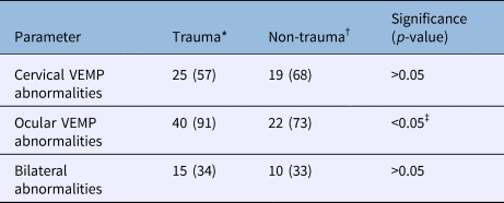
Regarding our current understanding of utricular and saccular function, it is not known what causes these waveforms. If it is assumed that the parameters measured (P1, N1, response amplitude and interaural amplitude difference) are independent of each other, a single result outside two standard deviations from the norm is expected in every fifth patient. Any rate of observed abnormalities above this is considered to be clinically significant, suggesting that a disease process (or trauma) has caused damage to the end organ to produce abnormalities. Table 4 (summarising data analysed in our companion paperReference Longridge and Mallinson20) shows that the rate of abnormalities in both groups of patients was significantly higher than this.
Abnormality rates of our trauma and non-trauma patients

Data represent numbers and percentages of patients. *n = 44; †n = 30. ‡Statistically significant result. VEMP = vestibular-evoked myogenic potential
If the cause of the waveforms is interrelated and interdependent, due to the vestibular insult, then it does not matter if one abnormality is detected or if multiple parameters are abnormal. It is an abnormal test. If the waveform parameters are independent of each other, then more than one abnormal result indicates more otolithic damage. Prospective clinical evaluation of symptoms and findings is needed to determine if there is any correlation between the number and type of abnormalities.
• Vestibular-evoked myogenic potentials are poorly understood, but provide a good measure of vestibular function
• Vestibular-evoked myogenic potentials provide information over and above that provided by standard vestibular (i.e. caloric) assessment
• It is important to try and standardise the assessment in order to increase its reproducibility
• This paper describes a new method suggested to increase vestibular-evoked myogenic potential testing efficiency
• It was possible to carry out accurate assessments on patients who had suffered traumatic vestibular injury
The importance of an air-conducted tone burst stimulus for ocular vestibular-evoked myogenic potentials is that a response is reliably obtained and, if absent, may indicate pathology. Using the described technique, it is rare not to be able to obtain an ocular vestibular-evoked myogenic potential response using a sound stimulus. In older people, this rate is less than 5 per cent, and in the young, this rate is near zero. In this small group with no response bilaterally, no conclusion can be drawn about the significance of the absent response. The increased specificity from using a sound stimulus significantly outweighs the disadvantage of failing to obtain any response in a small percentage of patients.
Conclusion
This paper describes a new cervical and ocular vestibular-evoked myogenic potential testing technique in which the patient adopts a single position that is easily maintained for both assessments. This reduces inconsistency and tiredness, as neck discomfort can prevent assessment completion. Measurement of latencies during vestibular-evoked myogenic potential testing results in the detection of a much higher rate of abnormalities than using an ‘amplitude-only’ comparison technique. Response amplitude can also decrease with age, making assessment of amplitudes less reliable than for latencies.
With our ocular vestibular-evoked myogenic potential technique, application of a hand to prevent slight head elevation in order to ease the uncomfortable task of eye elevation is an important aspect of our assessment. The technique we employ for recording cervical vestibular-evoked myogenic potentials also utilises a natural position and movement (Figure 1b); flexing the sternocleidomastoid by pushing down on a firm surface with the chin is a reasonably natural action. This technique also allows direct comparison of each side during cervical vestibular-evoked myogenic potential testing, minimising asymmetrical responses due to neck pain, particularly relevant in trauma patients.
Vestibular-evoked myogenic potentials allow at least a preliminary investigation of the otolith organs. With our present level of knowledge, the otolithic system abnormalities indicated by measured abnormalities outside the normal range are unknown, but by standardising techniques, careful clinical analysis should allow development of understanding in this field.
Competing interests
None declared